P22077 (P-22077; P 22077) is a potent, cell-permeable and selective inhibitor of ubiquitin-specific protease USP7 (ubiquitin-specific protease 7) with potential antitumor activity. It inhibits USP7 with an EC50 of 8.6 μM, and also inhibits the closely related USP47. P22077 potently induces apoptosis in NB cells with an intact USP7-HDM2-p53 axis but not in NB cells with mutant p53 or without human homolog of MDM2 (HDM2) expression. P22077 also significantly augmented the cytotoxic effects of doxorubicin (Dox) and etoposide (VP-16) in NB cells with an intact USP7-HDM2-p53 axis. Moreover, P22077 was found to be able to sensitize chemoresistant LA-N-6 NB cells to chemotherapy.
Physicochemical Properties
| Molecular Formula | C12H7F2NO3S2 | |
| Molecular Weight | 315.32 | |
| Exact Mass | 314.984 | |
| Elemental Analysis | C, 45.71; H, 2.24; F, 12.05; N, 4.44; O, 15.22; S, 20.34 | |
| CAS # | 1247819-59-5 | |
| Related CAS # |
|
|
| PubChem CID | 46931953 | |
| Appearance | Light yellow to yellow solid powder | |
| LogP | 4.811 | |
| Hydrogen Bond Donor Count | 0 | |
| Hydrogen Bond Acceptor Count | 7 | |
| Rotatable Bond Count | 3 | |
| Heavy Atom Count | 20 | |
| Complexity | 393 | |
| Defined Atom Stereocenter Count | 0 | |
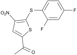
| SMILES | CC(C1=CC([N+]([O-])=O)=C(SC2=CC=C(F)C=C2F)S1)=O |
|
| InChi Key | RMAMGGNACJHXHO-UHFFFAOYSA-N | |
| InChi Code | InChI=1S/C12H7F2NO3S2/c1-6(16)11-5-9(15(17)18)12(20-11)19-10-3-2-7(13)4-8(10)14/h2-5H,1H3 | |
| Chemical Name | 1-(5-((2,4-difluorophenyl)thio)-4-nitrothiophen-2-yl)ethanone | |
| Synonyms | P22077; P-22077; 1247819-59-5; 1-[5-(2,4-Difluoro-phenylsulfanyl)-4-nitro-thiophen-2-yl]-ethanone; CHEMBL2159498; 1-(5-((2,4-Difluorophenyl)thio)-4-nitrothiophen-2-yl)ethanone; 1-(5-(2,4-difluorophenylthio)-4-nitrothiophen-2-yl)ethanone; 1-(5-((2,4-difluorophenyl)thio)-4-nitrothiophen-2-yl)ethan-1-one; P 22077. | |
| HS Tariff Code | 2934.99.9001 | |
| Storage |
Powder-20°C 3 years 4°C 2 years In solvent -80°C 6 months -20°C 1 month |
|
| Shipping Condition | Room temperature (This product is stable at ambient temperature for a few days during ordinary shipping and time spent in Customs) |
Biological Activity
| Targets |
USP7(EC50=8.01 μM);USP47(EC50=8.74 μM) P22077 specifically targets ubiquitin-specific protease 7 (USP7) (IC50 = 0.6 μM for USP7 deubiquitinating activity; Ki = 0.3 μM) [1] P22077 shows no significant inhibition of other DUBs (USP1, USP2, USP5, USP14, UCH-L1: IC50 > 50 μM) [1] |
| ln Vitro |
P 22077 has EC50 values of 8.01 μM and 8.74 μM, respectively, making it an inhibitor of DUB USP47 and USP7. A much smaller subset of DUBs is inhibited by P 22077 (15-45 μM). P 22077 at 25 μM inhibits DUBs in HEK293T cells[1].Neuroblastoma (NB) cells, such as IMR-32, NGP, CHLA-255, and SH-SY5Y cells, have significantly reduced cell viability when exposed to P 22077 (0–20 μM), but not NB-19 and SK-N-AS cells. P 22077 (10 μM) causes NB cells that express HDM2 and p53 wild-type cells to undergo apoptosis and increase p53 activity. P 22077 (5 μM) increases the cytotoxic effect of VP-16 and Dox on NB cells as well as the p53-mediated apoptosis that is induced by Dox and VP-16[2]. In recombinant USP7 enzyme assays, P22077 inhibited USP7-mediated deubiquitination with an IC50 of 0.6 μM and Ki of 0.3 μM, acting as a reversible competitive inhibitor. It exhibited high selectivity over other DUB family members [1] - In a panel of human neuroblastoma cell lines (SH-SY5Y, SK-N-SH, IMR-32, BE(2)-C), P22077 exhibited potent antiproliferative activity with IC50 values ranging from 1.2 to 3.5 μM. After 72 hours of treatment, the 2 μM concentration reduced cell viability by 55-75% across different cell lines [2] - In SH-SY5Y neuroblastoma cells, P22077 (1.5 μM) induced p53 stabilization (3.2-fold increase in protein levels vs. control) and MDM2 accumulation (2.8-fold vs. control) after 24 hours. It also upregulated p21 (3.5-fold) and Bax (2.6-fold), and downregulated Bcl-2 (0.4-fold vs. control) [2] - In SK-N-SH cells, P22077 (2 μM) induced apoptosis, with Annexin V-positive cells increasing from 3% (control) to 36% after 48 hours. It activated caspase-3/7 (3.1-fold vs. control) and caspase-9 (2.7-fold vs. control), and promoted PARP cleavage (3.3-fold vs. control) [2] - In IMR-32 cells, P22077 (2.5 μM) inhibited colony formation by 72% compared to control, indicating long-term antiproliferative effects [2] |
| ln Vivo |
In a xenograft mouse model with IMR-32-derived tumors, P22077 (15 mg/kg, i.p. for 21 days) demonstrates strong antitumor activities. P 22077 also shows antitumor effects in mice with tumors derived from SH-SY5Y and NGP after treatment at 10 mg/kg for 14 days and 20 mg/kg for 12 days, respectively[2]. P22077 significantly inhibits NB tumor growth in vivo [2] Next, researchers tested whether USP7 inhibition by P22077 could inhibit NB tumor growth in vivo. Researchers utilized an orthotopic NB mouse model by surgical injection of IMR-32 cells with luciferase expression into the left renal capsule of nude mice. Two weeks after injection, tumor signals were detected by bioluminescent imaging. Mice bearing tumors were randomly divided into two groups and treated with dimethyl sulfoxide (DMSO) (control) or P22077. P22077 was administered alone at 15 mg/kg daily for 3 weeks. Treatment with P22077 significantly inhibited tumor growth when compared with control (Figures 6a and b). Researchers observed similar results using other two NB cell lines, SH-SY5Y and NGP, in the orthotopic NB mouse models (Figures 6c–f). Of note, there were no obvious health problems or weight loss of mice in either the control or treatment group during the study (Supplementary Figure S4). The results demonstrate that P22077 is a potent antitumor drug for treating NB with an intact USP7-HDM2-p53 axis in the mouse model. In nude mice bearing SH-SY5Y neuroblastoma xenografts, intraperitoneal administration of P22077 (10 mg/kg, once daily for 21 days) significantly inhibited tumor growth. Tumor volume was reduced by 68% compared to vehicle-treated mice, and tumor weight decreased by 65% [2] - In the same xenograft model, P22077 (10 mg/kg) treatment led to stabilization of p53 (2.9-fold vs. vehicle) and MDM2 (2.5-fold vs. vehicle) proteins, upregulation of p21 (3.1-fold vs. vehicle), and activation of caspase-3 (cleaved caspase-3 levels increased by 2.8-fold) in tumor tissues [2] |
| Enzyme Assay |
Recombinant full length catalytic cores for SENP2, JOSD2, USP5, USP2, DEN1, PLpro, and USP7 are produced. Escherichia coli expresses amino terminal His6 tagged USP4, USP8, USP28, UCH-L1, UCH-L3, UCH-L5, and MMP13. Sf9 cells express USP15, USP20, and USP47 that are N-terminally His6 tagged. By using chromatography, all of the recombinant proteins are purified. A variety of substrates are prepared, including free catalytically active PLA2, SUMO3-PLA2 (SUMO3-CHOP), ISG15-PLA2 (ISG15-CHOP), NEDD8-PLA2 (NEDD8-CHOP), Ub-EKL (Ub-CHOP2), and amino terminal tagged His6 Ub-PLA2 (Ub-CHOP)[1]. USP7 deubiquitinating activity assay: Purified recombinant human USP7 was incubated with ubiquitin-AMC (fluorogenic substrate) and P22077 (0.01-10 μM) in assay buffer (50 mM Tris-HCl, pH 7.5, 150 mM NaCl, 1 mM DTT) at 37°C for 60 minutes. Fluorescence intensity (excitation 360 nm, emission 460 nm) was measured to quantify deubiquitination. IC50 values were calculated from dose-response inhibition curves, and Ki was derived using the Cheng-Prusoff equation [1] - DUB selectivity assay: Recombinant USP1, USP2, USP5, USP14, and UCH-L1 were incubated with their respective fluorogenic substrates and P22077 (0.1-100 μM) under optimal reaction conditions. Deubiquitinating activity was quantified to evaluate cross-reactivity [1] |
| Cell Assay |
Propidium iodide staining assay [2] Cells were exposed to different concentrations of P22077, Dox, VP-16, or DMSO for 24 h. Cells were trypsinized, resuspended in RPMI 1640 medium, centrifuged at 400 × g for 5 min at 4 °C. Cells were resuspended and washed with cold PBS twice. Finally, non-fixed cells were resuspended in 1 × binding buffer at a concentration of 1 × 10⁶ cells per ml. Five microliters of propidium iodide (PI) staining solution was added to each tube containing 100 μl of non-fixed suspended cells and incubated with cells for 15 min at RT. The samples were then analyzed by flow cytometry within 1 h after the addition of 400 μl of 1 × binding buffer. PI-positive cells were considered as apoptotic cells, which are permeable to PI owing to the loss of membrane integrity. Unstained cells were used as the negative control and untreated cells were used as a control to treated cells. Cytotoxic effect of P22077 on NB cell proliferation [2] Cells with or without luciferase expression were seeded in 48-well or 6-well plates at appropriate concentrations. After incubation for 24 h, cells were treated with 0, 10, or 20 μM of P22077 for 24 h at 37 °C. Cells were observed and photographed either by adding D-luciferin into cells followed by bioluminescence imaging or by the optical microscope. Cell viability assay [2] Cell viability assays were assessed using the Cell Counting Kit-8 (CCK-8, WST-8[2-(2-methoxy-4-nitrophenyl)-3-(4-nitrophenyl)-5-(2,4-disulfophenyl)-2 H-tetrazolium, monosodium salt]) following the manufacturer's instructions. Cells were seeded in 96-well flat-bottomed plates at the density of 1 × 10⁴ per well. After 24 h of incubation at 37 °C, increasing concentrations of P22077, Dox, VP-16, or their combinations were added to the wells. Twenty-four hours later, 10 μl of CCK-8 was added into each well and after 1 h of incubation, the absorbance was measure at 450 nm using the microplate reader. Each experiment was performed in replicates of six. Background reading of media only was used to normalize the results. The Cell Counting Kit-8 (CCK-8, WST-8[2-(2-methoxy-4-nitrophenyl)-3-(4-nitrophenyl)-5-(2,4-disulfophenyl)-2 H-tetrazolium, monosodium salt]) is used to evaluate cell viability assays. In 96-well plates with a flat bottom, cells are seeded at a density of 1 × 10^4 per well. Increasing concentrations of P22077, Dox, VP-16, or their combinations are added to the wells following a 24-hour incubation period at 37°C. After adding 10 μL of CCK-8 to each well, wait 24 hours. After an hour of incubation, use the microplate reader to measure the absorbance at 450 nm. Every experiment is run in six replicates. Only media background reading is utilized to normalize the outcomes. Antiproliferation assay: Neuroblastoma cell lines (SH-SY5Y, SK-N-SH, IMR-32, BE(2)-C) were seeded in 96-well plates at 3×10³ cells/well and cultured for 24 hours. P22077 was added at concentrations of 0.1-20 μM, and cells were incubated for 72 hours. Cell viability was assessed by MTT assay, and IC50 values were derived [2] - Protein stabilization assay: SH-SY5Y cells were seeded in 6-well plates at 2×10⁵ cells/well and treated with P22077 (1.5 μM) for 24 hours. Cells were lysed, and p53, MDM2, p21, Bax, and Bcl-2 levels were analyzed by Western blot using specific antibodies [2] - Apoptosis assay: SK-N-SH cells were treated with P22077 (2 μM) for 48 hours. Annexin V-FITC/PI staining was performed for flow cytometric analysis of apoptotic cells. Caspase-3/7 and caspase-9 activities were measured by luminescent assays, and PARP cleavage was detected by Western blot [2] - Colony formation assay: IMR-32 cells were seeded in 6-well plates at 500 cells/well and treated with P22077 (2.5 μM) or vehicle. After 14 days of culture, colonies were stained with crystal violet, and the number of colonies was counted to calculate inhibition rate [2] |
| Animal Protocol |
The assay makes use of the orthotopic Neuroblastoma (NB) mouse model. In brief, 5-week-old female NCR nude mice have their left renal capsule surgically injected with 1.5 × 10^6 human IMR-32, SH-SY5Y, or NGP cells expressing luciferase. After allowing the IMR-32, SH-SY5Y, and NGP-derived xenografts to grow for about two to three weeks, mice are randomized into two groups: one for control and the other for P22077 treatment. There are three or six mice per group. DMSO or P22077 is administered intraperitoneally (i.p.) to animals once a day for a duration of 12, 14, or 21 days. All of the mice are killed when the experiments are over. The right side control kidneys are removed, weighed, and photographed along with any tumors. Effect ofP22077 on NB growth in an orthotopic mouse model [2] The orthotopic NB mouse model was established as previously described. Briefly, 1.5 × 10⁶ human IMR-32, SH-SY5Y, or NGP cells with luciferase expression were surgically injected into the left renal capsule of 5-week-old female NCR nude mice. IMR-32, SH-SY5Y, and NGP-derived xenografts were allowed to grow for ∼2–3 weeks before randomizing the mice into a control group and a P22077 treatment group. Each group consisted of three or six mice. Animals were treated with DMSO or P22077 by intraperitoneal (i.p.) injection every day for 12, 14, or 21 days. At the end of the experiments, all mice were killed. Tumors and the right side control kidneys were resected, weighed, and photographed. All mice were housed in a pathogen-free environment and handled in strict accordance to institutional protocol. Nude mice (SH-SY5Y xenograft model): 6-8 weeks old nude mice were subcutaneously inoculated with SH-SY5Y neuroblastoma cells (5×10⁶ cells/mouse). When tumors reached a volume of ~100 mm³, mice were randomly divided into vehicle and P22077 groups. P22077 was dissolved in DMSO and diluted with saline (final DMSO concentration ≤5%) and administered intraperitoneally at 10 mg/kg once daily for 21 days. Vehicle-treated mice received DMSO/saline mixture. Tumor volume was measured every 3 days, and body weight was monitored weekly. At the end of the study, tumors were excised for Western blot analysis [2] |
| Toxicity/Toxicokinetics |
In the in vivo xenograft study, P22077 (10 mg/kg, ip, once daily for 21 days) did not cause significant body weight loss (≤6% change vs. baseline) or overt toxicity in nude mice [2] - In vitro, P22077 showed reduced toxicity to normal human fibroblasts (IC50 > 25 μM), indicating a therapeutic window between cancer cells and normal cells [2] - No significant changes in liver function (ALT, AST) or renal function (creatinine, BUN) were observed in P22077-treated mice compared to vehicle controls [2] - Plasma protein binding rate of P22077 is 92-94% in mice (in vitro plasma binding assay) [2] |
| References |
[1].Activity-based chemical proteomics accelerates inhibitor development for deubiquitylating enzymes. Chem Biol. 2011 Nov 23;18(11):1401-12. [2].USP7 inhibitor P22077 inhibits neuroblastoma growth via inducing p53-mediated apoptosis. Cell Death Dis. 2013 Oct 17;4:e867. |
| Additional Infomation |
Converting lead compounds into drug candidates is a crucial step in drug development, requiring early assessment of potency, selectivity, and off-target effects. We have utilized activity-based chemical proteomics to determine the potency and selectivity of deubiquitylating enzyme (DUB) inhibitors in cell culture models. Importantly, we characterized the small molecule PR-619 as a broad-range DUB inhibitor, and P22077 as a USP7 inhibitor with potential for further development as a chemotherapeutic agent in cancer therapy. A striking accumulation of polyubiquitylated proteins was observed after both selective and general inhibition of cellular DUB activity without direct impairment of proteasomal proteolysis. The repertoire of ubiquitylated substrates was analyzed by tandem mass spectrometry, identifying distinct subsets for general or specific inhibition of DUBs. This enabled identification of previously unknown functional links between USP7 and enzymes involved in DNA repair. [1] Neuroblastoma (NB) is a common pediatric cancer and contributes to more than 15% of all pediatric cancer-related deaths. Unlike adult tumors, recurrent somatic mutations in NB, such as tumor protein 53 (p53) mutations, occur with relative paucity. In addition, p53 downstream function is intact in NB cells with wild-type p53, suggesting that reactivation of p53 may be a viable therapeutic strategy for NB treatment. Herein, we report that the ubiquitin-specific protease 7 (USP7) inhibitor, P22077, potently induces apoptosis in NB cells with an intact USP7-HDM2-p53 axis but not in NB cells with mutant p53 or without human homolog of MDM2 (HDM2) expression. In this study, we found that P22077 stabilized p53 by inducing HDM2 protein degradation in NB cells. P22077 also significantly augmented the cytotoxic effects of doxorubicin (Dox) and etoposide (VP-16) in NB cells with an intact USP7-HDM2-p53 axis. Moreover, P22077 was found to be able to sensitize chemoresistant LA-N-6 NB cells to chemotherapy. In an in vivo orthotopic NB mouse model, P22077 significantly inhibited the xenograft growth of three NB cell lines. Database analysis of NB patients shows that high expression of USP7 significantly predicts poor outcomes. Together, our data strongly suggest that targeting USP7 is a novel concept in the treatment of NB. USP7-specific inhibitors like P22077 may serve not only as a stand-alone therapy but also as an effective adjunct to current chemotherapeutic regimens for treating NB with an intact USP7-HDM2-p53 axis. [2] In summary, a small molecule, P22077 inhibits the function of USP7 resulting in p53 reactivation in NB cells. Our preclinical studies provide the rationale for the development of de-ubiquitinase-based therapies for NB and specifically demonstrate the promise of therapeutics targeting USP7 to improve the outcome of NB patients. NB patients with an intact USP7-HDM2-p53 axis may benefit from P22077 treatment either as single antitumor drug or as an effective adjunct to current chemotherapeutic regimens[2]. P22077 is a potent, selective, reversible inhibitor of USP7, a deubiquitinating enzyme that regulates the stability of p53-MDM2 pathway proteins [1][2] - Its mechanism of action involves binding to the active site of USP7, inhibiting its deubiquitinating activity, leading to stabilization of p53 and MDM2, upregulation of pro-apoptotic proteins (Bax, p21), downregulation of anti-apoptotic protein Bcl-2, and induction of cancer cell apoptosis [2] - P22077 exhibits significant in vitro and in vivo antitumor activity against neuroblastoma, supporting USP7 as a potential therapeutic target for neuroblastoma treatment [2] - The compound is used as a tool compound for studying USP7 function in p53 signaling and cancer biology, particularly in tumors with wild-type p53 [1][2] |
Solubility Data
| Solubility (In Vitro) |
DMSO : 50~63 mg/mL ( 158.57~199.79 mM) Ethanol : ~1 mg/mL |
| Solubility (In Vivo) |
Solubility in Formulation 1: ≥ 2.5 mg/mL (7.93 mM) (saturation unknown) in 10% DMSO + 90% Corn Oil (add these co-solvents sequentially from left to right, and one by one), clear solution. For example, if 1 mL of working solution is to be prepared, you can add 100 μL of 25.0 mg/mL clear DMSO stock solution to 900 μL of corn oil and mix evenly. Solubility in Formulation 2: 2% DMSO+30% PEG300+2% Tween80+66% ddH2O: 3mg/ml Solubility in Formulation 3: 5 mg/mL (15.86 mM) in 50% PEG300 50% Saline (add these co-solvents sequentially from left to right, and one by one), suspension solution; with ultrasonication (<60°C). Preparation of saline: Dissolve 0.9 g of sodium chloride in 100 mL ddH₂ O to obtain a clear solution. (Please use freshly prepared in vivo formulations for optimal results.) |
| Preparing Stock Solutions | 1 mg | 5 mg | 10 mg | |
| 1 mM | 3.1714 mL | 15.8569 mL | 31.7138 mL | |
| 5 mM | 0.6343 mL | 3.1714 mL | 6.3428 mL | |
| 10 mM | 0.3171 mL | 1.5857 mL | 3.1714 mL |