Physicochemical Properties
| Molecular Formula | C23H28O12 |
| Molecular Weight | 496.461228370667 |
| Exact Mass | 496.158 |
| CAS # | 39011-91-1 |
| PubChem CID | 21631105 |
| Appearance | White to off-white solid powder |
| Density | 1.7±0.1 g/cm3 |
| Boiling Point | 737.1±60.0 °C at 760 mmHg |
| Flash Point | 254.6±26.4 °C |
| Vapour Pressure | 0.0±2.5 mmHg at 25°C |
| Index of Refraction | 1.708 |
| LogP | -0.17 |
| Hydrogen Bond Donor Count | 6 |
| Hydrogen Bond Acceptor Count | 12 |
| Rotatable Bond Count | 7 |
| Heavy Atom Count | 35 |
| Complexity | 879 |
| Defined Atom Stereocenter Count | 11 |
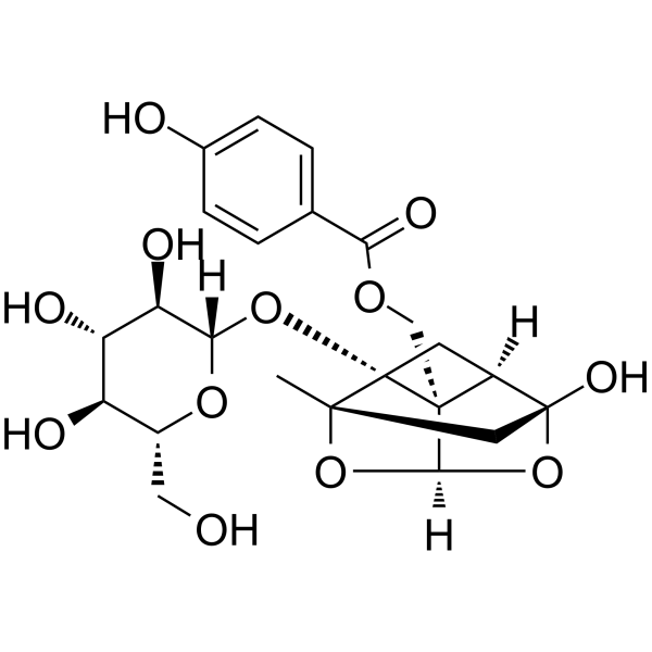
| SMILES | C[C@]12C[C@@]3([C@@H]4C[C@]1([C@@]4([C@H](O2)O3)COC(=O)C5=CC=C(C=C5)O)O[C@H]6[C@@H]([C@H]([C@@H]([C@H](O6)CO)O)O)O)O |
| InChi Key | FCHVXNVDFYXLIL-WRJNSLSBSA-N |
| InChi Code | InChI=1S/C23H28O12/c1-20-8-22(30)13-6-23(20,33-18-16(28)15(27)14(26)12(7-24)32-18)21(13,19(34-20)35-22)9-31-17(29)10-2-4-11(25)5-3-10/h2-5,12-16,18-19,24-28,30H,6-9H2,1H3/t12-,13-,14-,15+,16-,18+,19-,20+,21+,22-,23+/m1/s1 |
| Chemical Name | [(1R,2S,3R,5R,6R,8S)-6-hydroxy-8-methyl-3-[(2S,3R,4S,5S,6R)-3,4,5-trihydroxy-6-(hydroxymethyl)oxan-2-yl]oxy-9,10-dioxatetracyclo[4.3.1.02,5.03,8]decan-2-yl]methyl 4-hydroxybenzoate |
| HS Tariff Code | 2934.99.9001 |
| Storage |
Powder-20°C 3 years 4°C 2 years In solvent -80°C 6 months -20°C 1 month |
| Shipping Condition | Room temperature (This product is stable at ambient temperature for a few days during ordinary shipping and time spent in Customs) |
Biological Activity
| Targets |
The action target of Oxypaeoniflorin is the Sirt1/Foxo1 signaling pathway (in myocardial ischemia/reperfusion model) [1] |
| ln Vitro |
Hypoxia/reoxygenation (H/R)-induced reduction in cell viability and increase in H9c2 cell apoptosis were dramatically counteracted by oxypaeoniflorin (OPA; 0.1-10 µM; 8 hours). By triggering the Sirt1 (Silent Information Regulator 2-related Enzyme 1)/Foxo1 (Forkhead Transcription Factor FKHR) signaling pathway in cardiac tissue and H9c2 cells, oxypaeoniflorin prevents cell death [1]. By modifying the Toll-like receptor (TLR), extracellular signal-related kinase (ERK), and p38 mitogen-activated protein (MAP) kinase signaling pathways in LPS-stimulated RAW264.7 cells impact, oxypaeoniflorin (0-30 μM) decreases inflammation[2]. Protective effect on H9c2 cardiomyocytes under oxygen-glucose deprivation/reoxygenation (OGD/R): H9c2 cells were divided into control group, OGD/R model group, Oxypaeoniflorin 10 μM group, Oxypaeoniflorin 20 μM group, Oxypaeoniflorin 40 μM group. OGD was induced by culturing cells in glucose-free medium under 1% O2 for 4 hours, followed by reoxygenation in normal medium for 24 hours; Oxypaeoniflorin was added at the start of reoxygenation. MTT assay showed Oxypaeoniflorin increased cell viability (40 μM group: 78% vs. 45% in model group). Flow cytometry (Annexin V-FITC/PI staining) showed Oxypaeoniflorin reduced apoptosis rate (40 μM reduced apoptosis by ~52%). DCFH-DA staining showed Oxypaeoniflorin decreased reactive oxygen species (ROS) production (40 μM reduced ROS by ~48%). Western blot showed Oxypaeoniflorin upregulated Sirt1 and Foxo1 protein expression, and downregulated cleaved caspase-3 [1] |
| ln Vivo |
Treatment with oxypaeoniflorin (OPA; 10–40 mg/kg; intragastric injection; daily; for 30 days) can enhance fractional shortening (FS) and ejection fraction (EF) markers and dramatically lessen impairment to heart function. Myocardial infarction-related variables, such as cardiac troponin T (cTnT), cardiac troponin I (cTnI), and creatine kinase (CK-MB), can be greatly reduced by oxypaeoniflorin [1]. Protective effect in rat myocardial ischemia/reperfusion (I/R) model: Male Sprague-Dawley rats were subjected to left anterior descending coronary artery ligation for 30 minutes (ischemia) followed by 2 hours of reperfusion (I/R). Rats were divided into sham group, I/R model group, Oxypaeoniflorin 20 mg/kg group, Oxypaeoniflorin 40 mg/kg group. Oxypaeoniflorin was administered via tail vein injection 30 minutes before ischemia. TTC staining showed Oxypaeoniflorin reduced myocardial infarction volume (40 mg/kg group: 22% vs. 48% in model group). Echocardiography showed Oxypaeoniflorin improved cardiac function (40 mg/kg increased left ventricular ejection fraction (LVEF) to 62% vs. 38% in model group). Biochemical analysis showed Oxypaeoniflorin decreased serum creatine kinase-MB (CK-MB) and lactate dehydrogenase (LDH) levels (40 mg/kg reduced CK-MB by ~60%), and Western blot showed increased Sirt1/Foxo1 expression in myocardial tissue [1] |
| Enzyme Assay |
Sirt1 activity assay in H9c2 cells: H9c2 cells were treated with OGD/R and Oxypaeoniflorin (40 μM) for 24 hours. Total protein was extracted from cells, and Sirt1 activity was measured using a Sirt1 activity kit. The reaction system included cell lysate, Sirt1 substrate peptide, NAD+, and assay buffer; the mixture was incubated at 37°C for 1 hour. The fluorescent product was detected using a microplate reader (excitation: 340 nm, emission: 460 nm). Results showed Oxypaeoniflorin increased Sirt1 activity by ~45% compared to the OGD/R model group [1] |
| Cell Assay |
H9c2 cardiomyocyte OGD/R experiment: H9c2 cells were cultured in DMEM containing 10% fetal bovine serum and 1% penicillin-streptomycin. Cells were seeded in 96-well plates (for MTT assay, 5×10^3 cells/well) or 6-well plates (for Western blot/ROS/apoptosis, 5×10^5 cells/well) and incubated overnight. For OGD induction, medium was replaced with glucose-free DMEM, and cells were placed in a hypoxic chamber (1% O2, 5% CO2, 94% N2) for 4 hours. Reoxygenation was performed by replacing with normal DMEM and returning to a normoxic incubator; Oxypaeoniflorin (10-40 μM) was added at this step. After 24-hour reoxygenation: MTT reagent was added for 4 hours to measure cell viability at 570 nm; cells were stained with Annexin V-FITC/PI for flow cytometry to detect apoptosis; DCFH-DA was added for 30 minutes to measure ROS by flow cytometry; cells were lysed for Western blot to detect Sirt1, Foxo1, and cleaved caspase-3 [1] |
| Animal Protocol |
Animal/Disease Models: Myocardial ischemia/reperfusion (MI/R) injured C57BL/6 male mice (6-8 weeks old, 20-25 g) [1] Doses: 10 mg/kg, 20 mg/kg, 40 mg/kg Route of Administration: intragastric (po) (po)administration; daily; for 30 days. Experimental Results: Dramatically diminished damage to cardiac function, and improved ejection fraction (EF) and fractional shortening (FS) indicators. 1. Rat myocardial I/R model protocol: Male Sprague-Dawley rats (250-300 g) were anesthetized with pentobarbital sodium. A left thoracic incision was made to expose the heart, and the left anterior descending coronary artery was ligated with a 6-0 silk suture (ischemia). After 30 minutes, the suture was loosened for 2 hours of reperfusion (I/R). Sham group only had chest incision without ligation. Rats were divided into 4 groups (n=8/group): sham, I/R model, Oxypaeoniflorin 20 mg/kg, Oxypaeoniflorin 40 mg/kg. Oxypaeoniflorin was dissolved in normal saline and administered via tail vein injection (0.1 mL/10 g body weight) 30 minutes before ischemia. After reperfusion, rats were sacrificed; hearts were collected for TTC staining (infarction volume) and Western blot; blood was collected for serum CK-MB/LDH detection [1] 2. Rat pharmacokinetic experiment (oral administration of herbal extracts): Male Sprague-Dawley rats (200-220 g) were divided into two groups: Radix Paeoniae Rubra (RPR) extract group and Radix Paeoniae Alba (RPA) extract group (n=6/group). RPR and RPA extracts were dissolved in 0.5% carboxymethyl cellulose sodium solution; the dose was adjusted to contain 25 mg/kg Oxypaeoniflorin (calculated based on pre-determined Oxypaeoniflorin content in extracts). Rats were fasted for 12 hours before oral gavage of extracts. Blood samples (0.5 mL) were collected from the orbital vein at 0, 0.25, 0.5, 1, 2, 4, 6, 8, 12, 24 hours after administration. Plasma was separated by centrifugation (3000×g for 10 minutes) and stored at -80°C for subsequent HPLC analysis of Oxypaeoniflorin concentration [2] |
| ADME/Pharmacokinetics |
Pharmacokinetic parameters in rats after oral administration of RPR/RPA extracts: Plasma Oxypaeoniflorin concentration was detected by HPLC, and parameters were calculated using DAS 2.0 software. In RPR extract group: Cmax (peak plasma concentration) = 128.6 ± 15.3 ng/mL, Tmax (time to reach Cmax) = 1.2 ± 0.3 h, AUC0-t (area under concentration-time curve) = 586.4 ± 62.5 ng·h/mL, t1/2 (elimination half-life) = 4.8 ± 0.6 h. In RPA extract group: Cmax = 92.3 ± 11.7 ng/mL, Tmax = 1.5 ± 0.4 h, AUC0-t = 412.7 ± 58.3 ng·h/mL, t1/2 = 5.1 ± 0.7 h.[2] |
| References |
[1]. Oxypaeoniflorin improves myocardial ischemia/reperfusion injury by activating the Sirt1/Foxo1 signaling pathway. Acta Biochim Pol. 2020 Jun 18;67(2):239-245. [2]. Pharmacokinetic properties of paeoniflorin, albiflorin and oxypaeoniflorin after oral gavage of extracts of Radix Paeoniae Rubra and Radix Paeoniae Alba in rats. J Ethnopharmacol. 2010 Jul 20;130(2):407-13. |
| Additional Infomation |
Oxypaeoniflorin is a monoterpene glycoside with formula C23H28O12, isolated from several species of Paeoniae. It has a role as a plant metabolite. It is a cyclic acetal, a lactol, a bridged compound, a beta-D-glucoside, a 4-hydroxybenzoate ester and a monoterpene glycoside. Oxypaeoniflorin has been reported in Paeonia lactiflora and Phellodendron amurense with data available. See also: Paeonia lactiflora root (part of); Paeonia veitchii root (part of); Paeonia X suffruticosa root bark (part of). 1. Oxypaeoniflorin is a monoterpene glycoside isolated from the roots of Paeonia lactiflora Pall. (Radix Paeoniae), including Radix Paeoniae Rubra (RPR, with red roots) and Radix Paeoniae Alba (RPA, with white roots), which are widely used in traditional Chinese medicine for blood circulation promotion and anti-inflammatory effects [1][2] 2. Mechanism in myocardial protection: Oxypaeoniflorin alleviates myocardial ischemia/reperfusion injury by activating the Sirt1/Foxo1 signaling pathway, which enhances antioxidant capacity, inhibits cardiomyocyte apoptosis, and preserves cardiac function [1] 3. Pharmacokinetic characteristics: Oxypaeoniflorin shows better oral absorption (higher Cmax and AUC0-t) when administered in RPR extract compared to RPA extract, possibly due to differences in other components in the two extracts that affect its solubility or membrane permeability [2] |
Solubility Data
| Solubility (In Vitro) | DMSO : ~100 mg/mL (~201.43 mM) |
| Solubility (In Vivo) |
Solubility in Formulation 1: ≥ 2.5 mg/mL (5.04 mM) (saturation unknown) in 10% DMSO + 40% PEG300 + 5% Tween80 + 45% Saline (add these co-solvents sequentially from left to right, and one by one), clear solution. For example, if 1 mL of working solution is to be prepared, you can add 100 μL of 25.0 mg/mL clear DMSO stock solution to 400 μL PEG300 and mix evenly; then add 50 μL Tween-80 to the above solution and mix evenly; then add 450 μL normal saline to adjust the volume to 1 mL. Preparation of saline: Dissolve 0.9 g of sodium chloride in 100 mL ddH₂ O to obtain a clear solution. Solubility in Formulation 2: ≥ 2.5 mg/mL (5.04 mM) (saturation unknown) in 10% DMSO + 90% (20% SBE-β-CD in Saline) (add these co-solvents sequentially from left to right, and one by one), clear solution. For example, if 1 mL of working solution is to be prepared, you can add 100 μL of 25.0 mg/mL clear DMSO stock solution to 900 μL of 20% SBE-β-CD physiological saline solution and mix evenly. Preparation of 20% SBE-β-CD in Saline (4°C,1 week): Dissolve 2 g SBE-β-CD in 10 mL saline to obtain a clear solution. Solubility in Formulation 3: ≥ 2.5 mg/mL (5.04 mM) (saturation unknown) in 10% DMSO + 90% Corn Oil (add these co-solvents sequentially from left to right, and one by one), clear solution. For example, if 1 mL of working solution is to be prepared, you can add 100 μL of 25.0 mg/mL clear DMSO stock solution to 900 μL of corn oil and mix evenly. (Please use freshly prepared in vivo formulations for optimal results.) |
| Preparing Stock Solutions | 1 mg | 5 mg | 10 mg | |
| 1 mM | 2.0143 mL | 10.0713 mL | 20.1426 mL | |
| 5 mM | 0.4029 mL | 2.0143 mL | 4.0285 mL | |
| 10 mM | 0.2014 mL | 1.0071 mL | 2.0143 mL |