Optovin is a potent and reversible photoactivated TRPA1 activator that is able to activate human TRPA1 via structure dependent photochemical reactions. In dorsal root ganglia (DRG) sensory neurons isolated from mice, optovin significantly activated 33% (35/105) of DRG neurons, which was not occured in the dark. In HEK293T cells transfected with the human TRPA1, optovin activated cells. Optovin activated TRPA1 channels through a photoactivated intermediate that reacted with redox-sensitive cysteine residues in the channel.
Physicochemical Properties
| Molecular Formula | C15H13N3OS2 | |
| Molecular Weight | 315.41 | |
| Exact Mass | 315.049 | |
| CAS # | 348575-88-2 | |
| Related CAS # |
|
|
| PubChem CID | 1365101 | |
| Appearance | Light yellow to orange solid powder | |
| Density | 1.4±0.1 g/cm3 | |
| Index of Refraction | 1.718 | |
| LogP | 2.04 | |
| Hydrogen Bond Donor Count | 1 | |
| Hydrogen Bond Acceptor Count | 4 | |
| Rotatable Bond Count | 2 | |
| Heavy Atom Count | 21 | |
| Complexity | 480 | |
| Defined Atom Stereocenter Count | 0 | |
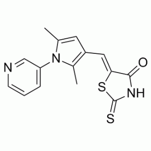
| SMILES | CC1=CC(=C(N1C2=CN=CC=C2)C)/C=C/3\C(=O)NC(=S)S3 |
|
| InChi Key | YISGMOZSGOGYOU-NTUHNPAUSA-N | |
| InChi Code | InChI=1S/C15H13N3OS2/c1-9-6-11(7-13-14(19)17-15(20)21-13)10(2)18(9)12-4-3-5-16-8-12/h3-8H,1-2H3,(H,17,19,20)/b13-7+ | |
| Chemical Name | (5E)-5-[(2,5-dimethyl-1-pyridin-3-ylpyrrol-3-yl)methylidene]-2-sulfanylidene-1,3-thiazolidin-4-one | |
| Synonyms |
|
|
| HS Tariff Code | 2934.99.9001 | |
| Storage |
Powder-20°C 3 years 4°C 2 years In solvent -80°C 6 months -20°C 1 month |
|
| Shipping Condition | Room temperature (This product is stable at ambient temperature for a few days during ordinary shipping and time spent in Customs) |
Biological Activity
| Targets |
Optovin targets transient receptor potential ankyrin 1 (TRPA1) channels (photochemically activated, EC50 = 1.3 μM for TRPA1-mediated current activation in DRG neurons under 470 nm light irradiation) [1] |
| ln Vitro |
With an EC50 of 2 μM, optovin (1-10 μM) enhances the motor activity of zebrafish embryos by more than 40 standard deviations above the mean of the control at photic stimuli[1]. Dorsal root ganglia (DRG) neurons are substantially activated by optovin (100 μM; 120 s) in 33% (35 /105) of cases[1]. Optovin (10 μM; 1 min) stimulates hTrpA1-transfected HEK293T cells[1]. Optovin (1 μM) induced TRPA1-dependent inward currents in mouse dorsal root ganglion (DRG) neurons under 470 nm light (1 mW/mm²) irradiation, with current amplitude reaching 320 ± 45 pA (n=25) [1] Optovin (0.5–5 μM) exhibited concentration-dependent activation of TRPA1 channels: EC50 = 1.3 μM, maximal activation at 3 μM (current amplitude 410 ± 52 pA) [1] Optovin (2 μM, 470 nm light) failed to activate TRPA1 knockout (TRPA1⁻/⁻) DRG neurons, confirming TRPA1 specificity; no significant activation of other TRP channels (TRPV1, TRPV4, TRPM8) at concentrations up to 10 μM [1] Optovin (1.5 μM) induced Ca²⁺ influx in HEK293T cells transfected with human TRPA1 (hTRPA1) under light irradiation, with Fluo-4 fluorescence intensity increasing by 3.8-fold; no Ca²⁺ response in non-transfected cells [1] Optovin showed no cytotoxicity to DRG neurons or HEK293T cells at concentrations up to 20 μM (cell viability >95% after 24 hours) [1] |
| ln Vivo |
In vivo, optovin (1-10 μM) induces motor activation that is reliant on light[1]. In adult mice, optovin (15 mM/20 μL); applied externally) causes nociceptive behaviors[1]. The appearance, touch response, heart rate, fin motions, morphology, and percentage survival of larval and adult zebrafish are unaffected by optovin (10 μM; 96 h)[1]. Optovin (50 μg, intraplantar injection) combined with 470 nm light irradiation (5 mW/mm², 10 seconds) induced acute nociceptive responses in wild-type mice: paw withdrawal latency reduced from 12.5 ± 1.8 seconds to 3.2 ± 0.6 seconds, and paw licking time increased to 45 ± 8 seconds (n=10) [1] Optovin (50 μg, intraplantar) failed to induce nociceptive responses in TRPA1⁻/⁻ mice under the same light conditions, confirming TRPA1 dependence [1] Optovin (100 μg, intraperitoneal injection) + 470 nm light irradiation (10 mW/mm², 20 seconds) activated visceral sensory neurons in mice, inducing abdominal constriction responses (12 ± 3 constrictions in 5 minutes) [1] |
| Enzyme Assay |
TRPA1 channel current assay: Mouse DRG neurons were cultured and whole-cell patch-clamp recordings were performed; Optovin (0.1–10 μM) was applied to the bath solution, and cells were irradiated with 470 nm light (1 mW/mm²) for 5 seconds; current responses were recorded at a holding potential of -60 mV, and EC50 was calculated via dose-response curves [1] TRP channel selectivity assay: HEK293T cells transfected with hTRPA1, hTRPV1, hTRPV4, or hTRPM8 were subjected to patch-clamp recordings; Optovin (5 μM) + 470 nm light irradiation was applied, and current amplitudes were compared to assess selectivity [1] |
| Cell Assay |
DRG neuron Ca²⁺ imaging assay: Mouse DRG neurons were isolated and loaded with Fluo-4 AM dye; Optovin (0.5–5 μM) was added, and cells were irradiated with 470 nm light (0.5 mW/mm²); fluorescence intensity was monitored by confocal microscopy to quantify Ca²⁺ influx [1] hTRPA1-transfected HEK293T cell activation assay: HEK293T cells transfected with hTRPA1 plasmid were seeded in 96-well plates; Optovin (0.1–10 μM) was added, followed by 470 nm light irradiation (1 mW/mm²) for 10 seconds; Ca²⁺ fluorescence was measured by microplate reader to assess TRPA1 activation [1] Cytotoxicity assay: DRG neurons and HEK293T cells were seeded in 96-well plates (5×10³ cells/well) and treated with Optovin (0.1–20 μM) + light irradiation (1 mW/mm², 10 seconds); cell viability was assessed by MTT assay (absorbance at 570 nm) after 24 hours [1] |
| Animal Protocol |
Dissolved in DMSO; 50 μM (Zebrafish); 15 mM (mice); Treated on the ear (mice); Embryos are incubated in Optovin solution (Zebrafish) Zebrafish and mice Nociceptive response model: Wild-type and TRPA1⁻/⁻ mice (6–8 weeks old) were randomly divided into groups; Optovin (50 μg in 20 μL saline) was injected intraplantarly into the hind paw; 10 minutes later, the injected paw was irradiated with 470 nm light (5 mW/mm²) for 10 seconds; paw withdrawal latency (hot plate test) and paw licking time were recorded within 5 minutes [1] Visceral sensory activation model: Wild-type mice were administered Optovin (100 μg in 100 μL saline) via intraperitoneal injection; 15 minutes later, the abdominal region was irradiated with 470 nm light (10 mW/mm²) for 20 seconds; abdominal constriction responses were counted over 5 minutes [1] |
| References |
[1]. Photochemical activation of TRPA1 channels in neurons and animals. Nat Chem Biol. 2013 Apr;9(4):257-63. |
| Additional Infomation |
Optovin is a member of the class of thiazolidinone that is rhodanine in which the two methylene hydrogens have been replaced by a 2,5-dimethyl-1-(pyridin-3-yl)-1H-pyrrol-3-yl]methylidene group. A TRPA1 ligand that can be reversibly photoactivated. It has a role as a TRPA1 channel agonist. It is a member of pyrroles, a member of pyridines, an olefinic compound and a thiazolidinone. It is functionally related to a rhodanine. Optovin is a small-molecule photoswitch that photochemically activates TRPA1 channels upon 470 nm blue light irradiation [1] Its activation mechanism involves light-induced conformational changes of the compound, which enhances binding to TRPA1 and triggers channel gating, leading to cation influx (Ca²⁺, Na⁺) [1] Optovin exhibits high specificity for TRPA1, with no cross-activation of other TRP channels at therapeutic concentrations [1] It enables spatiotemporally precise activation of TRPA1-expressing sensory neurons in vitro and in vivo, serving as a tool for studying TRPA1 function in pain signaling, neurophysiology, and sensory processing [1] The compound is inactive in the dark, requiring light irradiation (450–490 nm) for TRPA1 activation, allowing optical control of neural activity [1] |
Solubility Data
| Solubility (In Vitro) |
|
|||
| Solubility (In Vivo) |
Solubility in Formulation 1: ≥ 2.5 mg/mL (7.93 mM) (saturation unknown) in 10% DMSO + 40% PEG300 +5% Tween-80 + 45% Saline (add these co-solvents sequentially from left to right, and one by one), clear solution. For example, if 1 mL of working solution is to be prepared, you can add 100 μL of 25.0 mg/mL clear DMSO stock solution to 400 μL PEG300 and mix evenly; then add 50 μL Tween-80 + to the above solution and mix evenly; then add 450 μL normal saline to adjust the volume to 1 mL. Preparation of saline: Dissolve 0.9 g of sodium chloride in 100 mL ddH₂ O to obtain a clear solution. (Please use freshly prepared in vivo formulations for optimal results.) |
| Preparing Stock Solutions | 1 mg | 5 mg | 10 mg | |
| 1 mM | 3.1705 mL | 15.8524 mL | 31.7048 mL | |
| 5 mM | 0.6341 mL | 3.1705 mL | 6.3410 mL | |
| 10 mM | 0.3170 mL | 1.5852 mL | 3.1705 mL |