Oleandomycin (PA 775) is a classic macrolide antibiotic produced by strains of Streptomyces that demonstrates antimicrobial activity similar to penicillin and erythromycin. It is structurally closely related to Erythromycin and consists of a macrocyclic lactone ring of 14 carbon atoms with one sugar, oleandrose, and one amino sugar, desoxamine, attached to the lactone ring. The mechanism of its biosynthesis and development of resistance to its antibiotic activity have been studied in order to understand the reactive enzymes in these processes.
Physicochemical Properties
| Molecular Formula | C35H61NO12.C22H24N2O8.O4P-3 |
| Molecular Weight | 1227.2643 |
| Exact Mass | 687.419 |
| Elemental Analysis | C, 61.11; H, 8.94; N, 2.04; O, 27.91 |
| CAS # | 3922-90-5 |
| PubChem CID | 5284598 |
| Appearance | White, amorphous powder |
| Density | 1.2±0.1 g/cm3 |
| Boiling Point | 802.6±65.0 °C at 760 mmHg |
| Flash Point | 439.2±34.3 °C |
| Vapour Pressure | 0.0±6.4 mmHg at 25°C |
| Index of Refraction | 1.533 |
| LogP | 1.23 |
| Hydrogen Bond Donor Count | 3 |
| Hydrogen Bond Acceptor Count | 13 |
| Rotatable Bond Count | 6 |
| Heavy Atom Count | 48 |
| Complexity | 1090 |
| Defined Atom Stereocenter Count | 18 |
| SMILES | O=C1[C@H](C)[C@@H](O)[C@@H](C)[C@@H](C)OC([C@H](C)[C@@H](O[C@]2([H])O[C@@H](C)[C@H](O)[C@@H](OC)C2)[C@H](C)[C@@H](O[C@@]3([H])[C@H](O)[C@@H](N(C)C)C[C@@H](C)O3)[C@@H](C)C[C@@]14CO4)=O |
| InChi Key | RZPAKFUAFGMUPI-DDSISPHDSA-N |
| InChi Code | InChI=1S/C35H61NO12/c1-16-14-35(15-43-35)32(40)19(4)27(37)18(3)22(7)46-33(41)21(6)31(47-26-13-25(42-11)28(38)23(8)45-26)20(5)30(16)48-34-29(39)24(36(9)10)12-17(2)44-34/h16-31,34,37-39H,12-15H2,1-11H3/t16-,17-,18-,19+,20+,21+,22+,23-,24+,25-,26+,27-,28-,29+,30-,31-,34+,35-/m0/s1 |
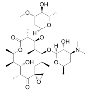
| Chemical Name | Oleandomycin(3S,5R,6S,7R,8R,11R,12S,13R,14S,15S)-14-(((2R,3R,4R,6S)-4-(dimethylamino)-3-hydroxy-6-methyltetrahydro-2H-pyran-2-yl)oxy)-6-hydroxy-12-(((2S,4S,5S,6S)-5-hydroxy-4-methoxy-6-methyltetrahydro-2H-pyran-2-yl)oxy)-5,7,8,11,13,15-hexamethyl-1,9-dioxaspiro[2.13]hexadecane-4,10-dione |
| Synonyms | PA 775; PA-775; PA775 |
| HS Tariff Code | 2934.99.03.00 |
| Storage |
Powder-20°C 3 years 4°C 2 years In solvent -80°C 6 months -20°C 1 month |
| Shipping Condition | Room temperature (This product is stable at ambient temperature for a few days during ordinary shipping and time spent in Customs) |
Biological Activity
| Targets | Macrolide |
| ln Vitro | Cell extracts of Streptomyces antibioticus, an oleandomycin producer, can inactivate oleandomycin in the presence of UDP-glucose. The inactivation can be detected through the loss of biological activity or by alteration in the chromatographic mobility of the antibiotic. This enzyme activity also inactivates other macrolides (rosaramicin, methymycin, and lankamycin) which contain a free 2'-OH group in a monosaccharide linked to the lactone ring (with the exception of erythromycin), but not those which contain a disaccharide (tylosin, spiramycin, carbomycin, josamycin, niddamycin, and relomycin). The culture supernatant contains another enzyme activity capable of reactivating the glycosylated oleandomycin and regenerating the biological activity through the release of a glucose molecule. It is proposed that these two enzyme activities could be an integral part of the oleandomycin biosynthetic pathway.[1] |
| Cell Assay |
In vitro inactivation of oleandomycin. Oleandomycin (6.6 ,ug/ml; 8.3 ,M) was incubated with 50 1.l of dialyzed cell extract in the presence of 1 mM UDP-glucose at 30°C in a final volume of 150 [lI. Both at zero time and after 6 h of incubation, samples were removed and boiled for 2 min. After cooling, the residual antibiotic activity was determined by bioassay against M. luteus . In some experiments, UDP-D-[6-3H]glucose (20 ,uCi/ml; 2 mM) was used instead ofthe nonlabelled compound, and the products of the reaction mixture were analyzed by paper chromatography .[1] In vitro reactivation of oleandomycin. For assays of reactivation of oleandomycin, tritiated inactive oleandomycin was used'as a substrate after preparation as follows. An inactivation assay was carried out using UDP-[3H]glucose as a cofactor and the labelled inactive and modified oleandomycin was eluted from the paper strips after paper chromatography by immersion in absolute methanol and incubation at room temperature overnight with gentle shaking. The eluate was lyophilized and dissolved in a small volume of methanol; then aliquots (approximately 8,000 cpm) were incubated with 40 to 50 pI of the ammonium sulfate precipitates from the culture supernatant (without addition of any cofactor) in a final volume of 100 ,u at 30°C for 2 h. The reaction products were analyzed by counting the radioactivity in 1-cm strips after paper chromatography.[1] Thin-layer chromatography. Samples of the inactivation assays (50 pul; 15 pug of oleandomycin) were applied to silica gel F254 plates (Merck) and subjected to ascending chromatography with methanol as the solvent. The plates were developed and then stained with a mixture of anisaldehyde concentrated sulfuric acid-ethanol (1:1:9) and heating at 100°C for 2 min (16). Parallel samples (without staining) were assayed by bioautography against M. luteus.[1] |
| ADME/Pharmacokinetics |
Absorption, Distribution and Excretion Macrolides become widely distributed in tissues, and concentrations are about the same as in plasma, or even higher in some instances. They actually accumulate within many cells, including macrophages, in which they may be > or = 20 times the plasma concentration. This accumulation accounts in part for the long dosing interval that characterizes some macrolides (eg, tilmicosin). ... Macrolides tend to concentrate in the spleen, liver, kidneys, and particularly the lungs. They enter pleural and ascitic fluids but not the CSF (only 2-13% of plasma concentration unless the meninges are inflamed). They concentrate in the bile and milk. Up to 75% of the dose is bound to plasma proteins, and they bind to alpha1-acid glycoproteins rather than to albumin. /Macrolides/ Macrolides are readily absorbed from the GI tract if not inactivated by gastric acid. ... Plasma levels peak within 1-2 hours in most cases, although absorption patterns may be erratic due to the presence of food and may depend on the salt or ester used. Absorption from the ruminoreticulum is usualy delayed and is unreliable. /Macrolides/ Macrolide antibiotics and their metabolites are excreted mainly in the bile (> 60%) and often undergo enterohepatic cycling. Urinary clearance may be slow and variable (often <10%) but my represent a more significant route of elimination after parenteral administration. The concentration of macrolides in milk often is several times greater than in plasma, especially in mastitis. /Macrolide/ The pharmacokinetics of oleandomycin (OLD) after intravenous and oral administration, both alone and after intramuscular pretreatment with metamizole or dexamethasone, were studied in healthy dogs. After intravenous injection of oleandomycin alone (10 mg/kg as bolus), the elimination half-life (t 1/2 beta, volume of distribution (Vd, area), body clearance (ClB) and area under the concentration time curve (AUC) were 1.60 hr, 1.11 L/kg. 7.36 (ml/kg)/min and 21.66 ug hr/ml, respectively. There were no statistically significant differences following pretreatment with metamizole or dexamethasone. After oral administration of oleandomycin alone, the t 1/2 beta, maximum plasma concentrations (Cmax), time of Cmax (tmax), mean absorption time and absolute bioavailability (Fabs) were 1.6 hr, 5.34 ug/ml, 1.5 hr, 1.34 hr and 84.29%, respectively. Pretreatment with metamizole caused a significantly decreased value for Cmax (2.93 ug/ml) but the mean absorption time value (2.23 hr) was significantly increased. Statistically significant changes in the pharmacokinetic parameters of oleandomycin following oral administration were also observed as a result of pretreatment with dexamethasone. The Cmax was increased (8.24 ug/ml) and the tmax (0.5 hr) and mean absorption time (0.45 hr) were lower. Knowledge of the disposition of macrolides in a single animal species has been insufficient for the prediction of the pharmacokinetics of macrolides in humans. To better understand the species differences in the pharmacokinetics of macrolide antibiotics, the disposition of erythromycin, oleandomycin, and tylosin in several mammalian species was examined. Generally, the serum concentration versus time profiles of these drugs after intravenous administration were described by two-compartment kinetic models and were similar within each species. These drugs were rapidly cleared, resulting in terminal half-lives of less than 2 h. Comparison of their pharmacokinetics showed greater variation in antibiotic disposition among animal species than noted for the differences within a species. When the pharmacokinetic data was fitted to an allometric model, the logarithms of volume of distribution, clearance, and half-life were linearly related to the logarithms of body weight. From these relationships, the human pharmacokinetics of erythromycin and oleandomycin were extrapolated and found to approximate observed human pharmacokinetics. Metabolism / Metabolites Metabolic inactivation of the macrolides is usually extensive, but the relative proportion depends on the route of administration and th particular antibiotic. ... /Macrolides/ Biological Half-Life The plasma half-lives of macrolides usually are 1-3 hr, ... /Macrolides/ |
| Toxicity/Toxicokinetics |
Interactions Macrolide antibiotics probably should not be used with chloramphenicol or the lincosamides because they may compete for the same 50 S ribosomal binding site, although the in vivo significance of this potential interaction is unclear. Activity of macrolides is depressed in acidic environments. Macrolide preparations for parenteral administration are incompatible with many other pharmaceutical preparations. ... /Macrolides/ ... /The ability to/ Induce phase III migrating myoelectric complex (MMC) activity in dogs and increase smooth muscle contractility ... is shared to varying extents by some macrolide antibiotics, including oleandomycin ... /Motilin: macrolides and erythromycin/ The combination effect of tetracycline (TC) and oleandomycin (OM) on acute infection of mice with four strains of Staphylococcus aureus including TC or OM resistant ones was examined by the quantitative determination of protective potencies of single and combined drugs. The grade of synergism was expressed by the synergistic ratio (SR), a ratio of experimentally determined potency of the combined drug over a hypothetical potency in which additive effect of the both drugs is assumed. With 3 out of the 4 strains of S. aureus synergism between TC and OM or triacetyloleandomycin (TAO) was demonstrated by the determination of the 50% effective dose and by statistical examination of the SR. The grade of synergistic protection by these drugs varied with the strains infected and it did not depend upon the sensitivity to antibiotics or grade of synergism in vitro. There was no synergistic enhancement of acute toxic action in the combined administration of TC and OM to mice. |
| References | [1]. Role of glycosylation and deglycosylation in biosynthesis of and resistance to oleandomycin in the producer organism, Streptomyces antibioticus. J Bacteriol. 1992 Jan;174(1):161-5. |
| Additional Infomation |
Oleandomycin is a macrolide antibiotic, though it is less effective than erythromycin. It is synthesized from strains of Streptomyces antibioticus. Oleandomycin is a macrolide antibiotic similar to erythromycin with antimicrobial activity. Oleandomycin targets and reversibly binds to the 50S subunit of bacterial ribosomes. This prevents translocation of peptidyl tRNA leading to an inhibition of protein synthesis. Antibiotic macrolide produced by Streptomyces antibioticus. See also: Oleandomycin (annotation moved to). Mechanism of Action The antimicrobial mechanism seems to be the same for all of the macrolides. They interfere with protein synthesis by reversibly binding to the 50 S subunit of the ribosome. They appear to bind at the donor site, thus preventing the translocation necessary to keep the peptide chain growing. The effect is essentially confined to rapidly dividing bacteria and mycoplasmas. Macrolides are regarded as being bacteriostatic, ... . Macrolides are significantly more active at higher pH ranges (7.8-8). /Macrolides/ Therapeutic Uses Mesh Heading: anti-bacterial agents MEDICATION: ... Used orally or intravenously to treat pyoderma, sepsis, meningitis, surgical and abdominal infections, respiratory tract and urinary tract infections, and other infections caused by Staphylococci, Streptococci, Corynebacterium, Neisseria, and Mycoplasma. MEDICATION (VET): Macrolide antibiotic. THERAP CAT: Antibacterial For more Therapeutic Uses (Complete) data for OLEANDOMYCIN (6 total), please visit the HSDB record page. Drug Warnings VET: Toxicity and side effects are uncommon for most macrolides ... , although pain and swelling may develop at injection sites. Hypersensitivity reactions have occasionally been seen. ... Horses are sensitive to macrolide-induced CI disturbances that can be serious and even fatal. ... |
Solubility Data
| Solubility (In Vitro) |
DMSO : ~100 mg/mL (~145.38 mM) H2O : ≥ 12.5 mg/mL (~18.17 mM) |
| Solubility (In Vivo) |
Solubility in Formulation 1: ≥ 2.5 mg/mL (3.63 mM) (saturation unknown) in 10% DMSO + 40% PEG300 + 5% Tween80 + 45% Saline (add these co-solvents sequentially from left to right, and one by one), clear solution. For example, if 1 mL of working solution is to be prepared, you can add 100 μL of 25.0 mg/mL clear DMSO stock solution to 400 μL PEG300 and mix evenly; then add 50 μL Tween-80 to the above solution and mix evenly; then add 450 μL normal saline to adjust the volume to 1 mL. Preparation of saline: Dissolve 0.9 g of sodium chloride in 100 mL ddH₂ O to obtain a clear solution. Solubility in Formulation 2: ≥ 2.5 mg/mL (3.63 mM) (saturation unknown) in 10% DMSO + 90% (20% SBE-β-CD in Saline) (add these co-solvents sequentially from left to right, and one by one), clear solution. For example, if 1 mL of working solution is to be prepared, you can add 100 μL of 25.0 mg/mL clear DMSO stock solution to 900 μL of 20% SBE-β-CD physiological saline solution and mix evenly. Preparation of 20% SBE-β-CD in Saline (4°C,1 week): Dissolve 2 g SBE-β-CD in 10 mL saline to obtain a clear solution. Solubility in Formulation 3: ≥ 2.5 mg/mL (3.63 mM) (saturation unknown) in 10% DMSO + 90% Corn Oil (add these co-solvents sequentially from left to right, and one by one), clear solution. For example, if 1 mL of working solution is to be prepared, you can add 100 μL of 25.0 mg/mL clear DMSO stock solution to 900 μL of corn oil and mix evenly. Solubility in Formulation 4: 10% DMSO+40% PEG300+5% Tween-80+45% Saline: ≥ 2.5 mg/mL (3.63 mM) (Please use freshly prepared in vivo formulations for optimal results.) |
| Preparing Stock Solutions | 1 mg | 5 mg | 10 mg | |
| 1 mM | 0.8148 mL | 4.0741 mL | 8.1482 mL | |
| 5 mM | 0.1630 mL | 0.8148 mL | 1.6296 mL | |
| 10 mM | 0.0815 mL | 0.4074 mL | 0.8148 mL |