Physicochemical Properties
| Molecular Formula | C47H80O17 |
| Molecular Weight | 917.13 |
| Exact Mass | 916.539 |
| CAS # | 155683-00-4 |
| PubChem CID | 91973814 |
| Appearance | White to off-white solid powder |
| Density | 1.4±0.1 g/cm3 |
| Boiling Point | 997.8±65.0 °C at 760 mmHg |
| Flash Point | 557.2±34.3 °C |
| Vapour Pressure | 0.0±0.6 mmHg at 25°C |
| Index of Refraction | 1.607 |
| LogP | 8.22 |
| Hydrogen Bond Donor Count | 11 |
| Hydrogen Bond Acceptor Count | 17 |
| Rotatable Bond Count | 12 |
| Heavy Atom Count | 64 |
| Complexity | 1630 |
| Defined Atom Stereocenter Count | 24 |
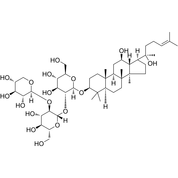
| SMILES | CC(=CCC[C@](C)([C@H]1CC[C@@]2([C@@H]1[C@@H](C[C@H]3[C@]2(CC[C@@H]4[C@@]3(CC[C@@H](C4(C)C)O[C@H]5[C@@H]([C@H]([C@@H]([C@H](O5)CO)O)O)O[C@H]6[C@@H]([C@H]([C@@H]([C@H](O6)CO)O)O)O[C@H]7[C@@H]([C@H]([C@@H](CO7)O)O)O)C)C)O)C)O)C |
| InChi Key | LLXVPTXOKTYXHU-UGGLCNOCSA-N |
| InChi Code | InChI=1S/C47H80O17/c1-22(2)10-9-14-47(8,58)23-11-16-46(7)31(23)24(50)18-29-44(5)15-13-30(43(3,4)28(44)12-17-45(29,46)6)62-41-38(35(55)33(53)26(19-48)60-41)64-42-39(36(56)34(54)27(20-49)61-42)63-40-37(57)32(52)25(51)21-59-40/h10,23-42,48-58H,9,11-21H2,1-8H3/t23-,24+,25+,26+,27+,28-,29+,30-,31-,32-,33+,34+,35-,36-,37+,38+,39+,40-,41-,42-,44-,45+,46+,47+/m0/s1 |
| Chemical Name | (2S,3R,4S,5R)-2-[(2S,3R,4S,5S,6R)-2-[(2R,3R,4S,5S,6R)-4,5-dihydroxy-2-[[(3S,5R,8R,9R,10R,12R,13R,14R,17S)-12-hydroxy-17-[(2R)-2-hydroxy-6-methylhept-5-en-2-yl]-4,4,8,10,14-pentamethyl-2,3,5,6,7,9,11,12,13,15,16,17-dodecahydro-1H-cyclopenta[a]phenanthren-3-yl]oxy]-6-(hydroxymethyl)oxan-3-yl]oxy-4,5-dihydroxy-6-(hydroxymethyl)oxan-3-yl]oxyoxane-3,4,5-triol |
| HS Tariff Code | 2934.99.9001 |
| Storage |
Powder-20°C 3 years 4°C 2 years In solvent -80°C 6 months -20°C 1 month Note: Please store this product in a sealed and protected environment (e.g. under nitrogen), avoid exposure to moisture. |
| Shipping Condition | Room temperature (This product is stable at ambient temperature for a few days during ordinary shipping and time spent in Customs) |
Biological Activity
| ln Vitro |
In human umbilical vein endothelial cells (HUVECs), Notoginsenoside Ft1 (0.25-2.5 µM) concentration-dependently stimulated tube formation in a Matrigel-based assay. It concentration-dependently increased HUVEC viability, with a minimal effective concentration of 1 µM. Treatment with 10 µM Ft1 time-dependently induced cell cycle progression from G1 to S phase, as evidenced by flow cytometry analysis, confirming its proliferative effect. The compound also concentration-dependently promoted HUVEC migration in a wound healing assay. Ft1 (10 µM) time-dependently increased VEGF secretion into the cell culture supernatant and elevated VEGF mRNA levels. It facilitated the translocation of HIF-1α protein from the cytoplasm to the nucleus in a time-dependent manner and enhanced the binding of HIF-1α to the VEGF promoter, as shown by chromatin immunoprecipitation assay. Western blot analysis demonstrated that Ft1 time-dependently induced phosphorylation (activation) of key proteins in the PI3K/AKT/mTOR/p70 S6 kinase pathway (PI3K, AKT, mTOR, p70 S6 kinase) and the Raf/MEK/ERK pathway (c-Raf, MEK1/2, ERK1/2). Pharmacological inhibition of PI3K (with LY294002 or wortmannin) or MEK1/2 (with PD98059) attenuated Ft1-induced cell proliferation, tube formation, VEGF mRNA expression, VEGF secretion, and HIF-1α nuclear translocation. Knockdown of mTOR using siRNA reduced Ft1-induced tube formation, cell proliferation, VEGF mRNA expression, and HIF-1α nuclear translocation. [1] |
| ln Vivo |
In a Matrigel plug assay in C57BL/6 mice, subcutaneous implants containing Notoginsenoside Ft1 (1, 5, 25 µM per plug) appeared redder than controls after 7 days, indicating functional blood vessel formation. Hemoglobin content in the plugs increased dose-dependently, and immunohistochemical staining for the endothelial marker CD31 revealed a dose-dependent increase in the number and size of blood vessels within the plugs. In a mouse ear wound healing model (BALB/c mice), intraperitoneal administration of Ft1 (0.25, 2.5, 25 mg/kg every other day) for 28 days accelerated wound closure compared to the control group. The wound hole diameter decreased significantly in the Ft1-treated groups in a dose-dependent manner. Additionally, VEGF mRNA expression was elevated in the marginal healing region of the ear tissue from treated mice. [1] |
| Cell Assay |
Cell viability was assessed using a colorimetric assay. HUVECs were seeded in 96-well plates and allowed to attach. After serum reduction, cells were treated with different concentrations of Notoginsenoside Ft1 for 48 hours. A tetrazolium salt reagent was added and converted to formazan by viable cells. The formazan crystals were dissolved, and the optical density was measured at 570 nm with a reference wavelength of 630 nm to quantify cell viability. [1] Cell cycle analysis was performed by flow cytometry. HUVECs were treated with Ft1, collected, and fixed in ethanol. Fixed cells were then stained with propidium iodide and RNase A solution, incubated, and analyzed by flow cytometry to determine the percentage of cells in G0-G1, S, and G2-M phases. [1] For the migration assay, a confluent monolayer of HUVECs grown in gelatin-coated plates was scratched to create a lesion. After washing, cells were incubated with medium containing different concentrations of Ft1. Images of the wound area were captured after 24 hours using an inverted microscope to assess cell migration into the scratched area. [1] The tube formation assay was performed by seeding HUVECs onto Matrigel-coated 96-well plates in medium containing Ft1. After 4 hours of incubation, images were taken using an inverted microscope. Tubes forming intact networks were counted manually in a blinded manner to quantify angiogenic activity. [1] VEGF secretion was quantified using an enzyme-linked immunosorbent assay (ELISA). HUVECs were cultured and treated with Ft1. Cell culture supernatants were collected and added to antibody-coated plates. After incubation and washing steps, a detection system was used, and optical density was measured at 450 nm. VEGF concentration was determined relative to a standard curve. [1] For gene silencing, HUVECs were transfected with mTOR-specific siRNA or a control siRNA using a lipid-based transfection reagent according to the manufacturer's protocol to knock down mTOR expression. [1] Western blot analysis was performed to detect protein expression and phosphorylation. After treatment, cells were lysed, and proteins were separated by SDS-PAGE, transferred to membranes, and probed with specific primary antibodies against target proteins or their phosphorylated forms, followed by incubation with horseradish peroxidase-conjugated secondary antibodies. Signals were detected using an appropriate chemiluminescent substrate. [1] Total RNA was extracted from treated cells using a phenol-based reagent. cDNA was synthesized from the RNA using reverse transcriptase. VEGF and GAPDH mRNA levels were amplified by PCR using specific primers. PCR products were visualized by agarose gel electrophoresis. [1] A chromatin immunoprecipitation assay was used to study protein-DNA interaction. HUVECs were treated, and chromatin was cross-linked and isolated. The chromatin was sheared and immunoprecipitated using an antibody against HIF-1α or a control IgG. The precipitated DNA was purified and analyzed by PCR using primers specific for the VEGF promoter region to assess HIF-1α binding. [1] |
| Animal Protocol |
For the Matrigel plug assay, liquid Matrigel at 4°C was mixed with heparin and Notoginsenoside Ft1 at final concentrations of 1, 5, or 25 µM. A volume of 0.5 ml of this mixture was injected subcutaneously into the abdominal region of C57BL/6 mice. After 7 days, mice were sacrificed, and the Matrigel plugs were excised for analysis of hemoglobin content and immunohistochemistry. [1] For the wound healing study, a full-thickness wound (1.5 mm diameter hole) was created in the center of both ears of BALB/c mice using a punch. Mice received intraperitoneal injections of Ft1 at doses of 0.25, 2.5, or 25 mg/kg every other day. The wound diameter was measured periodically using calipers over 28 days. At the endpoint, mice were sacrificed, and ear tissue from the wound margin was collected for VEGF mRNA analysis. [1] |
| Toxicity/Toxicokinetics | In the 28-day mouse ear wound healing study, there was no significant difference in body weight between mice treated with Notoginsenoside Ft1 (at doses up to 25 mg/kg every other day) and control mice, suggesting no overt systemic toxicity at these doses under the experimental conditions. [1] |
| References |
[1]. Notoginsenoside Ft1 promotes angiogenesis via HIF-1α mediated VEGF secretion and the regulation of PI3K/AKT and Raf/MEK/ERK signaling pathways. Biochem Pharmacol. 2012 Sep 15;84(6):784-92. [2]. Platelet P2Y?? receptors are involved in the haemostatic effect of notoginsenoside Ft1, a saponin isolated from Panax notoginseng. Br J Pharmacol. 2014 Jan;171(1):214-23. [3]. Notoginsenoside Ft1 activates both glucocorticoid and estrogen receptors to induce endothelium-dependent, nitric oxide-mediated relaxations in rat mesenteric arteries. Biochem Pharmacol. 2014 Mar 1;88(1):66-74. [4]. p38 MAPK and ERK1/2 pathways are involved in the pro-apoptotic effect of notoginsenoside Ft1 on human neuroblastoma SH-SY5Y cells. Life Sci. 2014 Jul 17;108(2):63-70. |
| Additional Infomation |
Notoginsenoside Ft1 has been reported in Centella asiatica with data available. Notoginsenoside Ft1 is a saponin isolated from the leaves of Panax notoginseng, a herb traditionally used in East Asia for treating trauma injuries. The study proposes that Ft1 promotes angiogenesis and wound healing by increasing HIF-1α nuclear translocation, which enhances VEGF transcription and secretion. This process is mediated through the activation of both the PI3K/AKT/mTOR/p70 S6 kinase and Raf/MEK/ERK signaling pathways, with mTOR serving as a critical integration point. These findings provide a potential mechanistic explanation for the traditional use of Panax notoginseng in wound management and suggest Ft1 as a candidate for development into an angiogenic therapeutic agent. [1] |
Solubility Data
| Solubility (In Vitro) | DMSO : ~6.67 mg/mL (~7.27 mM) |
| Solubility (In Vivo) |
Solubility in Formulation 1: ≥ 0.67 mg/mL (0.73 mM) (saturation unknown) in 10% DMSO + 40% PEG300 + 5% Tween80 + 45% Saline (add these co-solvents sequentially from left to right, and one by one), clear solution. For example, if 1 mL of working solution is to be prepared, you can add 100 μL of 6.7 mg/mL clear DMSO stock solution to 400 μL PEG300 and mix evenly; then add 50 μL Tween-80 to the above solution and mix evenly; then add 450 μL normal saline to adjust the volume to 1 mL. Preparation of saline: Dissolve 0.9 g of sodium chloride in 100 mL ddH₂ O to obtain a clear solution. Solubility in Formulation 2: ≥ 0.67 mg/mL (0.73 mM) (saturation unknown) in 10% DMSO + 90% (20% SBE-β-CD in Saline) (add these co-solvents sequentially from left to right, and one by one), clear solution. For example, if 1 mL of working solution is to be prepared, you can add 100 μL of 6.7 mg/mL clear DMSO stock solution to 900 μL of 20% SBE-β-CD physiological saline solution and mix evenly. Preparation of 20% SBE-β-CD in Saline (4°C,1 week): Dissolve 2 g SBE-β-CD in 10 mL saline to obtain a clear solution. Solubility in Formulation 3: ≥ 0.67 mg/mL (0.73 mM) (saturation unknown) in 10% DMSO + 90% Corn Oil (add these co-solvents sequentially from left to right, and one by one), clear solution. For example, if 1 mL of working solution is to be prepared, you can add 100 μL of 6.7 mg/mL clear DMSO stock solution to 900 μL of corn oil and mix evenly. (Please use freshly prepared in vivo formulations for optimal results.) |
| Preparing Stock Solutions | 1 mg | 5 mg | 10 mg | |
| 1 mM | 1.0904 mL | 5.4518 mL | 10.9036 mL | |
| 5 mM | 0.2181 mL | 1.0904 mL | 2.1807 mL | |
| 10 mM | 0.1090 mL | 0.5452 mL | 1.0904 mL |