Physicochemical Properties
| Molecular Formula | C18H19NO4 |
| Molecular Weight | 313.3478 |
| Exact Mass | 313.131 |
| CAS # | 23599-69-1 |
| Related CAS # | Norisoboldine hydrochloride;5083-84-1 |
| PubChem CID | 14539911 |
| Appearance | Brown to khaki solid powder |
| Density | 1.3±0.1 g/cm3 |
| Boiling Point | 553.0±50.0 °C at 760 mmHg |
| Flash Point | 288.3±30.1 °C |
| Vapour Pressure | 0.0±1.5 mmHg at 25°C |
| Index of Refraction | 1.643 |
| LogP | 1.43 |
| Hydrogen Bond Donor Count | 3 |
| Hydrogen Bond Acceptor Count | 5 |
| Rotatable Bond Count | 2 |
| Heavy Atom Count | 23 |
| Complexity | 433 |
| Defined Atom Stereocenter Count | 1 |
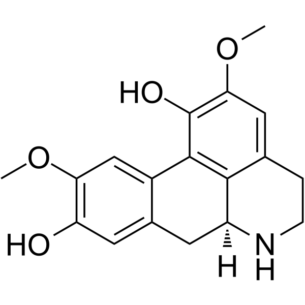
| SMILES | COC1=C(C2=C3[C@H](CC4=CC(=C(C=C42)OC)O)NCCC3=C1)O |
| InChi Key | HORZNQYQXBFWNZ-LBPRGKRZSA-N |
| InChi Code | InChI=1S/C18H19NO4/c1-22-14-8-11-10(6-13(14)20)5-12-16-9(3-4-19-12)7-15(23-2)18(21)17(11)16/h6-8,12,19-21H,3-5H2,1-2H3/t12-/m0/s1 |
| Chemical Name | (6aS)-2,10-dimethoxy-5,6,6a,7-tetrahydro-4H-dibenzo[de,g]quinoline-1,9-diol |
| HS Tariff Code | 2934.99.9001 |
| Storage |
Powder-20°C 3 years 4°C 2 years In solvent -80°C 6 months -20°C 1 month Note: This product requires protection from light (avoid light exposure) during transportation and storage. |
| Shipping Condition | Room temperature (This product is stable at ambient temperature for a few days during ordinary shipping and time spent in Customs) |
Biological Activity
| Targets |
Norisoboldine targets aryl hydrocarbon receptor (AhR) - selective agonist [2] Norisoboldine modulates glycolysis pathway and NAD+/SIRT1/SUV39H1/H3K9me3 signaling pathway [2] |
| ln Vitro |
In a hypoxic microenvironment, norisoboldine (1~30 μM; 0~24 hours; CD4+T cells) activates AhR and dramatically reduces the production of miR-31 mRNA [2]. In hypoxic settings, norisoboldine (30 μM; 0~24 hours; CD4+T cells) slows glycolysis [2]. In hypoxic environments, norisoboldine (1~30 μM; 0~72 hours; Treg cells) stimulates Treg differentiation [2]. The formation of the AhR/ARNT complex, nuclear translocation of AhR, and dissociation of the HSP90/AhR complex are all facilitated by norisoboldine (10, 30 μM). In the absence of miR-31, norisoboldine stimulates the production of Treg cells in hypoxic environments [2]. 1. Treg cell differentiation promotion: - Norisoboldine (10, 20, 40 μM) promotes differentiation of mouse splenic CD4+ T cells into regulatory T cells (Tregs) in a concentration-dependent manner, with Foxp3+ Treg ratio increasing from 5.2% (control) to 12.8% (40 μM) [2] - The effect is abolished by AhR antagonist CH223191, confirming AhR-dependent mechanism [2] 2. Glycolysis inhibition in T cells: - Norisoboldine (20, 40 μM) inhibits glycolysis in activated CD4+ T cells, reducing glucose consumption by 35% and lactate production by 42% at 40 μM [2] - It downregulates protein expression of glycolytic enzymes (HK2, PFK1, LDHA) and mRNA levels by 45-60% (RT-qPCR) [2] 3. Modulation of NAD+/SIRT1/SUV39H1/H3K9me3 pathway: - Norisoboldine (10-40 μM) increases intracellular NAD+ level and SIRT1 activity in CD4+ T cells, with 40 μM group showing 2.3-fold higher NAD+ and 1.8-fold higher SIRT1 activity than control [2] - It decreases SUV39H1 protein expression and H3K9me3 level (Western blot), which are reversed by SIRT1 inhibitor EX527 [2] |
| ln Vivo |
Norisoboldine (10~40 mg/kg; oral; 20 days) considerably reduced the severity of joint swelling and erythema during the experiment [1]. Norisoboldine (40 mg/kg; ig; 10 days) promotes elevated CYP1A1 expression and inhibits Glut1 and HK2 expression in the colon [2]. 1. Therapeutic effect on collagen-induced arthritis (CIA) in mice: - Norisoboldine (50, 100 mg/kg, p.o., once daily for 21 days) reduces CIA severity, with arthritis score decreasing from 8.3 (control) to 3.2 (100 mg/kg) [1] - It inhibits joint inflammation and destruction, as shown by reduced synovial hyperplasia, inflammatory cell infiltration, and cartilage erosion (histopathology) [1] - Serum levels of pro-inflammatory cytokines (TNF-α, IL-6, IL-1β) are reduced by 40-55%, and anti-inflammatory cytokine IL-10 is increased by 2.1-fold (100 mg/kg) [1] 2. Attenuation of DSS-induced colitis in mice: - Norisoboldine (20, 40 mg/kg, p.o., once daily for 7 days) alleviates DSS-induced colitis, with disease activity index (DAI) decreasing from 8.5 (control) to 3.1 (40 mg/kg) [2] - It increases colon length (from 4.2 cm to 6.1 cm) and reduces colonic mucosal damage, ulceration, and inflammatory cell infiltration [2] - Colonic Treg population is increased by 2.5-fold, and pro-inflammatory cytokines (TNF-α, IFN-γ) in colon tissue are reduced by 50-65% (40 mg/kg) [2] - It upregulates colonic SIRT1 expression and downregulates SUV39H1 and H3K9me3 levels, consistent with in vitro findings [2] |
| Enzyme Assay |
1. CD4+ T cell isolation and Treg differentiation assay: - CD4+ T cells are isolated from mouse spleen using magnetic bead sorting [2] - Cells are cultured in RPMI 1640 medium supplemented with anti-CD3, anti-CD28 antibodies, IL-2, and TGF-β1 to induce Treg differentiation [2] - Cells are treated with Norisoboldine (10, 20, 40 μM) with or without AhR antagonist CH223191 for 72 hours [2] - Foxp3+ Treg ratio is analyzed by flow cytometry (Foxp3-PE staining), and Foxp3 mRNA level is detected by RT-qPCR [2] 2. Glycolysis metabolism assay: - Activated CD4+ T cells are treated with Norisoboldine (20, 40 μM) for 48 hours [2] - Glucose consumption and lactate production in culture supernatant are measured using colorimetric kits [2] - HK2, PFK1, LDHA protein expression is detected by Western blot, and their mRNA levels by RT-qPCR [2] 3. Western blot for signaling pathway proteins: - Norisoboldine (10-40 μM)-treated CD4+ T cells are lysed with RIPA buffer containing protease and phosphatase inhibitors [2] - Protein concentrations are determined by BCA assay, and equal amounts of protein are separated by SDS-PAGE [2] - Membranes are probed with antibodies against SIRT1, SUV39H1, H3K9me3, and GAPDH (loading control), followed by secondary antibody incubation [2] - Chemiluminescent signals are detected and quantified using imaging software [2] |
| Cell Assay |
Western Blot analysis[2] Cell Types: CD4+T cells Tested Concentrations: 1~30 μM Incubation Duration: 24 hrs (hours) Experimental Results: Intracellular AhR was activated under hypoxic microenvironment. RT-PCR[2] Cell Types: CD4+T cells Tested Concentrations: 1~30 μM Incubation Duration: 24 hrs (hours) Experimental Results: miR-31 mRNA expression was Dramatically down-regulated. Immunofluorescence[2] Cell Types: CD4+T cells Tested Concentrations: 30 μM Incubation Duration: 24 hrs (hours) Experimental Results: Glycolysis is inhibited during hypoxia. Cell differentiation experiment [2] Cell Types: Treg cell Tested Concentrations: 1~30 μM Incubation Duration: 72 hrs (hours) Experimental Results: Treg cell differentiation was promoted under hypoxic conditions. 1. CD4+ T cell isolation and Treg differentiation assay: - CD4+ T cells are isolated from mouse spleen using magnetic bead sorting [2] - Cells are cultured in RPMI 1640 medium supplemented with anti-CD3, anti-CD28 antibodies, IL-2, and TGF-β1 to induce Treg differentiation [2] - Cells are treated with Norisoboldine (10, 20, 40 μM) with or without AhR antagonist CH223191 for 72 hours [2] - Foxp3+ Treg ratio is analyzed by flow cytometry (Foxp3-PE staining), and Foxp3 mRNA level is detected by RT-qPCR [2] 2. Glycolysis metabolism assay: - Activated CD4+ T cells are treated with Norisoboldine (20, 40 μM) for 48 hours [2] - Glucose consumption and lactate production in culture supernatant are measured using colorimetric kits [2] - HK2, PFK1, LDHA protein expression is detected by Western blot, and their mRNA levels by RT-qPCR [2] 3. Western blot for signaling pathway proteins: - Norisoboldine (10-40 μM)-treated CD4+ T cells are lysed with RIPA buffer containing protease and phosphatase inhibitors [2] - Protein concentrations are determined by BCA assay, and equal amounts of protein are separated by SDS-PAGE [2] - Membranes are probed with antibodies against SIRT1, SUV39H1, H3K9me3, and GAPDH (loading control), followed by secondary antibody incubation [2] - Chemiluminescent signals are detected and quantified using imaging software [2] |
| Animal Protocol |
Animal/Disease Models: Male ICR mice (18-22 grams) Doses: 10~40 mg/kg Route of Administration: Oral Experimental Results:The severity of joint swelling and erythema was Dramatically diminished during the experiment. Animal/Disease Models: Female C57BL/6 mice (18–22 g) Doses: 40 mg/kg Route of Administration: Ig Experimental Results: Induced enhanced CYP1A1 expression and inhibited Glut1 and HK2 expression in the colon. |
| References |
[1]. Therapeutic effect of norisoboldine, an alkaloid isolated from Radix Linderae, on collagen-induced arthritis in mice. Phytomedicine. 2010;17(10):726-731. [2]. Norisoboldine, a natural AhR agonist, promotes Treg differentiation and attenuates colitis via targeting glycolysis and subsequent NAD+/SIRT1/SUV39H1/H3K9me3 signaling pathway. Cell Death Dis. 2018;9(3):258. Published 2018 Feb 15. |
| Additional Infomation |
Norisoboldine has been reported in Stephania cephalantha, Fissistigma oldhamii, and other organisms with data available. - Norisoboldine is a natural alkaloid isolated from the roots of Radix Linderae (Lindera aggregata) [1][2] - It exerts anti-inflammatory effects through dual mechanisms: activating AhR to promote Treg differentiation, and inhibiting glycolysis via NAD+/SIRT1/SUV39H1/H3K9me3 signaling pathway [2] - It shows therapeutic potential for autoimmune diseases, including rheumatoid arthritis and inflammatory bowel disease [1][2] - The anti-arthritic effect is associated with regulation of pro-inflammatory and anti-inflammatory cytokine balance [1] |
Solubility Data
| Solubility (In Vitro) | DMSO : ≥ 62.5 mg/mL (~199.46 mM) |
| Solubility (In Vivo) |
Solubility in Formulation 1: ≥ 2.08 mg/mL (6.64 mM) (saturation unknown) in 10% DMSO + 40% PEG300 + 5% Tween80 + 45% Saline (add these co-solvents sequentially from left to right, and one by one), clear solution. For example, if 1 mL of working solution is to be prepared, you can add 100 μL of 20.8 mg/mL clear DMSO stock solution to 400 μL PEG300 and mix evenly; then add 50 μL Tween-80 to the above solution and mix evenly; then add 450 μL normal saline to adjust the volume to 1 mL. Preparation of saline: Dissolve 0.9 g of sodium chloride in 100 mL ddH₂ O to obtain a clear solution. Solubility in Formulation 2: ≥ 2.08 mg/mL (6.64 mM) (saturation unknown) in 10% DMSO + 90% (20% SBE-β-CD in Saline) (add these co-solvents sequentially from left to right, and one by one), clear solution. For example, if 1 mL of working solution is to be prepared, you can add 100 μL of 20.8 mg/mL clear DMSO stock solution to 900 μL of 20% SBE-β-CD physiological saline solution and mix evenly. Preparation of 20% SBE-β-CD in Saline (4°C,1 week): Dissolve 2 g SBE-β-CD in 10 mL saline to obtain a clear solution. (Please use freshly prepared in vivo formulations for optimal results.) |
| Preparing Stock Solutions | 1 mg | 5 mg | 10 mg | |
| 1 mM | 3.1913 mL | 15.9566 mL | 31.9132 mL | |
| 5 mM | 0.6383 mL | 3.1913 mL | 6.3826 mL | |
| 10 mM | 0.3191 mL | 1.5957 mL | 3.1913 mL |