Physicochemical Properties
| Molecular Formula | C12H7CL2NO3 |
| Molecular Weight | 284.09 |
| Exact Mass | 282.98 |
| CAS # | 1836-75-5 |
| PubChem CID | 15787 |
| Appearance |
Crystalline solid White solid Crystals Yellow crystalline solid Free-flowing solid, dark brown color |
| Density | 1.5±0.1 g/cm3 |
| Boiling Point | 360.6±32.0 °C at 760 mmHg |
| Melting Point | 69-70°C |
| Flash Point | 171.9±25.1 °C |
| Vapour Pressure | 0.0±0.8 mmHg at 25°C |
| Index of Refraction | 1.623 |
| LogP | 4.92 |
| Hydrogen Bond Donor Count | 0 |
| Hydrogen Bond Acceptor Count | 3 |
| Rotatable Bond Count | 2 |
| Heavy Atom Count | 18 |
| Complexity | 290 |
| Defined Atom Stereocenter Count | 0 |
| SMILES | ClC1C([H])=C(C([H])=C([H])C=1OC1C([H])=C([H])C(=C([H])C=1[H])[N+](=O)[O-])Cl |
| InChi Key | XITQUSLLOSKDTB-UHFFFAOYSA-N |
| InChi Code | InChI=1S/C12H7Cl2NO3/c13-8-1-6-12(11(14)7-8)18-10-4-2-9(3-5-10)15(16)17/h1-7H |
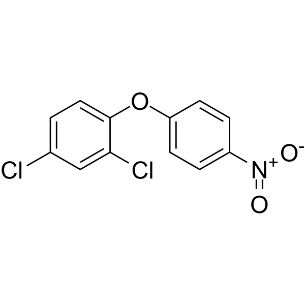
| Chemical Name | 2,4-dichloro-1-(4-nitrophenoxy)benzene |
| HS Tariff Code | 2934.99.9001 |
| Storage |
Powder-20°C 3 years 4°C 2 years In solvent -80°C 6 months -20°C 1 month |
| Shipping Condition | Room temperature (This product is stable at ambient temperature for a few days during ordinary shipping and time spent in Customs) |
Biological Activity
| ADME/Pharmacokinetics |
Absorption, Distribution and Excretion ... NO RESIDUES OF NITROFEN & ITS METABOLITE ... WERE FOUND IN MILK, URINE, OR FECES OF A COW FED 5 PPM OF THE HERBICIDE. ... STUDIED /NITROFEN/ ABSORPTION & TRANSLOCATION USING RAPE, REDROOT PIGWEED, & GREEN FOXTAIL. ... FOUND ONLY SLIGHT TRANSLOCATION OF FOLIARLY APPLIED (14)C-NITROFEN & DIFFERENT ANATOMICAL EFFECTS BETWEEN THE WEEDS & RAPE. ... NITROFEN HAD ALMOST NO EFFECT ON TOPS OF BARNYARD GRASS WHEN HERBICIDE WAS APPLIED TO SOIL ONLY UNDER WEED SEEDS. LOW TRANSLOCATION & LACK OF LIGHT UNDER SOIL MAY BE REASONS FOR THIS ... WEEDS ABSORB THE CHEM DURING THEIR GERMINATION & ELONGATION THROUGH TREATED SOILS, & AFTER EXPOSURE TO SUNLIGHT THEY DIE. NOT READILY ABSORBED FROM SOIL INTO PLANTS. ... NOT SIGNIFICANTLY TRANSLOCATED IN CROPS OR WEEDS. (14)C Labelled nitrofen was administered orally to a sheep at a dose of 40 mg/kg body weight. After 99 hr, 76.2% of the applied dose was accounted for in the excreta. After 100 hr, radioactivity was highest in fat (23 mg/kg); the liver, thyroid, mammary gland, adrenal gland, kidney, lung, muscle, skin and spleen contained levels of 1-3 mg/kg. Oral exposure of pregnant Long Evans rats on day 11 of gestation to nitrofen which was uniformly labeled with (14)C in the nitrophenyl ring, resulted in the accumulation of radioactivity in maternal fat with lesser amounts found in liver, kidney, other tissues, and in the embryonic compartment. The peak concn of radioactivity occurred 7-9 hr after dosing and the half-life of the label in maternal blood was approx 8 days. In the embryonic compartment, radioactivity was first detected at 2 hr after dosing, peaked at 4-6 hr, and declined to half of that initially seen by 24 hr. Metabolism / Metabolites THE FATE OF /LABELED/ NITROFEN IN ... PLANTS/ ... WAS STUDIED ... /BY INVESTIGATORS/, WHO REPORTED THAT RAPE ... REDROOT PIGWEED ... & GREEN FOXTAIL ... LEAVES TREATED WITH 2 DROpS OF (14)C-NITROFEN ... UNDER HIGH LIGHT CONDITIONS, PRODUCED SEVERAL LABELED CMPD OF DIFFERENT MOLECULAR SIZE & CHROMATOGRAPHIC PROPERTIES. THEY THEORIZED THAT AT LEAST 2 OF THESE CMPD WERE LIPID-NITROFEN CONJUGATES OR NITROFEN POLYMERS, WHILE THE OTHERS MIGHT BE FORMED BY CLEAVAGE OF NITROFEN AT THE ETHER LINKAGE. IDENTIFICATION OF THE DERIVATIVES WERE NOT POS ... . ALL RADIOACTIVITY RECOVERED FROM PLANTS GROWN UNDER CONDITIONS OF LOW LIGHT INTENSITY & HIGH TEMP WAS FOUND ONLY IN THE (14)C-NITROFEN PEAK. HOWEVER, 2 RADIOACTIVE CMPD, IN ADDITION TO (14)-C NITROFEN, WERE DETECTED IN PLANT EXTRACTS 2 DAYS AFTER TREATMENT UNDER HIGHER LIGHT INTENSITY & LOWER TEMP. ... /IT WAS/ ... PROPOSED THAT A LIGHT-CONTROLLED BIOCHEM PROCESS APPARENTLY CAUSES (14)-C-NITROFEN TO POLYMERIZE OR ENABLES IT TO COMBINE WITH UNIDENTIFIED CELL LIPIDS. ... NITROFEN IN FRESH RUMEN FLUID IN VITRO /SHOWED/ ... A METABOLITE RAPIDLY PRODUCED WHICH WAS ... P-AMINOPHENYL 2,4-DICHLOROPHENYL ETHER, A REDUCED COMPD OF NITROFEN. Rat liver homogenates incubated with amino derivatives of nitrofen, an NADPH-generating system and erythrocytes from rats or humans metabolized the amino derivatives to methemoglobin forming metabolites. ... (14)C Labelled nitrofen was administered orally to a sheep at a dose of 40 mg/kg body weight. ... The predominant metabolites were 2,4-dichlorophenyl 4-aminophenyl ether, 2,4-dichloro-5-hydroxyphenyl-4-nitrodiphenyl ether, 2,4-dichlorophenol, 2-chlorophenyl-4-nitrophenyl ether & conjugates. Oral exposure of pregnant Long Evans rats on day 11 of gestation to nitrofen which was uniformly labeled with (14)C in the nitrophenyl ring, resulted in the accumulation of radioactivity in maternal fat with lesser amounts found in liver, kidney, other tissues, and in the embryonic compartment. High performance liquid chromatography of embryo placental extracts revealed 4 metabolites in addition to the parent cmpd: 4'-amino and 4'-acetylamino derivatives plus 2 hydroxylated derivatives. A similar metabolic profiles was observed in maternal blood and liver. For more Metabolism/Metabolites (Complete) data for NITROFEN (6 total), please visit the HSDB record page. |
| Toxicity/Toxicokinetics |
Toxicity Data LCLo (cats) = 620 mg/m3/4h Interactions ... The aim of this study was to examine whether antenatal treatment with vitamin A can increase lung growth and reduce the incidence of CDH in a nitrofen-treated rat model. The animals were randomly assigned to four groups: control, vitamin A, nitrofen, and nitrofen/vitamin A (NIP/Vit A). The incidence of CDH /congenital diaphragmatic hernia/ in the NIP/Vit A group (54%) was markedly lower than that in the nitrofen-treated group (85%). Although lung weight was decreased in the nitrofen-treated and NIP/vitamin A groups, the fetal lung weight-to-body weight ratio was slightly increased in the NIP/vitamin A group, compared to the nitrofen-treated group. The mRNA levels of lung surfactant proteins were decreased in the NIP/vitamin A group. We conclude that antenatal treatment with vitamin A reduced the incidence of CDH without lung maturation in the nitrofen-induced rat model. Vitamin A (vit A) alleviates the effects of nitrofen in exposed rat pups. The present study examines the effects of early exposure to vitamin A on the neural-crest-related cardiovascular, thymic, parathyroid, and thyroid malformations previously reported in the rat model of congenital diaphragmatic hernia (CDH). Pregnant rats were exposed on gestational day 9.5 to 100 mg 2-4-dichlorophenyl-p-nitrophenyl ether (nitrofen) alone or followed by 15,000 IU vit A. Controls were treated only with oil or oil + vit A. The fetuses were recovered near term and diaphragmatic, lung, heart, and thymic malformations were sought after dissection. The parathyroids and thyroid were histologically investigated. The hearts were also examined for protein, DNA, and proportion of proliferating cells. None of the control fetuses had malformations, whereas 41% of nitrofen and 27% of nitrofen + vit A fetuses had CDH. Anomalies of the heart outflow tract and pharyngeal arteries were seen in 64% and 43%, respectively, in both groups. Heart and thymic hypoplasia, which were severe in the nitrofen group with significant decreases of total DNA and percent proliferating cells, were significantly improved in the nitrofen + vit A group. The hypoplastic thymus was malformed in 53% and 27% of fetuses, respectively, and the parathyroids were abnormal in 48% and 35%, respectively. Only minimal anomalies of the thyroid were found. The significant improvement of heart and thymic hypoplasia associated with vit A was not seen for the other variables studied, but there was a trend in this direction for all of them. Vit A definitely improved heart hypoplasia induced by nitrofen by stimulating myogenesis. It also improved thymic hypoplasia, but had limited beneficial effects on malformations of the cardiac outflow tract and pharyngeal derivatives that accompany CDH in rats exposed to nitrofen. ... DIURON & SIMETRYNE DECR ACTIVITY OF NITROFEN, BUT THERE WAS A POSSIBILITY THAT CLOSURE OF LEAF STOMATA BY /THEM/ ... MIGHT DECR NITROFEN UPTAKE & ITS ACTIVITY. Non-Human Toxicity Values LD50 Rabbit dermal >5000 mg/kg bw LD50 Rat dermal 5000 mg/kg bw LD50 Rat dermal >2000 mg/kg bw LC50 Rat inhalation 205 mg/L /1 hr For more Non-Human Toxicity Values (Complete) data for NITROFEN (17 total), please visit the HSDB record page. |
| References |
[1]. Siqin Zhaorigetu, et al. Perturbations in Endothelial Dysfunction-Associated Pathways in the Nitrofen-Induced Congenital Diaphragmatic Hernia Model. J Vasc Res. 2018;55(1):26-34. [2]. J M Jacobs, et al. Effects of Diphenyl Ether Herbicides on Porphyrin Accumulation by Cultured Hepatocytes. J Biochem Toxicol. Summer 1992;7(2):87-95. |
| Additional Infomation |
Nitrofen (Technical Grade) can cause cancer according to an independent committee of scientific and health experts. Nitrofen appears as colorless crystals or black solid. Used as a pre- or post-emergence herbicide. Nitrofen is an organic molecular entity. It has a role as an EC 1.3.3.4 (protoporphyrinogen oxidase) inhibitor and a herbicide. Nitrofen is a white, crystalline, solid, combustible, chlorinated compound. Nitrofen was used as an herbicide, but is no longer used or manufactured in the United States. Exposure to nitrofen irritates the skin, eyes and respiratory tract and affects the blood and central nervous system. This substance is teratogenic and carcinogenic in animals and is reasonably anticipated to be a human carcinogen. (NCI05) Mechanism of Action Pregnant rats were exposed to nitrofen or vehicle on gestational day 9 (D9). Embryos were sacrificed on D15, D18 and D21 and divided into nitrofen- and control group. Pulmonary RNA was extracted and mRNA levels of EDNRA and EDNRB were determined by real-time PCR. Immunohistochemistry for protein expression of both receptors was performed. mRNA levels of EDNRA and EDNRB were significantly increased in the nitrofen group on D15, D18 and D21. Immunohistochemistry revealed increased pulmonary vascular expression of EDNRA and EDNRB /Endothelin Receptors A and B/ compared to controls. ... The present studies were performed to begin to address the cellular mechanisms of these nitrofen-induced effects. Heart fibroblasts were isolated and treated with varying doses of nitrofen in vitro. Experiments were performed to determine the effects of this herbicide on important cellular processes including migration, proliferation and apoptosis. These studies illustrated a dose-dependent decrease in collagen gel contraction and proliferation in response to nitrofen. Assays were also performed to determine the effects of nitrofen on fibroblast gene expression. Increased expression of collagen type I and specific integrins were seen following nitrofen exposure. These studies illustrate that nitrofen has direct effects on cardiac fibroblast proliferation and extracellular matrix remodeling, cellular events important in valvuloseptal development. Lactate dehydrogenase (LDH) release test, 3H-thymidine (3H-TdR) and 3H-leucine (3H-Leu) incorporation tests and flow cytometric analysis (FCM) of cell cycle were employed to elucidate cellular and molecular mechanism of nitrofen-induced toxicity in cultured keratinocytes. The results showed that cell morphologic damages were observed after exposure to 1.0 mmol/L and 10.0 mmol/L nitrofen. LDH release increased in a dose- and time-dependent manner. Depressions in 3H-TdR and 3H-Leu incorporation were found even at 0.01 mmol/L, and increased with the exposure dose. Cell cycle was analyzed from the DNA- histogram with propidium iodide stain. The results showed that there was no pronounced alteration in cell cycle after cells exposed to 0.01 and 0.1 mmol/L nitrofen. At dose of 1.0 mmol/L, S phase cells increased 2 times of that of control. With the increase of dose, G2/M phase cells became to increase about 5 times of that of the control. At 1.0 mmol/L, time course of cell cycle after exposure was observed. At the beginning of exposure, cells in S phase and G2/M phase were about 8.7% and 11%. Following 24 hr incubation with nitrofen, cells in S phase increased to 18.0% with almost no change in G2/M. 72 hr after exposure, G2/M phase cells increased to 63.3%. The above results demonstrated that S phase and G2/M phase blockage in cultured keratinocytes after exposed to nitrofen seems of importance in the mechanism of nitrofen-induced toxicity. ... Nitrofen induces a time-dependent cell death of P19 cells that is associated with increases in TUNEL-positivity and caspase-3 cleavage suggesting that nitrofen induces P19 cell apoptosis. In addition, the increase in TUNEL-positive cells was inhibited with zVAD-fmk, suggesting that nitrofen induces a caspase-dependent apoptosis. Nitrofen treatment was associated with increased p38 MAP kinase activity, though pretreatment of cells with multiple p38 inhibitors did not affect nitrofen-mediated caspase-3 cleavage, suggesting caspase-3 cleavage is p38-independent. Nitrofen induced a dose-dependent increase in reactive oxygen species (ROS), which was accompanied by a decrease in the ratio of reduced/oxidized glutathione, indicating that nitrofen alters the cellular redox state of these cells. Furthermore, pretreatment of cells with N-acetyl cysteine gave a dose- and time-dependent reduction of caspase-3 cleavage, supporting the observations that caspase-3 cleavage is cell-redox-dependent. Therefore, nitrofen induces P19 cell apoptosis that is cell-redox-dependent and is associated with increases in p38 activity and ROS and may play a role in nitrofen-mediated birth defects. For more Mechanism of Action (Complete) data for NITROFEN (8 total), please visit the HSDB record page. |
Solubility Data
| Solubility (In Vitro) | May dissolve in DMSO (in most cases), if not, try other solvents such as H2O, Ethanol, or DMF with a minute amount of products to avoid loss of samples |
| Solubility (In Vivo) |
Note: Listed below are some common formulations that may be used to formulate products with low water solubility (e.g. < 1 mg/mL), you may test these formulations using a minute amount of products to avoid loss of samples. Injection Formulations (e.g. IP/IV/IM/SC) Injection Formulation 1: DMSO : Tween 80: Saline = 10 : 5 : 85 (i.e. 100 μL DMSO stock solution → 50 μL Tween 80 → 850 μL Saline) *Preparation of saline: Dissolve 0.9 g of sodium chloride in 100 mL ddH ₂ O to obtain a clear solution. Injection Formulation 2: DMSO : PEG300 :Tween 80 : Saline = 10 : 40 : 5 : 45 (i.e. 100 μL DMSO → 400 μLPEG300 → 50 μL Tween 80 → 450 μL Saline) Injection Formulation 3: DMSO : Corn oil = 10 : 90 (i.e. 100 μL DMSO → 900 μL Corn oil) Example: Take the Injection Formulation 3 (DMSO : Corn oil = 10 : 90) as an example, if 1 mL of 2.5 mg/mL working solution is to be prepared, you can take 100 μL 25 mg/mL DMSO stock solution and add to 900 μL corn oil, mix well to obtain a clear or suspension solution (2.5 mg/mL, ready for use in animals). Injection Formulation 4: DMSO : 20% SBE-β-CD in saline = 10 : 90 [i.e. 100 μL DMSO → 900 μL (20% SBE-β-CD in saline)] *Preparation of 20% SBE-β-CD in Saline (4°C,1 week): Dissolve 2 g SBE-β-CD in 10 mL saline to obtain a clear solution. Injection Formulation 5: 2-Hydroxypropyl-β-cyclodextrin : Saline = 50 : 50 (i.e. 500 μL 2-Hydroxypropyl-β-cyclodextrin → 500 μL Saline) Injection Formulation 6: DMSO : PEG300 : castor oil : Saline = 5 : 10 : 20 : 65 (i.e. 50 μL DMSO → 100 μLPEG300 → 200 μL castor oil → 650 μL Saline) Injection Formulation 7: Ethanol : Cremophor : Saline = 10: 10 : 80 (i.e. 100 μL Ethanol → 100 μL Cremophor → 800 μL Saline) Injection Formulation 8: Dissolve in Cremophor/Ethanol (50 : 50), then diluted by Saline Injection Formulation 9: EtOH : Corn oil = 10 : 90 (i.e. 100 μL EtOH → 900 μL Corn oil) Injection Formulation 10: EtOH : PEG300:Tween 80 : Saline = 10 : 40 : 5 : 45 (i.e. 100 μL EtOH → 400 μLPEG300 → 50 μL Tween 80 → 450 μL Saline) Oral Formulations Oral Formulation 1: Suspend in 0.5% CMC Na (carboxymethylcellulose sodium) Oral Formulation 2: Suspend in 0.5% Carboxymethyl cellulose Example: Take the Oral Formulation 1 (Suspend in 0.5% CMC Na) as an example, if 100 mL of 2.5 mg/mL working solution is to be prepared, you can first prepare 0.5% CMC Na solution by measuring 0.5 g CMC Na and dissolve it in 100 mL ddH2O to obtain a clear solution; then add 250 mg of the product to 100 mL 0.5% CMC Na solution, to make the suspension solution (2.5 mg/mL, ready for use in animals). Oral Formulation 3: Dissolved in PEG400 Oral Formulation 4: Suspend in 0.2% Carboxymethyl cellulose Oral Formulation 5: Dissolve in 0.25% Tween 80 and 0.5% Carboxymethyl cellulose Oral Formulation 6: Mixing with food powders Note: Please be aware that the above formulations are for reference only. InvivoChem strongly recommends customers to read literature methods/protocols carefully before determining which formulation you should use for in vivo studies, as different compounds have different solubility properties and have to be formulated differently. (Please use freshly prepared in vivo formulations for optimal results.) |
| Preparing Stock Solutions | 1 mg | 5 mg | 10 mg | |
| 1 mM | 3.5200 mL | 17.6001 mL | 35.2001 mL | |
| 5 mM | 0.7040 mL | 3.5200 mL | 7.0400 mL | |
| 10 mM | 0.3520 mL | 1.7600 mL | 3.5200 mL |