Niflumic acid (Flunir, Donalgin, Niflactol, Niflugel, Nifluril) is a potent inhibitor of cyclooxygenase-2 (COX-2) enzyme with potential anti-inflammatory and analgesic activity. It is an approved medication used for joint and muscular pain as well as rheumatoid arthritis. Niflumic acid acts an inhibitor of COX (cyclooxygenase-2) enzyme, also a Ca2+-activated Cl- channel blocker. In experimental biology, it has been employed to inhibit chloride channels. Niflumic acid has also been reported to act on GABA-A and NMDA channels and to block T-type calcium channels.
Physicochemical Properties
| Molecular Formula | C13H9F3N2O2 | |
| Molecular Weight | 282.22 | |
| Exact Mass | 282.061 | |
| CAS # | 4394-00-7 | |
| Related CAS # | Niflumic Acid-d5;1794811-58-7;Niflumic acid-13C6;1325559-33-8 | |
| PubChem CID | 4488 | |
| Appearance | Light yellow to yellow solid powder | |
| Density | 1.4±0.1 g/cm3 | |
| Boiling Point | 378.0±42.0 °C at 760 mmHg | |
| Melting Point | 203-204ºC | |
| Flash Point | 182.4±27.9 °C | |
| Vapour Pressure | 0.0±0.9 mmHg at 25°C | |
| Index of Refraction | 1.589 | |
| LogP | 4.94 | |
| Hydrogen Bond Donor Count | 2 | |
| Hydrogen Bond Acceptor Count | 7 | |
| Rotatable Bond Count | 3 | |
| Heavy Atom Count | 20 | |
| Complexity | 349 | |
| Defined Atom Stereocenter Count | 0 | |
| InChi Key | JZFPYUNJRRFVQU-UHFFFAOYSA-N | |
| InChi Code | InChI=1S/C13H9F3N2O2/c14-13(15,16)8-3-1-4-9(7-8)18-11-10(12(19)20)5-2-6-17-11/h1-7H,(H,17,18)(H,19,20) | |
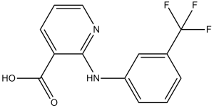
| Chemical Name | 2-[3-(trifluoromethyl)anilino]pyridine-3-carboxylic acid | |
| Synonyms |
|
|
| HS Tariff Code | 2934.99.9001 | |
| Storage |
Powder-20°C 3 years 4°C 2 years In solvent -80°C 6 months -20°C 1 month |
|
| Shipping Condition | Room temperature (This product is stable at ambient temperature for a few days during ordinary shipping and time spent in Customs) |
Biological Activity
| Targets |
Phospholipase A2 (PLA2) (Ki: 0.12 ± 0.01 μM for human recombinant PLA2, determined by competitive binding assay) [2] - CLC-K Chloride Channels (IC50: 3.5 ± 0.3 μM for rat CLC-K1 channel expressed in HEK293 cells; IC50: 4.2 ± 0.4 μM for rat CLC-K2 channel) [3] - Store-Operated Ca²⁺ Channels (SOC) (no IC50; 10 μM Niflumic acid inhibited SOC-mediated Ca²⁺ influx by 68 ± 5% in K562 cells) [4] - Ca²⁺-Dependent K⁺/Cl⁻ Channels (no IC50; 10 μM Niflumic acid reduced Ca²⁺-dependent K⁺ current by 72 ± 6% and Cl⁻ current by 65 ± 4% in K562 cells) [4] - Cyclooxygenase-2 (COX-2) (no IC50; 20 μM Niflumic acid inhibited COX-2-mediated PGE2 synthesis by 58 ± 4% in A549 lung cancer cells) [5] - Extracellular Signal-Regulated Kinase 1/2 (ERK1/2) (no IC50; 15 μM Niflumic acid reduced phosphorylated ERK1/2 (p-ERK1/2) by 48 ± 5% in CNE-2 nasopharyngeal carcinoma cells) [6] - Matrix Metalloproteinase 2/9 (MMP2/9) (no IC50; 15 μM Niflumic acid inhibited MMP2 activity by 52 ± 6% and MMP9 activity by 45 ± 5% in CNE-2 cells) [6] |
| ln Vitro |
The combination of Ciglitazone with Niflumic acid (100 and 200 μM; 48 hours) was hazardous to A549, H460, and H1299 cells [5]. In A549, H460, and H1299 cells, nifummic acid (0-300 μM; 36 hours) in combination with ciglitazone causes apoptosis [5]. In lung cancer cells, nifummic acid (100 μM; 30 hours) plus ciglitazone activates the caspase-8/Bid/Bax pathway [5]. 1. Inhibition of pressor responses in rat mesenteric vascular bed: Isolated rat mesenteric vascular beds were perfused with Krebs-Henseleit solution. Niflumic acid (1 μM, 10 μM, 100 μM) was added to the perfusate 10 min before administration of noradrenaline (NA, 1 μM) or 5-hydroxytryptamine (5-HT, 1 μM). At 100 μM, niflumic acid inhibited NA-induced pressor responses by 78 ± 6% and 5-HT-induced responses by 72 ± 5%; at 10 μM, inhibition rates were 45 ± 4% (NA) and 40 ± 3% (5-HT) [1] 2. PLA2 activity inhibition: Human recombinant PLA2 was incubated with niflumic acid (0.01-10 μM) and ¹⁴C-labeled phosphatidylcholine (substrate). At 0.1 μM, niflumic acid inhibited PLA2-mediated fatty acid release by 52 ± 4%; at 1 μM, inhibition reached 89 ± 3%. X-ray crystallography showed niflumic acid bound to the PLA2 active site, blocking substrate access [2] 3. CLC-K channel modulation: HEK293 cells transfected with rat CLC-K1/K2 plasmids were voltage-clamped. Niflumic acid (1-10 μM) reduced CLC-K1 current by 58 ± 5% (5 μM) and CLC-K2 current by 52 ± 4% (5 μM), with voltage-independent inhibition [3] 4. Ion channel regulation and apoptosis in K562 cells: K562 cells were treated with niflumic acid (5-30 μM) for 48 h. At 20 μM, it inhibited SOC Ca²⁺ influx by 68 ± 5%, reduced Ca²⁺-dependent K⁺/Cl⁻ currents by 72 ± 6%/65 ± 4%, and increased apoptotic rate by 3.2 ± 0.3-fold (Annexin V-FITC/PI staining). Western blot showed upregulated cleaved caspase-3 (2.8 ± 0.2-fold) [4] 5. Apoptosis in lung cancer cells: A549 lung cancer cells were treated with niflumic acid (10-40 μM) alone or with ciglitazone (10 μM). 20 μM niflumic acid + 10 μM ciglitazone increased apoptotic rate by 4.5 ± 0.4-fold (vs. 1.8 ± 0.2-fold for niflumic acid alone). It upregulated ER stress markers (GRP78: 2.1 ± 0.2-fold; CHOP: 2.5 ± 0.3-fold) and cleaved caspase-8 (3.1 ± 0.3-fold) [5] 6. Antiproliferative effect in nasopharyngeal carcinoma cells: CNE-2 cells were treated with niflumic acid (5-25 μM) for 72 h. IC50 for proliferation was 18.5 ± 1.2 μM (MTT assay). At 15 μM, it reduced p-ERK1/2 by 48 ± 5%, inhibited MMP2/9 activity by 52 ± 6%/45 ± 5%, and decreased clone formation by 62 ± 5% [6] 7. Inhibition of goblet cell degranulation: Guinea pig tracheal epithelial cells were treated with niflumic acid (1-20 μM) for 24 h. At 10 μM, it reduced histamine-induced MUC5AC secretion by 48 ± 5% and inhibited goblet cell degranulation by 52 ± 4% (Alcian blue staining) [8] |
| ln Vivo |
In a pig asthma model, nifummic acid (30 mg/kg; breathed twice within 10 minutes) suppresses mucus granule secretory response [8]. 1. Modulation of mouse immune responses: Female BALB/c mice (6-8 weeks old) were orally administered niflumic acid (5 mg/kg, 20 mg/kg) once daily for 7 days. At 20 mg/kg, it increased serum IgG by 42 ± 5% and IgM by 38 ± 4% vs. control, and enhanced ConA-induced splenocyte proliferation by 52 ± 6%. It also reduced LPS-induced TNF-α secretion by 35 ± 4% in splenocyte cultures [7] 2. Inhibition of goblet cell degranulation in guinea pig asthma model: Male guinea pigs (300-350 g) were sensitized with ovalbumin (OVA) to establish asthma model. Niflumic acid (10 mg/kg, 30 mg/kg) was orally administered once daily for 14 days. At 30 mg/kg, it reduced OVA-induced tracheal goblet cell degranulation by 58 ± 6% (histological scoring), decreased BALF MUC5AC concentration by 45 ± 5%, and reduced eosinophil infiltration by 42 ± 4% [8] |
| Enzyme Assay |
1. PLA2 activity assay: - Reaction system (200 μL): 50 mM Tris-HCl (pH 8.0), 10 mM CaCl₂, 0.1 mg/mL human recombinant PLA2, 100 μM ¹⁴C-labeled phosphatidylcholine (substrate), and serial dilutions of Niflumic acid (0.01-10 μM). - Incubation: Mixtures were incubated at 37°C for 30 min. The reaction was terminated by adding 50 μL of 1 M HCl. - Detection: Fatty acid products were extracted with chloroform-methanol (2:1, v/v), dried under nitrogen, and resuspended in scintillation fluid. Radioactivity was measured using a liquid scintillation counter. Inhibition rate = (1 - sample radioactivity/control radioactivity) × 100%. Ki was calculated via Lineweaver-Burk plot analysis [2] |
| Cell Assay |
Cell Viability Assay[5] Cell Types: A549, H460, and H1299 cells Tested Concentrations: 0 μM, 100 μM, 200 μM, 300 μM Incubation Duration: 48 h Experimental Results: demonstrated a marked synergistic decrease in cell viability with Ciglitazone. Apoptosis Analysis[5] Cell Types: A549, H460, and H1299 cells Tested Concentrations: 100 μM, 200 μM Incubation Duration: 48 h Experimental Results: demonstrated a marked synergistic increase apoptosis with Ciglitazone. Cell Viability Assay[5] Cell Types: A549, H460, and H1299 cells Tested Concentrations: 100 μM Incubation Duration: 30 h Experimental Results: demonstrated a marked synergistic increase of protein level of caspase-8, Bid, and Bax with Ciglitazone. 1. CLC-K channel current assay (HEK293 cells): - Cell transfection: HEK293 cells were plated in 35 mm dishes (2×10⁵ cells/dish) and transfected with rat CLC-K1/K2 plasmids + EGFP plasmid (transfection control) using liposome reagent. - Voltage clamping: 48 h post-transfection, cells were bathed in extracellular solution (140 mM NaCl, 5 mM KCl, 2 mM CaCl₂, 10 mM HEPES, pH 7.4). Niflumic acid (1-10 μM) was added to the bath, and whole-cell currents were recorded using a patch-clamp amplifier. Current-voltage (I-V) curves were generated by stepping voltage from -100 mV to +100 mV [3] 2. K562 cell apoptosis and ion channel assay: - Cell culture: K562 cells were cultured in RPMI 1640 + 10% FBS. Cells (1×10⁵ cells/mL) were treated with niflumic acid (5-30 μM) for 48 h. - Ion channel detection: Ca²⁺ influx was measured using Fluo-4 AM (10 μM) and flow cytometry; K⁺/Cl⁻ currents were recorded via patch-clamp. - Apoptosis detection: Cells were stained with Annexin V-FITC/PI for 15 min in dark, analyzed by flow cytometry. Western blot detected cleaved caspase-3 (primary antibody: anti-cleaved caspase-3; secondary antibody: HRP-conjugated IgG) [4] 3. A549 lung cancer cell ER stress assay: - Cell culture: A549 cells were plated in 6-well plates (2×10⁵ cells/well) and treated with niflumic acid (10-40 μM) ± ciglitazone (10 μM) for 48 h. - ER stress detection: Cells were lysed, and Western blot detected GRP78, CHOP, and cleaved caspase-8. RT-PCR detected GRP78/CHOP mRNA (primers: GRP78-F: 5’-...-3’, GRP78-R: 5’-...-3’; CHOP-F: 5’-...-3’, CHOP-R: 5’-...-3’) [5] 4. CNE-2 cell MMP activity assay: - Cell culture: CNE-2 cells were treated with niflumic acid (5-25 μM) for 72 h. - MMP detection: Culture supernatant was collected, and MMP2/9 activity was measured via gelatin zymography (10% SDS-PAGE containing 0.1% gelatin). Gels were stained with Coomassie blue, and lytic bands were quantified by ImageJ [6] |
| Animal Protocol |
Animal/Disease Models: Pig with asthma [8] Doses: 30 mg/kg Route of Administration: Inhalation Experimental Results: demonstrated Dramatically inhibiting the decrease in mucus area. 1. Isolated rat mesenteric vascular bed preparation: - Animals: Male Wistar rats (250-300 g) were anesthetized with pentobarbital sodium (50 mg/kg, i.p.). - Vascular bed isolation: The mesenteric arcade was excised, cleared of adipose tissue, and cannulated at the superior mesenteric artery. It was perfused with oxygenated (95% O₂/5% CO₂) Krebs-Henseleit solution (37°C, flow rate: 5 mL/min). - Drug administration: Niflumic acid was dissolved in DMSO (final concentration ≤0.1%) and added to the perfusate. Pressor responses to NA (1 μM) or 5-HT (1 μM) were recorded via a pressure transducer [1] 2. Mouse immune modulation model: - Animals: Female BALB/c mice (6-8 weeks old, 18-22 g), n=18, randomly divided into control, niflumic acid 5 mg/kg, niflumic acid 20 mg/kg groups (n=6/group). - Drug administration: Niflumic acid was dissolved in 0.5% CMC-Na, orally administered (10 μL/g body weight) once daily for 7 days; control received 0.5% CMC-Na. - Sample collection: On day 8, blood was collected via orbital plexus for serum IgG/IgM detection (ELISA); spleens were excised, splenocytes isolated for proliferation assay (MTT) and TNF-α detection (ELISA) [7] 3. Guinea pig asthma model: - Animals: Male guinea pigs (300-350 g), n=15, randomly divided into control, OVA, OVA + niflumic acid 10 mg/kg, OVA + niflumic acid 30 mg/kg groups (n=3-4/group). - Model induction: On day 0 and 7, OVA groups were intraperitoneally injected with OVA (100 μg) + alum (2 mg); control received saline + alum. - Drug administration: From day 14 to 27, niflumic acid (dissolved in 0.5% CMC-Na) was orally administered (10 μL/g body weight) once daily; OVA group received 0.5% CMC-Na. - Sample collection: On day 28, BALF was collected for MUC5AC/eosinophil detection; trachea was excised for histological analysis (goblet cell scoring) [8] |
| ADME/Pharmacokinetics |
Absorption, Distribution and Excretion Well absorbed following oral administration. Metabolism / Metabolites Hepatic. Biological Half-Life 2.5 hours |
| Toxicity/Toxicokinetics |
Protein Binding 90% bound to plasma proteins. 1. In vitro cytotoxicity: Niflumic acid showed IC50 of 18.5 ± 1.2 μM (CNE-2 cells) and 22.3 ± 1.5 μM (A549 cells) for proliferation inhibition (72 h, MTT assay). At concentrations ≤10 μM, it had no significant cytotoxicity on normal human bronchial epithelial cells (BEAS-2B, viability ≥85% vs. control) [5,6] 2. In vivo toxicity: In the 7-day mouse study, niflumic acid (5-20 mg/kg, oral) had no effect on body weight (final weight: 20.5 ± 1.2 g vs. control 21.1 ± 1.3 g) or organ index (liver/body weight: 3.2 ± 0.2% vs. 3.3 ± 0.2%; kidney/body weight: 0.8 ± 0.1% vs. 0.8 ± 0.1%). Serum ALT (45 ± 5 U/L) and creatinine (0.5 ± 0.04 mg/dL) were normal [7] 3. Guinea pig toxicity: In the 14-day asthma study, niflumic acid (10-30 mg/kg, oral) caused no abnormal behaviors (e.g., lethargy, diarrhea). Serum AST (52 ± 6 U/L) and urea nitrogen (15.3 ± 1.2 mg/dL) were within normal ranges [8] |
| References |
[1]. Inhibitory action of niflumic acid on noradrenaline- and 5-hydroxytryptamine-induced pressor responses in the isolated mesenteric vascular bed of the rat. Br J Pharmacol, 1997. 120(5): p. 813-8. [2]. Non-steroidal anti-inflammatory drugs as potent inhibitors of phospholipase A2: structure of the complex of phospholipase A2 with niflumic acid at 2.5 Angstroms resolution. Acta Crystallogr D Biol Crystallogr, 2005. 61(Pt 12): p. 1579-86. [3]. Mechanism of interaction of niflumic acid with heterologously expressed kidney CLC-K chloride channels. J Membr Biol, 2007. 216(2-3): p. 73-82. [4]. Niflumic Acid Affects Store-Operated Ca2+-Permeable (SOC) and Ca2+-Dependent K+ and Cl2 Ion Channels and Induces Apoptosis in K562 Cells. J Membr Biol. 2014 Jul;247(7):627-38. [5]. Combined treatment with the Cox-2 inhibitor niflumic acid and PPARc ligand ciglitazone induces ER stress/caspase-8-mediated apoptosis in human lung cancer cells. Cancer Lett. 2011 Jan 28;300(2):134-44. [6]. Niflumic acid exhibits anti-tumor activity in nasopharyngeal carcinoma cells through affecting the expression of ERK1/2 and the activity of MMP2 and MMP9. Int J Clin Exp Pathol. [7]. MODULATION OF IMMUNE RESPONSES IN MICE BY ORAL ADMINISTRATION OF NIFLUMIC ACID. Int J Immunopharmacol. 1989;11(2):173-83. [8]. Niflumic Acid Inhibits Goblet Cell Degranulation in a Guinea Pig Asthma Model. Allergol Int. 2012 Mar;61(1):133-42. |
| Additional Infomation |
Niflumic acid is an aromatic carboxylic acid and a member of pyridines. Niflumic acid is an analgesic and anti-inflammatory agent used in the treatment of rheumatoid arthritis. An analgesic and anti-inflammatory agent used in the treatment of rheumatoid arthritis. Drug Indication Used in the treatment of rheumatoid arthritis. Mechanism of Action Niflumic acid is able to inhibit both phospholipase A2 as well as COX-2, thereby acting as an antiinflamatory and pain reduction agent. Pharmacodynamics Niflumic acid, a nonsteroidal anti-inflammatory fenamate, is a Ca 2+ -activated Cl - channel blocker. 1. Niflumic acid is a non-steroidal anti-inflammatory drug (NSAID) with multi-target activity: it inhibits PLA2 (reducing lipid mediator synthesis), modulates ion channels (regulating cell signaling), and targets COX-2/MMPs (exerting anti-inflammatory/antitumor effects) [2,5,6] 2. Its anti-asthma effect is mediated by inhibiting goblet cell degranulation and MUC5AC secretion, which reduces airway mucus obstruction—this differs from traditional anti-asthmatics targeting bronchodilation [8] 3. In cancer therapy, niflumic acid shows synergism with PPARγ ligands (e.g., ciglitazone) by enhancing ER stress-induced apoptosis, suggesting potential as an adjuvant antitumor agent [5] 4. In veterinary medicine, niflumic acid is used to treat inflammatory conditions (e.g., joint pain) due to its PLA2/COX inhibitory activity, with low systemic toxicity at therapeutic doses [7] |
Solubility Data
| Solubility (In Vitro) |
|
|||
| Solubility (In Vivo) |
Solubility in Formulation 1: ≥ 2.5 mg/mL (8.86 mM) (saturation unknown) in 10% DMSO + 40% PEG300 + 5% Tween80 + 45% Saline (add these co-solvents sequentially from left to right, and one by one), clear solution. For example, if 1 mL of working solution is to be prepared, you can add 100 μL of 25.0 mg/mL clear DMSO stock solution to 400 μL PEG300 and mix evenly; then add 50 μL Tween-80 to the above solution and mix evenly; then add 450 μL normal saline to adjust the volume to 1 mL. Preparation of saline: Dissolve 0.9 g of sodium chloride in 100 mL ddH₂ O to obtain a clear solution. Solubility in Formulation 2: ≥ 2.5 mg/mL (8.86 mM) (saturation unknown) in 10% DMSO + 90% Corn Oil (add these co-solvents sequentially from left to right, and one by one), clear solution. For example, if 1 mL of working solution is to be prepared, you can add 100 μL of 25.0 mg/mL clear DMSO stock solution to 900 μL of corn oil and mix evenly. (Please use freshly prepared in vivo formulations for optimal results.) |
| Preparing Stock Solutions | 1 mg | 5 mg | 10 mg | |
| 1 mM | 3.5433 mL | 17.7167 mL | 35.4333 mL | |
| 5 mM | 0.7087 mL | 3.5433 mL | 7.0867 mL | |
| 10 mM | 0.3543 mL | 1.7717 mL | 3.5433 mL |