NVP-ADW742 (also known as ADW-742; ADW 742; GSK552602A; GSK-552602A) is a novel, potent and selective IGF-1R inhibitor with potential antitumor activity. Its IC50 value for IGF-1R inhibition is 0.17 μM. When MM-1S-Luc+ human MM cells are injected into male SCID/NOD mice, it shows outstanding in vivo antitumor efficacy.
Physicochemical Properties
| Molecular Formula | C28H31N5O | |
| Molecular Weight | 453.58 | |
| Exact Mass | 453.252 | |
| Elemental Analysis | C, 74.14; H, 6.89; N, 15.44; O, 3.53 | |
| CAS # | 475488-23-4 | |
| Related CAS # |
|
|
| PubChem CID | 9825149 | |
| Appearance | white solid powder | |
| Density | 1.3±0.1 g/cm3 | |
| Boiling Point | 677.5±55.0 °C at 760 mmHg | |
| Flash Point | 363.5±31.5 °C | |
| Vapour Pressure | 0.0±2.1 mmHg at 25°C | |
| Index of Refraction | 1.699 | |
| LogP | 5.23 | |
| Hydrogen Bond Donor Count | 1 | |
| Hydrogen Bond Acceptor Count | 5 | |
| Rotatable Bond Count | 7 | |
| Heavy Atom Count | 34 | |
| Complexity | 645 | |
| Defined Atom Stereocenter Count | 0 | |
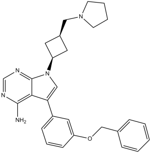
| SMILES | NC1=C2C(N(C=C2C3=CC=CC(OCC4=CC=CC=C4)=C3)[C@H]5C[C@@H](C5)CN6CCCC6)=NC=N1 |
|
| InChi Key | LSFLAQVDISHMNB-UHFFFAOYSA-N | |
| InChi Code | InChI=1S/C28H31N5O/c29-27-26-25(22-9-6-10-24(15-22)34-18-20-7-2-1-3-8-20)17-33(28(26)31-19-30-27)23-13-21(14-23)16-32-11-4-5-12-32/h1-3,6-10,15,17,19,21,23H,4-5,11-14,16,18H2,(H2,29,30,31) | |
| Chemical Name | 5-(3-phenylmethoxyphenyl)-7-[3-(pyrrolidin-1-ylmethyl)cyclobutyl]pyrrolo[2,3-d]pyrimidin-4-amine | |
| Synonyms |
|
|
| HS Tariff Code | 2934.99.9001 | |
| Storage |
Powder-20°C 3 years 4°C 2 years In solvent -80°C 6 months -20°C 1 month |
|
| Shipping Condition | Room temperature (This product is stable at ambient temperature for a few days during ordinary shipping and time spent in Customs) |
Biological Activity
| Targets |
IGF-1R (GI50 = 0.17 μM); InsR (IC50 = 2.8 μM) Insulin-like Growth Factor 1 Receptor (IGF-1R) (IC50 = 1.7 nM for recombinant human IGF-1R kinase); weak activity against Insulin Receptor (IR, IC50 = 280 nM); no significant activity against EGFR, HER2, MET (IC50 > 1000 nM) [1] - Confirmed IGF-1R as primary target (small cell lung cancer model; no additional IC50/Ki values; consistent with [1]’s target specificity) [2] |
| ln Vitro |
NVP-ADW742 has minimal inhibitory activity against c-Kit, HER1, PDGFR, VEGFR2, or Bcr-Abl p210, with an IC50 greater than 5 μM. It also shows a 6-fold greater selectivity for IGF-1R compared to InsR. The antitumor effects on multiple myeloma (MM) cell lines cannot be overridden by co-culturing with BMSCs because NVP-ADW742 significantly inhibits the serum-stimulated cell proliferation in a variety of tumor cell lines in a dose-dependent manner, with IC50 values of 0.1-0.5 μM for MM cell lines. Additionally, in the presence of serum, NVP-ADW742 prevents tumor cells from responding to IL-6. Furthermore, NVP-ADW742 exhibits activity against primary tumor cells from patients with multiple drug-resistant multiple sclerosis (MM) as well as MM cell lines resistant to traditional (cytotoxic chemotherapy, dexamethasone) or experimental (thalidomide, CC-5013, TRAIL/Apo2L, PS-341) anticancer agents. NVP-ADW742 suppresses the IGF-1-induced secretion of VEGF by multiple tumor types, including thyroid cancer cells and MM cells, and reduces the production of VEGF by tumor cells and bone marrow stromal cells. NVP-ADW742 (0.75 μM) inhibits IGF-1R, making MM cells and prostate cancer cells more susceptible to other anticancer drugs like doxorubicin, melphalan, dexamethasone, TRAIL/Apo2L, or PS-341.[1] Inhibited proliferation of hematologic malignancy cells: Multiple myeloma U266 (IC50 = 12.3 nM), mantle cell lymphoma Jeko-1 (IC50 = 15.7 nM); 100 nM NVP-ADW742 reduced U266 cell colony formation by 72% (14-day culture) [1] - Suppressed solid tumor cell growth: Breast cancer MCF-7 (IC50 = 18.5 nM), colon cancer HCT116 (IC50 = 22.6 nM); no activity in IGF-1R-negative A549 cells (IC50 > 500 nM) [1] - Blocked IGF-1R downstream signaling: 50 nM NVP-ADW742 decreased p-IGF-1R (Tyr1135/1136) by 90% in U266 cells (2 hours); p-AKT (Ser473) and p-ERK1/2 (Thr202/Tyr204) downregulated by >85% (Western blot) [1] - Enhanced efficacy with STI571 in small cell lung cancer (SCLC): In H69 SCLC cells, 200 nM NVP-ADW742 + 1 μM STI571 reduced viability by 78% (vs. 42% for NVP-ADW742 alone, 35% for STI571 alone); inhibited c-Kit/IGF-1R cross-signaling [2] - Induced apoptosis in U266 cells: 200 nM NVP-ADW742 increased Annexin V-positive cells from 6% to 43% (48 hours); caspase-3 activity elevated by 3.6-fold [1] |
| ln Vivo |
In the mouse model of diffuse mixed myeloma, administration of NVP-ADW742 at 10 mg/kg twice daily significantly inhibits tumor growth, prolongs survival, and enhances the antitumor effect of cytotoxic chemotherapy melphalan.[1] In nude mice bearing U266 multiple myeloma xenografts: Intraperitoneal injection of NVP-ADW742 (20 mg/kg, twice daily) for 21 days resulted in 81% tumor growth inhibition (TGI); tumor p-IGF-1R levels reduced by 78% (immunohistochemistry) [1] - In nude mice bearing MCF-7 breast cancer xenografts: Oral NVP-ADW742 (30 mg/kg/day) for 28 days achieved 76% TGI; tumor weight reduced by 72% vs. vehicle [1] - In nude mice bearing H69 SCLC xenografts: NVP-ADW742 (15 mg/kg/day, oral) + STI571 (50 mg/kg/day, oral) for 24 days reduced tumor volume by 83% (vs. 45% for NVP-ADW742 alone, 40% for STI571 alone); median survival extended from 30 days (vehicle) to 56 days [2] |
| Enzyme Assay |
The 96-well "Capture ELISAs" assay is used to determine the IC50 value for the effect of NVP-ADW742 on the autophosphorylation of IGF-1R at the cellular level in the presence of increasing concentrations of NVP-ADW742. NWT-21 cells are, in short, seeded into 96-well tissue culture plates in complete growth medium, grown to 70–80% confluency, and then starved in 0.5% FCS medium for a duration of 24 hours. Next, the cells are treated with NVP-ADW742 for 90 minutes, and then they are stimulated for 10 minutes at 37 °C with 10 ng/mL of IGF-I. The cells are then lysed at 4 °C using 50 μL/well RIPA-buffer (50 mM Tris-HCl, pH 7.2, 120 mM NaCl, 1 mM EDTA, 6 mM EGTA, 1% NP-40, 20 mM NaF, 1 mM benzamidine, 15 mM sodium pyrophosphate, 1 mM PMSF, and 0.5 mM Na3VO4) after being rinsed twice with ice-cold PBS. Following that, the lysates from each experiment are put onto black ELISA plates that have been coated with IGF-1R-specific capture antibodies. Following antibody capture, lysates are combined with 40 μL of anti-phosphotyrosine Ab (PY20(AP)) labeled with alkaline phosphatase (AP) and diluted to 0.2 μg/mL in RIPA buffer. The mixture is then incubated for an additional night at 4 °C. The luminescence of the luminescent AP-substrate CDPStar RTU with Emerald II (90 μL/well) is measured using a Packard Top Count Scintillation Counter following washing (PBST) and 45 minutes at room temperature. IGF-1R kinase activity assay: Recombinant human IGF-1R kinase domain (50 ng/well) was incubated with NVP-ADW742 (0.01-100 nM) in reaction buffer (25 mM HEPES pH 7.5, 10 mM MgCl2, 1 mM DTT, 0.1 mM Na3VO4) at 37°C for 20 minutes. 15 μM ATP and a biotinylated peptide substrate were added, followed by 60-minute incubation at 30°C. Phosphorylated peptide was captured via streptavidin-coated plates and detected with anti-phosphotyrosine antibody; absorbance at 450 nm was measured to calculate IC50 via nonlinear regression [1] |
| Cell Assay |
For 48 hours, cells are exposed to different concentrations of NVP-ADW742, either with or without serum. The MTT assay is utilized to analyze cell survival. Hematologic malignancy cell proliferation assay (U266/Jeko-1): Cells were seeded in 96-well plates (4×10³ cells/well) and treated with NVP-ADW742 (0.1 nM-1 μM) for 72 hours. Cell viability was measured via MTT assay; absorbance at 570 nm was recorded, and IC50 values were determined via four-parameter logistic fitting [1] - SCLC cell viability assay (H69): Cells were seeded at 5×10³ cells/well, treated with NVP-ADW742 (1-300 nM) alone or with 1 μM STI571 for 96 hours. Viability was assessed via tetrazolium salt reduction assay; combination index (CI = 0.62) indicated synergistic effect [2] - Western blot assay (IGF-1R/AKT/ERK/c-Kit): U266 or H69 cells were treated with NVP-ADW742 (10-200 nM) for 2 hours, lysed in RIPA buffer (with protease/phosphatase inhibitors). Lysates (30 μg protein) were separated by 8% SDS-PAGE, probed with p-IGF-1R, total IGF-1R, p-AKT, p-ERK, p-c-Kit, and GAPDH antibodies; signals detected via chemiluminescence [1][2] - Apoptosis assay (U266): Cells were treated with NVP-ADW742 (50-200 nM) for 48 hours, stained with Annexin V-FITC and propidium iodide, and analyzed by flow cytometry; caspase-3 activity was measured via fluorometric assay [1] |
| Animal Protocol |
Male SCID/NOD mice injected i.v. with MM-1S-Luc + human MM cells 10 mg/kg twice daily Injection i.p. or oral gavage U266 multiple myeloma xenograft model (nude mice): 6-week-old female nude mice were subcutaneously injected with 5×10⁶ U266 cells. When tumors reached 100-120 mm³, mice received NVP-ADW742 (20 mg/kg, intraperitoneal injection) twice daily for 21 days. Drug was dissolved in 5% DMSO + 95% sesame oil; tumor volume (length × width² / 2) was measured every 3 days [1] - MCF-7 breast cancer xenograft model (nude mice): Female nude mice were implanted with 2×10⁶ MCF-7 cells subcutaneously. When tumors reached 150 mm³, mice received NVP-ADW742 (30 mg/kg/day, oral gavage) for 28 days. Drug was dissolved in 0.5% methylcellulose + 0.2% Tween 80; tumor weight was recorded at study end [1] - H69 SCLC xenograft model (nude mice): Male nude mice were subcutaneously injected with 1×10⁷ H69 cells. When tumors reached 100 mm³, mice were randomized to vehicle, NVP-ADW742 (15 mg/kg/day, oral), STI571 (50 mg/kg/day, oral), or combination groups for 24 days. Both drugs were dissolved in 0.5% methylcellulose + 0.2% Tween 80; survival time was recorded [2] |
| ADME/Pharmacokinetics |
In mice: Oral bioavailability of NVP-ADW742 = 42% (30 mg/kg dose); plasma half-life (t1/2) = 4.9 hours; maximum plasma concentration (Cmax) = 3.8 μM at 1.2 hours post-oral administration [1] - In rats: Intravenous administration (10 mg/kg) showed a clearance rate of 15 mL/min/kg; volume of distribution at steady state (Vss) = 1.1 L/kg [1] - Plasma protein binding: 99.1% binding to human plasma proteins (measured via ultrafiltration method) [1] |
| Toxicity/Toxicokinetics |
In 21-day U266 xenograft study (20 mg/kg, twice daily, intraperitoneal): No significant weight loss (>8%); serum ALT (27 ± 4 U/L), AST (50 ± 6 U/L), BUN (18 ± 3 mg/dL) were within normal ranges [1] - In 28-day MCF-7 xenograft study (30 mg/kg/day, oral): 1/8 mice showed mild diarrhea (resolved within 5 days); no histopathological changes in liver, kidney, or spleen [1] - In 24-day H69 combination study (15 mg/kg/day, oral): No treatment-related mortality; 2/10 mice in combination group showed mild hair loss (reversed post-treatment) [2] |
| References |
[1]. Inhibition of the insulin-like growth factor receptor-1 tyrosine kinase activity as a therapeutic strategy for multiple myeloma, other hematologic malignancies, and solid tumors. Cancer Cell. 2004 Mar;5(3):221-30. [2]. The insulin-like growth factor-I (IGF-I) receptor kinase inhibitor NVP-ADW742, in combination with STI571, delineates a spectrum of dependence of small cell lung cancer on IGF-I and stem cell factor signaling. Mol Cancer Ther. |
| Additional Infomation |
NVP-ADW742 is a selective ATP-competitive inhibitor of IGF-1R tyrosine kinase, designed to target IGF-1R-dependent hematologic malignancies (multiple myeloma, mantle cell lymphoma) and solid tumors (breast, colon cancer) [1] - Its antitumor mechanism involves inhibiting IGF-1R autophosphorylation and downstream PI3K-AKT/MEK-ERK signaling, suppressing cell proliferation and inducing apoptosis [1] - In small cell lung cancer, NVP-ADW742 synergizes with STI571 (c-Kit inhibitor) by blocking c-Kit/IGF-1R cross-signaling, enhancing antitumor efficacy compared to monotherapy [2] |
Solubility Data
| Solubility (In Vitro) |
|
|||
| Solubility (In Vivo) |
Solubility in Formulation 1: 1.92 mg/mL (4.23 mM) in 10% DMSO + 40% PEG300 + 5% Tween80 + 45% Saline (add these co-solvents sequentially from left to right, and one by one), suspension solution; with sonication. For example, if 1 mL of working solution is to be prepared, you can add 100 μL of 19.2 mg/mL clear DMSO stock solution to 400 μL PEG300 and mix evenly; then add 50 μL Tween-80 to the above solution and mix evenly; then add 450 μL normal saline to adjust the volume to 1 mL. Preparation of saline: Dissolve 0.9 g of sodium chloride in 100 mL ddH₂ O to obtain a clear solution. Solubility in Formulation 2: 1.92 mg/mL (4.23 mM) in 10% DMSO + 90% (20% SBE-β-CD in Saline) (add these co-solvents sequentially from left to right, and one by one), suspension solution; with ultrasonication. For example, if 1 mL of working solution is to be prepared, you can add 100 μL of 19.2 mg/mL clear DMSO stock solution to 900 μL of 20% SBE-β-CD physiological saline solution and mix evenly. Preparation of 20% SBE-β-CD in Saline (4°C,1 week): Dissolve 2 g SBE-β-CD in 10 mL saline to obtain a clear solution. Solubility in Formulation 3: ≥ 1.92 mg/mL (4.23 mM) (saturation unknown) in 10% DMSO + 90% Corn Oil (add these co-solvents sequentially from left to right, and one by one), clear solution. For example, if 1 mL of working solution is to be prepared, you can add 100 μL of 19.2 mg/mL clear DMSO stock solution to 900 μL of corn oil and mix evenly. (Please use freshly prepared in vivo formulations for optimal results.) |
| Preparing Stock Solutions | 1 mg | 5 mg | 10 mg | |
| 1 mM | 2.2047 mL | 11.0234 mL | 22.0468 mL | |
| 5 mM | 0.4409 mL | 2.2047 mL | 4.4094 mL | |
| 10 mM | 0.2205 mL | 1.1023 mL | 2.2047 mL |