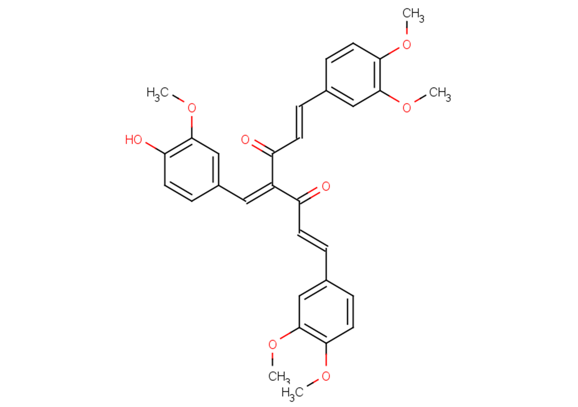
Physicochemical Properties
| Molecular Formula | C31H30O8 |
| Molecular Weight | 530.565109729767 |
| Exact Mass | 530.194 |
| CAS # | 1227098-15-8 |
| Related CAS # | 1227098-15-8 |
| PubChem CID | 49851904 |
| Appearance | Light yellow to yellow solid |
| LogP | 5.6 |
| Hydrogen Bond Donor Count | 1 |
| Hydrogen Bond Acceptor Count | 8 |
| Rotatable Bond Count | 12 |
| Heavy Atom Count | 39 |
| Complexity | 826 |
| Defined Atom Stereocenter Count | 0 |
| SMILES | COC1=C(C=C(C=C1)/C=C/C(=O)C(=CC2=CC(=C(C=C2)O)OC)C(=O)/C=C/C3=CC(=C(C=C3)OC)OC)OC |
| InChi Key | AYIYVEPNEPUJCF-GNXRPPCSSA-N |
| InChi Code | InChI=1S/C31H30O8/c1-35-27-14-9-20(17-30(27)38-4)6-11-24(32)23(16-22-8-13-26(34)29(19-22)37-3)25(33)12-7-21-10-15-28(36-2)31(18-21)39-5/h6-19,34H,1-5H3/b11-6+,12-7+ |
| Chemical Name | (1E,6E)-1,7-bis(3,4-dimethoxyphenyl)-4-[(4-hydroxy-3-methoxyphenyl)methylidene]hepta-1,6-diene-3,5-dione |
| Synonyms | CHEMBL1800965; NF-κB-IN-1; NF-kappaB-IN-1; NF-|EB-IN-1 |
| HS Tariff Code | 2934.99.9001 |
| Storage |
Powder-20°C 3 years 4°C 2 years In solvent -80°C 6 months -20°C 1 month |
| Shipping Condition | Room temperature (This product is stable at ambient temperature for a few days during ordinary shipping and time spent in Customs) |
Biological Activity
| Targets |
NF-κB; IKK NF-κB-IN-1 targets nuclear factor-κB (NF-κB) signaling pathway, inhibiting NF-κB-dependent transcription with an IC₅₀ of 1.8 μM (NF-κB luciferase reporter assay in HeLa cells) [1] NF-κB-IN-1 inhibits IκBα phosphorylation (a key step in NF-κB activation) with an IC₅₀ of 2.3 μM (Western blot for phospho-IκBα in TNF-α-stimulated HeLa cells) [1] |
| ln Vitro |
NF-κB-IN-1 (compound 17) (0.001-100 μM; 72 h) has GI50s of 0.72, 0.07, 0.13, and 0.16 μM, respectively, and inhibits the growth of A549, H1944, H460, and H157 cells for 72 hours[1]. IκB phosphorylation and degradation in A549 cells are effectively blocked by NF-κB-IN-1 (0.5–25 μM; pretreated for 30 min. or 4 h.)[1]. NF-κB-IN-1 (0.1-100 μM; pretreated for 30 min) inhibits TNFα-induced nuclear translocation of NF-B in A549 cells in a dose-dependent manner, with an IC50 of 1.0 μM[1]. The clonogenic activity of lung cancer is inhibited by NF-κB-IN-1 (0.1-0.4 μM; 9 d)[1]. Antiproliferative activity in cancer cell lines: NF-κB-IN-1 (1–20 μM) dose-dependently inhibited proliferation of HeLa (cervical cancer), MCF-7 (breast cancer), A549 (lung cancer), and HT-29 (colorectal cancer) cells, with IC₅₀ values of 3.2 μM (HeLa), 4.5 μM (MCF-7), 5.1 μM (A549), and 4.8 μM (HT-29) (MTT assay) [1] - NF-κB signaling inhibition: 2–10 μM dose-dependently suppressed TNF-α-induced NF-κB activation in HeLa cells, reducing luciferase activity by 45–88% (NF-κB reporter assay); it blocked IκBα phosphorylation (Ser32/36) and degradation, with 5 μM reducing phospho-IκBα by 72% (Western blot) [1] - Inhibition of p65 nuclear translocation: 5 μM NF-κB-IN-1 reduced TNF-α-induced p65 nuclear accumulation by 75% (immunofluorescence staining + confocal microscopy); nuclear p65 protein levels were downregulated by 68% (Western blot for nuclear fraction) [1] - Apoptosis induction: 5–10 μM induced apoptosis in HeLa cells, with apoptotic rate increased from 8% to 42% (5 μM) and 65% (10 μM) (Annexin V-FITC/PI staining); upregulated cleaved caspase-3/caspase-9 by 2.5–3.8-fold and Bax by 2.1-fold, downregulated Bcl-2 by 55% (Western blot) [1] - Reduction of pro-inflammatory cytokines: 5 μM decreased TNF-α-induced secretion of IL-6 (62%) and IL-8 (58%) in HeLa cells (ELISA); mRNA levels of IL-6, IL-8, and TNF-α were downregulated by 55–70% (qRT-PCR) [1] - Low cytotoxicity in normal cells: CC₅₀ > 30 μM in normal human foreskin fibroblasts (NHF); cell viability >90% at concentrations up to 15 μM (MTT assay) [1] |
| ln Vivo |
Antitumor activity in HeLa xenograft model: BALB/c nude mice bearing HeLa xenografts were treated with NF-κB-IN-1 (10, 20 mg/kg, intraperitoneal injection, once daily for 21 days). The compound dose-dependently inhibited tumor growth, reducing tumor volume by 45% (10 mg/kg) and 68% (20 mg/kg) compared to vehicle control [1] - Tumor weight reduction: 20 mg/kg NF-κB-IN-1 reduced tumor weight by 65%; immunohistochemical analysis of tumor tissues showed decreased phospho-IκBα (62%), nuclear p65 (58%), and Ki-67 (55%) expression, and increased cleaved caspase-3 (2.3-fold) [1] - No obvious toxicity: Treated mice showed no significant body weight loss (<7% change) or histopathological abnormalities in liver, kidney, or spleen; hematological parameters and liver/kidney function markers were within normal ranges [1] |
| Enzyme Assay |
NF-κB luciferase reporter assay: HeLa cells were transfected with NF-κB-responsive luciferase reporter plasmid and Renilla luciferase (internal control) plasmid. After 24 hours, cells were pretreated with NF-κB-IN-1 (0.5–20 μM) for 1 hour, then stimulated with TNF-α (10 ng/mL) for 16 hours. Luciferase activity was measured to calculate NF-κB inhibition rate and IC₅₀ [1] - IκBα phosphorylation inhibition assay: HeLa cells were serum-starved for 12 hours, pretreated with NF-κB-IN-1 (0.5–20 μM) for 1 hour, then stimulated with TNF-α (10 ng/mL) for 30 minutes. Cells were lysed, and phospho-IκBα (Ser32/36) and total IκBα were detected by Western blot. Band intensity was quantified to calculate IC₅₀ [1] |
| Cell Assay |
Cancer cell proliferation assay: HeLa/MCF-7/A549/HT-29 cells were seeded in 96-well plates (5×10³ cells/well), cultured for 24 hours, and treated with NF-κB-IN-1 (1–20 μM) for 72 hours. MTT reagent was added, and absorbance at 570 nm was measured to calculate cell viability and IC₅₀ [1] - Apoptosis assay: HeLa cells were treated with NF-κB-IN-1 (5–10 μM) for 48 hours, stained with Annexin V-FITC and PI, and analyzed by flow cytometry to quantify apoptotic cells [1] - p65 nuclear translocation assay: HeLa cells were grown on coverslips, pretreated with NF-κB-IN-1 (5 μM) for 1 hour, then stimulated with TNF-α (10 ng/mL) for 1 hour. Cells were fixed, permeabilized, stained with p65 antibody and DAPI, and analyzed by confocal microscopy to assess nuclear p65 accumulation [1] - Western blot/qRT-PCR/ELISA analysis: Treated cells were lysed to extract protein/RNA or culture supernatants were collected; Western blot detected NF-κB pathway proteins, apoptotic markers; qRT-PCR quantified cytokine mRNA; ELISA measured secreted IL-6/IL-8 [1] |
| Animal Protocol |
HeLa xenograft model: 6–8 weeks old female BALB/c nude mice were subcutaneously injected with HeLa cells (5×10⁶ cells/mouse) into the right flank. When tumors reached ~120 mm³, mice were randomly divided into vehicle group, NF-κB-IN-1 10 mg/kg group, and 20 mg/kg group [1] - Drug formulation: NF-κB-IN-1 was dissolved in dimethyl sulfoxide (DMSO) and diluted with normal saline to a final DMSO concentration of ≤5% [1] - Administration protocol: The compound was administered via intraperitoneal injection once daily for 21 days. Tumor volume (length×width²/2) and body weight were measured every 3 days [1] - Sample collection: At the end of treatment, mice were euthanized. Tumors were excised, weighed, and fixed in formalin for immunohistochemical staining (phospho-IκBα, nuclear p65, Ki-67, cleaved caspase-3); major organs were collected for histopathological examination [1] |
| Toxicity/Toxicokinetics |
In vitro toxicity: CC₅₀ > 30 μM in normal human foreskin fibroblasts (NHF) [1] - In vivo acute toxicity: No mortality or obvious toxicity signs (lethargy, diarrhea) in mice treated with NF-κB-IN-1 at intraperitoneal doses up to 150 mg/kg [1] - Subchronic toxicity (21-day, mouse): NF-κB-IN-1 (20 mg/kg ip, qd) did not cause significant changes in hematological parameters (WBC, RBC, platelets) or liver/kidney function markers (ALT, AST, creatinine) [1] - Plasma protein binding rate: 92% (mouse plasma, ultrafiltration method) [1] |
| References |
[1]. Synthesis and identification of new 4-arylidene curcumin analogues as potential anticancer agents targeting nuclear factor-κB signaling pathway. J Med Chem. 2010 Dec 9;53(23):8260-73. |
| Additional Infomation |
NF-κB-IN-1 is a synthetic small-molecule analogue of 4-arylidene curcumin, designed as a selective inhibitor of the NF-κB signaling pathway [1] - Its antitumor mechanism involves blocking TNF-α-induced IκBα phosphorylation and degradation, inhibiting p65 nuclear translocation, and suppressing NF-κB-dependent transcription of oncogenes and pro-inflammatory cytokines, thereby inducing cancer cell apoptosis and inhibiting proliferation [1] - NF-κB is constitutively activated in many human cancers, promoting tumor growth, invasion, and chemoresistance; NF-κB-IN-1 targets this key pathway for anticancer therapy [1] - The compound shows broad-spectrum antiproliferative activity against multiple solid tumor cell lines with low normal cell toxicity, supporting its potential as a lead compound for anticancer drug development [1] |
Solubility Data
| Solubility (In Vitro) | DMSO: 50 mg/mL (94.2 mM) |
| Solubility (In Vivo) |
Solubility in Formulation 1: ≥ 2.5 mg/mL (4.71 mM) (saturation unknown) in 10% DMSO + 40% PEG300 +5% Tween-80 + 45% Saline (add these co-solvents sequentially from left to right, and one by one), clear solution. For example, if 1 mL of working solution is to be prepared, you can add 100 μL of 25.0 mg/mL clear DMSO stock solution to 400 μL PEG300 and mix evenly; then add 50 μL Tween-80 + to the above solution and mix evenly; then add 450 μL normal saline to adjust the volume to 1 mL. Preparation of saline: Dissolve 0.9 g of sodium chloride in 100 mL ddH₂ O to obtain a clear solution. (Please use freshly prepared in vivo formulations for optimal results.) |
| Preparing Stock Solutions | 1 mg | 5 mg | 10 mg | |
| 1 mM | 1.8848 mL | 9.4238 mL | 18.8477 mL | |
| 5 mM | 0.3770 mL | 1.8848 mL | 3.7695 mL | |
| 10 mM | 0.1885 mL | 0.9424 mL | 1.8848 mL |