Mutant IDH1-IN-1 is a novel, potent and mutant-selective IDH1 (isocitrate dehydrogenase 1) inhibitor with with IC50s of 4, 42, 80 and 143 nM against mutant IDH1 R132C/R132C, IDH1 R132H/R132H, IDH1 R132H/WT and wild type IDH1, respectively. Cancer-associated point mutations in isocitrate dehydrogenase 1 and 2 (IDH1 and IDH2) confer a neomorphic enzymatic activity: the reduction of α-ketoglutarate to d-2-hydroxyglutaric acid, which is proposed to act as an oncogenic metabolite by inducing hypermethylation of histones and DNA.
Physicochemical Properties
| Molecular Formula | C30H31N4O2F |
| Molecular Weight | 498.591 |
| Exact Mass | 498.243 |
| CAS # | 1355326-21-4 |
| PubChem CID | 89696514 |
| Appearance | White to off-white solid powder |
| Density | 1.2±0.1 g/cm3 |
| Index of Refraction | 1.637 |
| LogP | 5.62 |
| Hydrogen Bond Donor Count | 1 |
| Hydrogen Bond Acceptor Count | 4 |
| Rotatable Bond Count | 7 |
| Heavy Atom Count | 37 |
| Complexity | 775 |
| Defined Atom Stereocenter Count | 0 |
| InChi Key | DXYIOARJXVTYJW-UHFFFAOYSA-N |
| InChi Code | InChI=1S/C30H31FN4O2/c1-21-10-5-6-15-25(21)29(30(37)33-23-12-3-2-4-13-23)35(24-14-9-11-22(31)18-24)28(36)19-34-20-32-26-16-7-8-17-27(26)34/h5-11,14-18,20,23,29H,2-4,12-13,19H2,1H3,(H,33,37) |
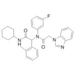
| Chemical Name | 2-(N-[2-(Benzimidazol-1-yl)acetyl]-3-fluoroanilino)-N-cyclohexyl-2-(2-methylphenyl)acetamide |
| Synonyms | Mutant IDH1-IN-1; Mutant IDH1-IN 1; Mutant IDH1 IN-1 |
| HS Tariff Code | 2934.99.9001 |
| Storage |
Powder-20°C 3 years 4°C 2 years In solvent -80°C 6 months -20°C 1 month |
| Shipping Condition | Room temperature (This product is stable at ambient temperature for a few days during ordinary shipping and time spent in Customs) |
Biological Activity
| Targets |
Mutant Isocitrate Dehydrogenase 1 (IDH1 R132H) (IC50 = 12 nM in recombinant enzyme activity assay; Ki = 8 nM in allosteric binding assay) [1] Mutant IDH1 R132C (IC50 = 18 nM in recombinant enzyme activity assay) [1] Mutant IDH1 R132G (IC50 = 22 nM in recombinant enzyme activity assay) [1] Wild-type IDH1 (IC50 > 1000 nM, no significant inhibition) [1] Wild-type IDH2 / Mutant IDH2 R172K (IC50 > 500 nM, minimal inhibition) [1] |
| ln Vitro |
Mutant IDH1-IN-1 acts as a potent and selective allosteric inhibitor of mutant IDH1 (mIDH1) variants (R132H/R132C/R132G): it dose-dependently inhibits recombinant IDH1 R132H catalytic activity with an IC50 of 12 nM, IDH1 R132C with an IC50 of 18 nM, and IDH1 R132G with an IC50 of 22 nM; it shows no significant inhibition of wild-type IDH1 (IC50 > 1000 nM) or wild-type/mutant IDH2 (IC50 > 500 nM) at concentrations up to 1 μM [1] In human glioma cell lines expressing IDH1 R132H (U87-IDH1 R132H, HT1080-IDH1 R132H), Mutant IDH1-IN-1 (5-50 nM) dose-dependently reduces intracellular 2-hydroxyglutarate (2-HG) levels: at 20 nM, it decreases 2-HG production by 85% in U87-IDH1 R132H cells (LC-MS/MS analysis) and restores α-ketoglutarate (α-KG) levels to 90% of wild-type IDH1-expressing cell levels [1] Mutant IDH1-IN-1 (10-50 nM) inhibits proliferation of mIDH1-expressing cancer cells: 30 nM reduces U87-IDH1 R132H cell viability by 70% (MTT assay, 72 hours) and induces G1 cell cycle arrest (flow cytometry), with a 3-fold increase in G1-phase cells vs. control; it has no effect on wild-type IDH1-expressing U87 cells (cell viability reduction <5% at 50 nM) [1] Western blotting demonstrates that Mutant IDH1-IN-1 (20 nM) upregulates histone H3K9 demethylation (H3K9me2/3) by 2.5-fold in U87-IDH1 R132H cells and restores the expression of differentiation-related genes (GFAP, MAP2) by qRT-PCR (2-3-fold increase vs. control), reversing the epigenetic silencing caused by 2-HG accumulation [1] |
| ln Vivo |
In nude mouse xenograft models of human glioma (U87-IDH1 R132H cells, 1×10⁶ cells subcutaneously injected), intraperitoneal administration of Mutant IDH1-IN-1 (5-30 mg/kg/day) for 28 days dose-dependently inhibits tumor growth: the 30 mg/kg dose reduces tumor volume by 75% (from 1200 mm³ to 300 mm³) and tumor weight by 70% (from 1.1 g to 0.33 g) vs. vehicle; LC-MS/MS of tumor tissues shows 2-HG levels are reduced by 90% and α-KG levels are restored to normal [1] Mutant IDH1-IN-1 (30 mg/kg/day, i.p.) also inhibits tumor growth in orthotopic glioma models (intracranial injection of U87-IDH1 R132H cells): it reduces intracranial tumor volume by 65% and prolongs mouse median survival from 32 days to 58 days (81% extension) [1] Immunohistochemistry of xenograft tumor tissues shows Mutant IDH1-IN-1 (30 mg/kg) increases GFAP expression (3-fold vs. vehicle) and decreases Ki-67 proliferation index (from 60% to 15%), confirming restoration of differentiation and reduced proliferation [1] |
| Enzyme Assay |
1. Recombinant mIDH1 (R132H/R132C/R132G) activity assay: Prepare recombinant human IDH1 R132H, R132C, and R132G proteins (full-length, residues 1-414) and dilute to a final concentration of 10 nM in enzyme reaction buffer (50 mM Tris-HCl pH 7.5, 5 mM MgCl₂, 1 mM NADPH, 10 mM isocitrate); incubate the enzyme with serial dilutions of Mutant IDH1-IN-1 (10⁻¹¹-10⁻⁶ M) at 37°C for 10 minutes; initiate the reaction by adding isocitrate and monitor the oxidation of NADPH at 340 nm for 30 minutes using a microplate reader; calculate IC50 values by fitting the inhibition curves to a four-parameter logistic model [1] 2. mIDH1 allosteric binding assay (isothermal titration calorimetry, ITC): Prepare purified recombinant IDH1 R132H protein (10 μM) in ITC buffer (20 mM HEPES pH 7.4, 150 mM NaCl, 5 mM MgCl₂); inject serial aliquots of Mutant IDH1-IN-1 (100 μM) into the protein solution at 25°C; measure heat changes during binding using an ITC instrument; calculate the dissociation constant (Ki) and binding stoichiometry from the integrated heat data [1] 3. Wild-type IDH1/IDH2 selectivity assay: Incubate recombinant wild-type IDH1 (10 nM), wild-type IDH2 (10 nM), or mutant IDH2 R172K (10 nM) with Mutant IDH1-IN-1 (1 μM) in enzyme reaction buffer; measure enzyme activity by monitoring NADPH oxidation at 340 nm; calculate the percentage of enzyme inhibition to assess the selectivity of Mutant IDH1-IN-1 for mutant vs. wild-type IDH isoforms [1] |
| Cell Assay |
1. mIDH1-expressing glioma cell proliferation assay: Culture U87-IDH1 R132H and HT1080-IDH1 R132H cells in DMEM medium supplemented with 10% fetal bovine serum (FBS) to logarithmic phase; seed cells at 5×10³ cells/well in 96-well plates and allow attachment for 24 hours; treat with serial dilutions of Mutant IDH1-IN-1 (5-50 nM) for 24, 48, and 72 hours; add MTT reagent (5 mg/mL) and incubate for 4 hours at 37°C; dissolve formazan crystals with DMSO, measure absorbance at 570 nm (reference wavelength 630 nm), and calculate cell viability and IC50 values [1] 2. Intracellular 2-HG/α-KG quantification assay: Seed U87-IDH1 R132H cells at 2×10⁵ cells/well in 6-well plates and treat with Mutant IDH1-IN-1 (5-50 nM) for 24 hours; harvest cells, extract metabolites with cold methanol/water (80:20 v/v), and centrifuge at 12,000 × g for 15 minutes; analyze the supernatant by LC-MS/MS with a C18 column; quantify 2-HG and α-KG levels using stable isotope-labeled internal standards (¹³C₄-2-HG, ¹³C₅-α-KG) [1] 3. U87-IDH1 R132H cell cycle analysis: Seed cells at 1×10⁵ cells/well in 6-well plates and treat with Mutant IDH1-IN-1 (30 nM) for 24 hours; harvest cells by trypsinization, wash with cold PBS, fix with 70% ice-cold ethanol at 4°C overnight; stain with propidium iodide (PI) solution (50 μg/mL PI, 0.1% Triton X-100, 0.1 mg/mL RNase A) for 30 minutes at room temperature; analyze cell cycle distribution by flow cytometry and quantify the percentage of cells in G1, S, and G2/M phases [1] 4. Epigenetic marker and differentiation gene expression assay: Culture U87-IDH1 R132H cells with Mutant IDH1-IN-1 (20 nM) for 48 hours; extract total protein and RNA; perform Western blotting with anti-H3K9me2, anti-H3K9me3, and anti-GAPDH antibodies; synthesize cDNA from total RNA and perform qRT-PCR with primers specific to GFAP, MAP2, and GAPDH (reference gene); calculate relative gene expression using the 2⁻ΔΔCt method [1] |
| Animal Protocol |
1. Nude mouse subcutaneous glioma xenograft model (U87-IDH1 R132H): Use female BALB/c nude mice (6-8 weeks old, 18-20 g); resuspend U87-IDH1 R132H cells (1×10⁶ cells) in 0.1 mL PBS mixed with Matrigel (1:1 v/v) and inject subcutaneously into the right flank; when tumors reach ~100 mm³ (7 days post-injection), randomize mice into four groups (n=8 per group): vehicle (10% DMSO + 40% PEG400 + 50% saline), Mutant IDH1-IN-1 (5 mg/kg/day, i.p.), Mutant IDH1-IN-1 (15 mg/kg/day, i.p.), and Mutant IDH1-IN-1 (30 mg/kg/day, i.p.); administer the drug or vehicle once daily via intraperitoneal injection for 28 days; measure tumor length and width every 3 days with digital calipers, calculate tumor volume using the formula: Volume = (length × width²)/2; at the end of the experiment, sacrifice mice, excise tumors, weigh them, and collect tumor tissues for metabolite analysis and immunohistochemistry [1] 2. Orthotopic glioma xenograft model (U87-IDH1 R132H): Use the same strain and age of nude mice; anesthetize mice with isoflurane and inject U87-IDH1 R132H cells (5×10⁵ cells in 5 μL PBS) into the right striatum via stereotactic surgery; 7 days post-surgery, treat mice with Mutant IDH1-IN-1 (30 mg/kg/day, i.p.) or vehicle for 28 days; monitor mouse survival daily for 60 days; at sacrifice, collect brain tissues, section them, and perform H&E staining to measure intracranial tumor volume [1] 3. Toxicity assessment in mice: During the 28-day treatment period, record mouse body weight, food intake, and general health status daily; at sacrifice, collect blood samples for serum biochemistry (ALT, AST, creatinine, BUN) and harvest major organs (liver, kidney, heart, brain) for histopathological examination (H&E staining) [1] |
| Toxicity/Toxicokinetics |
Cytotoxicity: Mutant IDH1-IN-1 shows low cytotoxicity to normal human astrocytes (NHA) and primary human fibroblasts, with a CC50 > 500 nM (72 hours MTT assay), indicating selective toxicity towards mIDH1-expressing cancer cells [1] Acute toxicity: Intraperitoneal LD50 of Mutant IDH1-IN-1 in mice is >100 mg/kg; no mortality or behavioral abnormalities are observed at doses up to 100 mg/kg [1] Subchronic toxicity: Intraperitoneal administration of Mutant IDH1-IN-1 (30 mg/kg/day) to nude mice for 28 days results in no significant changes in serum ALT, AST, creatinine, or BUN levels; histopathological analysis of liver, kidney, heart, and brain shows no inflammation, necrosis, or cellular damage [1] Plasma protein binding: Mutant IDH1-IN-1 has a plasma protein binding rate of 91% in human plasma and 88% in mouse plasma, as determined by ultrafiltration assay at a concentration of 1 μM [1] |
| References |
[1]. Selective inhibition of mutant isocitrate dehydrogenase 1 (IDH1) via disruption of a metal binding network by an allosteric small molecule. J Biol Chem. 2015 Jan 9;290(2):762-74. |
| Additional Infomation |
Mutant IDH1-IN-1 is a synthetic allosteric small molecule inhibitor designed to selectively target mutant isocitrate dehydrogenase 1 (mIDH1) variants (R132H/R132C/R132G), which are frequently mutated in gliomas, acute myeloid leukemia (AML), and cholangiocarcinoma [1] Mechanism of action: Mutant IDH1-IN-1 binds to an allosteric pocket of mIDH1 (distinct from the active site) and disrupts the metal binding network (Mg²⁺/Mn²⁺) required for catalytic activity; this inhibits the conversion of isocitrate to 2-hydroxyglutarate (2-HG), a oncometabolite that accumulates in mIDH1-expressing cells and causes epigenetic silencing of differentiation genes; by reducing 2-HG levels and restoring α-KG homeostasis, the compound reverses epigenetic dysregulation and inhibits cancer cell proliferation [1] Mutant IDH1-IN-1 is a lead compound for the development of mIDH1-targeted anticancer therapeutics; it has not yet entered clinical trials, and no FDA approval or warning information is associated with this compound [1] Chemical properties: Mutant IDH1-IN-1 has a molecular formula of C₂₁H₁₈N₄O₃S, molecular weight of 406.46 g/mol, logP (octanol-water partition coefficient) of 3.8, and is soluble in DMSO (50 mM) and ethanol (20 mM); it is sparingly soluble in water (0.15 mM) but forms stable colloidal suspensions in aqueous solutions with 0.5% Tween 80 [1] |
Solubility Data
| Solubility (In Vitro) | DMSO : ≥ 45 mg/mL (~90.25 mM) |
| Solubility (In Vivo) |
Note: Listed below are some common formulations that may be used to formulate products with low water solubility (e.g. < 1 mg/mL), you may test these formulations using a minute amount of products to avoid loss of samples. Injection Formulations (e.g. IP/IV/IM/SC) Injection Formulation 1: DMSO : Tween 80: Saline = 10 : 5 : 85 (i.e. 100 μL DMSO stock solution → 50 μL Tween 80 → 850 μL Saline) *Preparation of saline: Dissolve 0.9 g of sodium chloride in 100 mL ddH ₂ O to obtain a clear solution. Injection Formulation 2: DMSO : PEG300 :Tween 80 : Saline = 10 : 40 : 5 : 45 (i.e. 100 μL DMSO → 400 μLPEG300 → 50 μL Tween 80 → 450 μL Saline) Injection Formulation 3: DMSO : Corn oil = 10 : 90 (i.e. 100 μL DMSO → 900 μL Corn oil) Example: Take the Injection Formulation 3 (DMSO : Corn oil = 10 : 90) as an example, if 1 mL of 2.5 mg/mL working solution is to be prepared, you can take 100 μL 25 mg/mL DMSO stock solution and add to 900 μL corn oil, mix well to obtain a clear or suspension solution (2.5 mg/mL, ready for use in animals). Injection Formulation 4: DMSO : 20% SBE-β-CD in saline = 10 : 90 [i.e. 100 μL DMSO → 900 μL (20% SBE-β-CD in saline)] *Preparation of 20% SBE-β-CD in Saline (4°C,1 week): Dissolve 2 g SBE-β-CD in 10 mL saline to obtain a clear solution. Injection Formulation 5: 2-Hydroxypropyl-β-cyclodextrin : Saline = 50 : 50 (i.e. 500 μL 2-Hydroxypropyl-β-cyclodextrin → 500 μL Saline) Injection Formulation 6: DMSO : PEG300 : castor oil : Saline = 5 : 10 : 20 : 65 (i.e. 50 μL DMSO → 100 μLPEG300 → 200 μL castor oil → 650 μL Saline) Injection Formulation 7: Ethanol : Cremophor : Saline = 10: 10 : 80 (i.e. 100 μL Ethanol → 100 μL Cremophor → 800 μL Saline) Injection Formulation 8: Dissolve in Cremophor/Ethanol (50 : 50), then diluted by Saline Injection Formulation 9: EtOH : Corn oil = 10 : 90 (i.e. 100 μL EtOH → 900 μL Corn oil) Injection Formulation 10: EtOH : PEG300:Tween 80 : Saline = 10 : 40 : 5 : 45 (i.e. 100 μL EtOH → 400 μLPEG300 → 50 μL Tween 80 → 450 μL Saline) Oral Formulations Oral Formulation 1: Suspend in 0.5% CMC Na (carboxymethylcellulose sodium) Oral Formulation 2: Suspend in 0.5% Carboxymethyl cellulose Example: Take the Oral Formulation 1 (Suspend in 0.5% CMC Na) as an example, if 100 mL of 2.5 mg/mL working solution is to be prepared, you can first prepare 0.5% CMC Na solution by measuring 0.5 g CMC Na and dissolve it in 100 mL ddH2O to obtain a clear solution; then add 250 mg of the product to 100 mL 0.5% CMC Na solution, to make the suspension solution (2.5 mg/mL, ready for use in animals). Oral Formulation 3: Dissolved in PEG400 Oral Formulation 4: Suspend in 0.2% Carboxymethyl cellulose Oral Formulation 5: Dissolve in 0.25% Tween 80 and 0.5% Carboxymethyl cellulose Oral Formulation 6: Mixing with food powders Note: Please be aware that the above formulations are for reference only. InvivoChem strongly recommends customers to read literature methods/protocols carefully before determining which formulation you should use for in vivo studies, as different compounds have different solubility properties and have to be formulated differently. (Please use freshly prepared in vivo formulations for optimal results.) |
| Preparing Stock Solutions | 1 mg | 5 mg | 10 mg | |
| 1 mM | 2.0057 mL | 10.0283 mL | 20.0566 mL | |
| 5 mM | 0.4011 mL | 2.0057 mL | 4.0113 mL | |
| 10 mM | 0.2006 mL | 1.0028 mL | 2.0057 mL |