Physicochemical Properties
| Molecular Formula | C13H13N8NAO2 |
| Molecular Weight | 336.28 |
| Exact Mass | 336.105 |
| Elemental Analysis | C, 49.68; H, 4.49; N, 35.65; O, 10.18 |
| CAS # | 1375799-59-9 |
| Related CAS # | 1375799-59-9 (Sodium); 1154028-82-6 |
| PubChem CID | 69669724 |
| Appearance | Typically exists as White to off-white solid at room temperature |
| Density | 1.558 at 20℃ |
| Hydrogen Bond Donor Count | 0 |
| Hydrogen Bond Acceptor Count | 8 |
| Rotatable Bond Count | 3 |
| Heavy Atom Count | 24 |
| Complexity | 402 |
| Defined Atom Stereocenter Count | 0 |
| InChi Key | VYRQLKYGGSWDNH-UHFFFAOYSA-M |
| InChi Code | InChI=1S/C13H14N8O2.Na/c22-13-10(20-2-1-16-18-20)8-17-21(13)12-7-11(14-9-15-12)19-3-5-23-6-4-19;/h1-2,7-9,22H,3-6H2;/q;+1/p-1 |
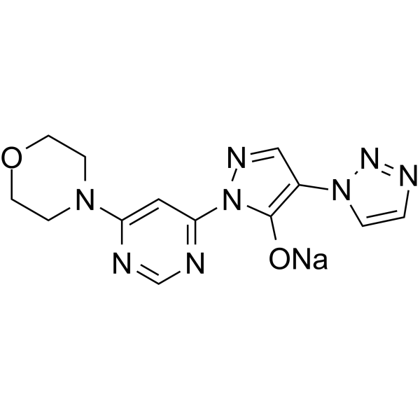
| Chemical Name | sodium;2-(6-morpholin-4-ylpyrimidin-4-yl)-4-(triazol-1-yl)pyrazol-3-olate |
| Synonyms | Molidustat; 1154028-82-6; BAY 85-3934; Molidustat sodium; Molidustat (sodium); 1375799-59-9; BAY-1053048; BAY-85-3934 SODIUM; CI0NE7C96T; sodium;2-(6-morpholin-4-ylpyrimidin-4-yl)-4-(triazol-1-yl)pyrazol-3-olate; 1-(6-(MORPHOLIN-4-YL)PYRIMIDIN-4-YL)-4-(1H-1,2,3-TRIAZOL-1-YL)-1H-PYRAZOL-5-OL, SODIUM SALT (1:1); Molidustat [INN]; UNII-9JH486CZ13; BAY85-3934; |
| HS Tariff Code | 2934.99.9001 |
| Storage |
Powder-20°C 3 years 4°C 2 years In solvent -80°C 6 months -20°C 1 month |
| Shipping Condition | Room temperature (This product is stable at ambient temperature for a few days during ordinary shipping and time spent in Customs) |
Biological Activity
| Targets | IC50: 480 nM (PHD1), 280 nM (PHD2), 450 nM (PHD3)[1] |
| ln Vitro | Molidustat sodium (5 μM; 20 min) is sufficient to induce detectable levels of HIF-1α in HeLa cells. In a cell-based reporter assay, molidustat sodium induced expression of a firefly luciferase reporter gene under the control of a hypoxia response element promoter with a mean (± SD) EC50 of 8.4 μM (n=4)[1]. |
| ln Vivo | Molidustat sodium (0.5-5 mg/kg; oral; once a day for 26 days) in Wistar rats and (0.5-1.5 mg/kg; once a day for 5 days) in cynomolgus monkeys induces dose-dependent erythropoietin (EPO) production in healthy Wistar rats and cynomolgus monkeys by stabilizing hypoxia-inducible factor (HIF)[1]. Molidustat sodium (0.5-10 mg/kg; oral; five times a week) normalizes hypertensive blood pressure in a rat model of chronic kidney disease (CKD) and effectively alleviates renal anemia in rats with impaired renal function[1]. |
| Enzyme Assay |
Prolyl hydroxylase assay[1]
The prolyl hydroxylase assay was performed as described previously with minor modifications. Biotinylated HIF-1α 556–574 (biotinyl-DLDLEMLAPYIPMDDDFQL) was bound to white 96-well NeutrAvidin high binding capacity plates, which were pre-blocked with Blocker Casein and subsequently blocked with 1 mM biotin. The immobilized peptide substrate was incubated with the appropriate amount of HIF-PH in buffer containing 20 mM Tris (pH 7.5), 5 mM KCl, 1.5 mM MgCl2, 20 µM 2-oxoglutarate, 10 µM FeSO4, 2 mM ascorbate, 4% protease inhibitors without EDTA in a final volume of 100 µl, with or without test compound added at appropriate concentrations. The reaction time was 60 min. To stop the reaction, plates were washed three times with wash buffer.[1] Hydroxylated biotinyl-HIF-1α 556–574 was incubated with Eu-VBC in 100 µl binding buffer (50 mM Tris [pH 7.5], 120 mM NaCl) for 60 min at room temperature. After washing six times with DELFIA wash buffer and adding 100 µl enhancer solution, the amount of bound VBC was determined by measuring time-resolved fluorescence with a Tecan infinite M200 plate reader. Measurements were taken in triplicate or more, and results were expressed as means ± SEM. IC50 values were determined after curve fitting using GraphPad Prism software applying the four-parameter logistic equation to the data sets. When adjustment of the concentration of free Fe2+ was necessary, the reaction buffer was supplemented with appropriate amounts of ammonium iron(II) sulfate ((NH4)2Fe(SO4)2.6H2O, Mohr’s salt). |
| Cell Assay |
Cell lines, cell culture media, and luciferase reporter assay[1] A549 and HeLa carcinoma cell lines (American Type Culture Collection) were cultured in DMEM/F-12, and Hep3B cells in RPMI medium, both supplemented with antibiotics, L-glutamine and 10% fetal calf serum. A549 cells stably transfected with the HIF-RE2-luc HIF reporter construct (constructed in pGL3) were seeded on 384-well plates at a density of 2500 cells/well in a volume of 25 µl complete cell culture medium, and re-incubated for 16–24 h before the test. Test compounds were added at appropriate dilutions in a volume of 10 µl, and cells were re-incubated for 6 h before measurement. Luciferase activity was determined in a luminometer after addition of cell lysis/luciferase buffer. Cell line identities were verified by STR DNA typing. Western blot analysis[1] For western blot analysis, cell lysates were separated on 4–12% SDS polyacrylamide gradient gels. Proteins were blotted onto polyvinylidene difluoride (PVDF) membranes. HIF-1α protein was detected using a HIF-1α specific monoclonal antibody at a dilution of 1∶250. HIF-2α protein was detected using a HIF-2α specific polyclonal antibody at a dilution of 1∶1000. Anti-β-actin antibody served as a loading control. Binding of the antibodies was visualized by binding of a horseradish peroxidase-conjugated anti-mouse IgG antibody, and subsequently enhanced using chemiluminescence, according to the manufactureŕs instructions. Novex Sharp Pre-stained Protein Standard was used as molecular weight marker. |
| Animal Protocol |
Studies in rats[1] Male Wistar rats (240–340 g in body weight) were housed with five animals per cage for at least 1 week before experimentation. Blood samples from rats were collected under anesthesia (2% isoflurane in air) by puncturing the retro-orbital vein plexus with a glass capillary. In a repeat-dose, 26-day experiment, animals were administered vehicle or Molidustat (BAY 85-3934) at doses of 0.5 mg/kg, 1.25 mg/kg, 2.5 mg/kg, and 5 mg/kg. PCV was determined at baseline and at weekly intervals after centrifugation in a hematocrit capillary tube (Brand) for 10 min at full speed in a Haemofuge centrifuge (Heraeus). The number of reticulocytes in 5 µl blood was counted after staining with thiazol orange (Becton Dickinson) according to the manufacturer’s instructions by FACS analysis on a BD FACSCalibur system (Becton Dickinson). The efficacy of BAY 85-3934 (2.5 mg/kg, once-daily, oral) was also compared with that of rhEPO (25 IU/kg, 50 IU/kg, and 100 IU/kg, twice-weekly, s.c. injection). The time-course of induction of EPO mRNA expression and plasma EPO was determined at baseline and 0.5 h, 1 h, 2 h, 4 h, 6 h, and 8 h after oral administration of a single dose of BAY 85-3934 (5 mg/kg). Studies in cynomolgus monkeys[1] Male and female cynomolgus monkeys (2.8–5.6 kg in body weight) were used, which were housed two per cage. Blood samples from conscious cynomolgus monkeys were taken by puncturing a superficial vein. In a 5-day, repeat-dose study of plasma EPO response, Molidustat (BAY 85-3934) was administered at doses of 0.5 mg/kg and 1.5 mg/kg at 0 h, 24 h, 48 h, 72 h, and 96 h. Blood samples were taken at 7 h, 31 h, 55 h, 79 h, 103 h, and 168 h. Erythropoietic parameters were also evaluated after a 2-week treatment period with s.c. administration of rhEPO (100 IU/kg twice weekly at days 1, 4, 8, and 11) and BAY 85-3934 (1.5 mg/kg) once daily. Gentamicin-induced kidney failure model[1] Male Wistar rats were treated once daily with gentamicin (Gibco/Invitrogen) at a dose of 100 mg/kg body weight via i.p. injection on 14 consecutive days. Control animals received injections of an equal volume of 0.9% saline. After gentamicin treatment, PCV was determined and animals were distributed to the vehicle or treatment groups with respect to equal mean PCV. On day 15, Molidustat (BAY 85-3934) was given orally once daily at doses of 1 mg/kg, 2.5 mg/kg, 5.0 mg/kg, and 10.0 mg/kg, five times weekly. PG-PS-induced inflammatory anemia model[1] Female Lewis rats, with a body weight of 155–181 g were used. Body weight, ankle diameter, hematocrit, and blood cell count were determined at baseline and thereafter at regular intervals. PG-PS from Streptococcus pyogenes was dissolved in sterile saline and administered via i.p. injection at 15 mg/kg. Animals that did not show an inflammatory response were not studied further. Two weeks after injection, animals were distributed into treatment groups in equal proportions based on their hematocrit levels. On day 15, Molidustat (BAY 85-3934) was given orally once daily at doses of 2.5 mg/kg and 5.0 mg/kg. At the end of the study, animals were sacrificed and kidney and liver samples were processed for qRT-PCR analysis. Subtotal nephrectomy model[1] Subtotal nephrectomy was conducted in adult male Wistar rats. Body weight, blood pressure, hematocrit, and blood cell counts were determined at baseline and thereafter at weekly intervals. At baseline, rats were randomly distributed into two groups: those that underwent subtotal nephrectomy and those that underwent a sham procedure without reduction of renal mass. Surgery was performed in deeply anesthetized (2% isoflurane in air) animals. Kidneys were accessed via a dorsolateral incision of the body wall of about 2 cm in length. The right kidney was removed after ligature of the renal peduncle, and subsequently the upper and lower pole of the left kidney were removed, followed by careful hemostasis. Approximately one third of the initial kidney mass remained (removed tissue was weighed to check this was achieved). In the sham-treated animals, both kidneys were exposed before closure of the wound. Three weeks after surgery, animals were allocated to each group in equal proportions with respect to systolic blood pressure and hematocrit values. For 5 weeks, animals were treated twice weekly with rhEPO (100 IU/kg), or once daily with BAY 85–3936 sodium (2.5 mg/kg or 5.0 mg/kg) or vehicle. In experiments using enalapril or a combination of Molidustat (BAY 85-3934) sodium and enalapril, study drugs were administered with drinking water. BAY 85-3934 sodium and enalapril were administered in drinking water at concentrations of 80 ppm and 30 ppm, respectively. This was equivalent to approximately 2 mg/kg/day for enalapril and 5 mg/kg/day for BAY 85-3934. Systolic blood pressure and heart rate were determined using the tail-cuff method (a semi-automatic, non-invasive blood pressure monitor; TSE Systems), with three repeated measurements per animal. |
| References |
[1]. Mimicking hypoxia to treat anemia: HIF-stabilizer BAY 85-3934 (Molidustat) stimulates erythropoietin production without hypertensive effects. PLoS One. 2014 Nov 13;9(11):e111838. [2]. Molidustat for Renal Anemia in Nondialysis Patients Previously Treated with Erythropoiesis-Stimulating Agents: A Randomized, Open-Label, Phase 3 Study. Am J Nephrol. 2021;52(10-11):884-893. |
| Additional Infomation |
See also: Molidustat (has active moiety). Drug Indication Treatment of anaemia due to chronic disorders |
Solubility Data
| Solubility (In Vitro) | May dissolve in DMSO (in most cases), if not, try other solvents such as H2O, Ethanol, or DMF with a minute amount of products to avoid loss of samples |
| Solubility (In Vivo) |
Note: Listed below are some common formulations that may be used to formulate products with low water solubility (e.g. < 1 mg/mL), you may test these formulations using a minute amount of products to avoid loss of samples. Injection Formulations (e.g. IP/IV/IM/SC) Injection Formulation 1: DMSO : Tween 80: Saline = 10 : 5 : 85 (i.e. 100 μL DMSO stock solution → 50 μL Tween 80 → 850 μL Saline) *Preparation of saline: Dissolve 0.9 g of sodium chloride in 100 mL ddH ₂ O to obtain a clear solution. Injection Formulation 2: DMSO : PEG300 :Tween 80 : Saline = 10 : 40 : 5 : 45 (i.e. 100 μL DMSO → 400 μLPEG300 → 50 μL Tween 80 → 450 μL Saline) Injection Formulation 3: DMSO : Corn oil = 10 : 90 (i.e. 100 μL DMSO → 900 μL Corn oil) Example: Take the Injection Formulation 3 (DMSO : Corn oil = 10 : 90) as an example, if 1 mL of 2.5 mg/mL working solution is to be prepared, you can take 100 μL 25 mg/mL DMSO stock solution and add to 900 μL corn oil, mix well to obtain a clear or suspension solution (2.5 mg/mL, ready for use in animals). Injection Formulation 4: DMSO : 20% SBE-β-CD in saline = 10 : 90 [i.e. 100 μL DMSO → 900 μL (20% SBE-β-CD in saline)] *Preparation of 20% SBE-β-CD in Saline (4°C,1 week): Dissolve 2 g SBE-β-CD in 10 mL saline to obtain a clear solution. Injection Formulation 5: 2-Hydroxypropyl-β-cyclodextrin : Saline = 50 : 50 (i.e. 500 μL 2-Hydroxypropyl-β-cyclodextrin → 500 μL Saline) Injection Formulation 6: DMSO : PEG300 : castor oil : Saline = 5 : 10 : 20 : 65 (i.e. 50 μL DMSO → 100 μLPEG300 → 200 μL castor oil → 650 μL Saline) Injection Formulation 7: Ethanol : Cremophor : Saline = 10: 10 : 80 (i.e. 100 μL Ethanol → 100 μL Cremophor → 800 μL Saline) Injection Formulation 8: Dissolve in Cremophor/Ethanol (50 : 50), then diluted by Saline Injection Formulation 9: EtOH : Corn oil = 10 : 90 (i.e. 100 μL EtOH → 900 μL Corn oil) Injection Formulation 10: EtOH : PEG300:Tween 80 : Saline = 10 : 40 : 5 : 45 (i.e. 100 μL EtOH → 400 μLPEG300 → 50 μL Tween 80 → 450 μL Saline) Oral Formulations Oral Formulation 1: Suspend in 0.5% CMC Na (carboxymethylcellulose sodium) Oral Formulation 2: Suspend in 0.5% Carboxymethyl cellulose Example: Take the Oral Formulation 1 (Suspend in 0.5% CMC Na) as an example, if 100 mL of 2.5 mg/mL working solution is to be prepared, you can first prepare 0.5% CMC Na solution by measuring 0.5 g CMC Na and dissolve it in 100 mL ddH2O to obtain a clear solution; then add 250 mg of the product to 100 mL 0.5% CMC Na solution, to make the suspension solution (2.5 mg/mL, ready for use in animals). Oral Formulation 3: Dissolved in PEG400 Oral Formulation 4: Suspend in 0.2% Carboxymethyl cellulose Oral Formulation 5: Dissolve in 0.25% Tween 80 and 0.5% Carboxymethyl cellulose Oral Formulation 6: Mixing with food powders Note: Please be aware that the above formulations are for reference only. InvivoChem strongly recommends customers to read literature methods/protocols carefully before determining which formulation you should use for in vivo studies, as different compounds have different solubility properties and have to be formulated differently. (Please use freshly prepared in vivo formulations for optimal results.) |
| Preparing Stock Solutions | 1 mg | 5 mg | 10 mg | |
| 1 mM | 2.9737 mL | 14.8686 mL | 29.7371 mL | |
| 5 mM | 0.5947 mL | 2.9737 mL | 5.9474 mL | |
| 10 mM | 0.2974 mL | 1.4869 mL | 2.9737 mL |