Physicochemical Properties
| Molecular Formula | C48H78O20 |
| Molecular Weight | 975.1209 |
| Exact Mass | 974.508 |
| CAS # | 34540-22-2 |
| PubChem CID | 161823 |
| Appearance | White to off-white solid powder |
| Density | 1.5±0.1 g/cm3 |
| Boiling Point | 1043.6±65.0 °C at 760 mmHg |
| Melting Point | 220 - 223 °C |
| Flash Point | 295.8±27.8 °C |
| Vapour Pressure | 0.0±0.6 mmHg at 25°C |
| Index of Refraction | 1.637 |
| LogP | 1.88 |
| Hydrogen Bond Donor Count | 13 |
| Hydrogen Bond Acceptor Count | 20 |
| Rotatable Bond Count | 10 |
| Heavy Atom Count | 68 |
| Complexity | 1860 |
| Defined Atom Stereocenter Count | 0 |
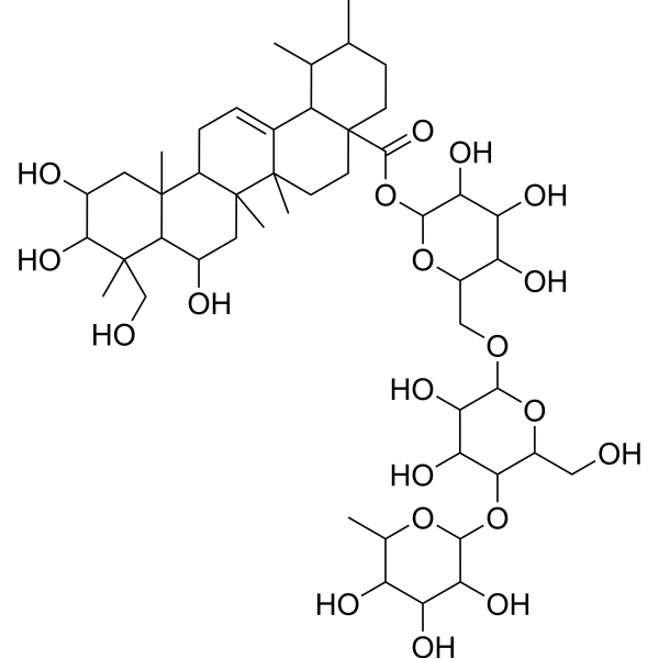
| SMILES | C[C@@H]1CC[C@@]2(CC[C@@]3(C(=CC[C@H]4[C@]3(C[C@H]([C@@H]5[C@@]4(C[C@H]([C@@H]([C@@]5(C)CO)O)O)C)O)C)[C@@H]2[C@H]1C)C)C(=O)O[C@H]6[C@@H]([C@H]([C@@H]([C@H](O6)CO[C@H]7[C@@H]([C@H]([C@@H]([C@H](O7)CO)O[C@H]8[C@@H]([C@@H]([C@H]([C@@H](O8)C)O)O)O)O)O)O)O)O |
| InChi Key | BNMGUJRJUUDLHW-HCZMHFOYSA-N |
| InChi Code | InChI=1S/C48H78O20/c1-19-10-11-48(13-12-46(6)22(28(48)20(19)2)8-9-27-44(4)14-24(52)39(61)45(5,18-50)38(44)23(51)15-47(27,46)7)43(62)68-42-35(59)32(56)30(54)26(66-42)17-63-40-36(60)33(57)37(25(16-49)65-40)67-41-34(58)31(55)29(53)21(3)64-41/h8,19-21,23-42,49-61H,9-18H2,1-7H3/t19-,20+,21+,23-,24-,25-,26-,27-,28+,29+,30-,31-,32+,33-,34-,35-,36-,37-,38-,39+,40-,41+,42+,44-,45+,46-,47-,48+/m1/s1 |
| Chemical Name | [(2S,3R,4S,5S,6R)-6-[[(2R,3R,4R,5S,6R)-3,4-dihydroxy-6-(hydroxymethyl)-5-[(2S,3R,4R,5R,6S)-3,4,5-trihydroxy-6-methyloxan-2-yl]oxyoxan-2-yl]oxymethyl]-3,4,5-trihydroxyoxan-2-yl] (1S,2R,4aS,6aR,6aS,6bR,8R,8aR,9R,10R,11R,12aR,14bS)-8,10,11-trihydroxy-9-(hydroxymethyl)-1,2,6a,6b,9,12a-hexamethyl-2,3,4,5,6,6a,7,8,8a,10,11,12,13,14b-tetradecahydro-1H-picene-4a-carboxylate |
| HS Tariff Code | 2934.99.9001 |
| Storage |
Powder-20°C 3 years 4°C 2 years In solvent -80°C 6 months -20°C 1 month |
| Shipping Condition | Room temperature (This product is stable at ambient temperature for a few days during ordinary shipping and time spent in Customs) |
Biological Activity
| Targets |
PKC, cMET, ERK1/2, COX-2 [1] PPAR-γ [2] Nrf2, HO-1 [10] class III PI3K, Beclin-1, Bcl-2 [9] |
| ln Vitro |
- In HGF-induced human hepatocellular carcinoma (HCC) cells (HepG2, Huh7), Madecassoside (10, 20, 40 μM) dose-dependently inhibited cell proliferation (MTT assay) and invasiveness (Transwell assay). It suppressed the PKC-cMET-ERK1/2-COX-2-PGE2 pathway: downregulated phosphorylated PKC, cMET, ERK1/2, and COX-2 protein expression, and reduced PGE2 secretion (Western blot, ELISA) [1] - In human umbilical vein endothelial cells (HUVECs) exposed to H2O2 (200 μM), Madecassoside (5, 10, 20 μM) dose-dependently increased cell viability (MTT assay), reduced ROS production (DCFH-DA staining), and decreased apoptosis (Annexin V-FITC/PI staining). It upregulated antioxidant enzymes (SOD, CAT) activity and downregulated caspase-3 activation (Western blot) [5] - In H2O2-induced human melanocytes, Madecassoside (10, 20, 40 μM) alleviated oxidative stress: reduced ROS generation, increased SOD activity, and inhibited excessive autophagy (decreased LC3-II/LC3-I ratio, upregulated p62 expression, Western blot). It also improved cell viability (MTT assay) [7] - In doxorubicin (DOX)-treated HK-2 renal tubular epithelial cells, Madecassoside (5, 10, 20 μM) dose-dependently protected cells from DOX-induced cytotoxicity (MTT assay), reduced ROS production, and inhibited apoptosis (downregulated Bax/Bcl-2 ratio, cleaved caspase-3 expression, Western blot) [3] - In Aβ(25-35)-treated SH-SY5Y neuronal cells, Madecassoside (5, 10, 20 μM) suppressed inflammatory responses (reduced TNF-α, IL-1β mRNA expression, qPCR) and inhibited abnormal autophagy (regulated class III PI3K/Beclin-1/Bcl-2 pathway, Western blot). It improved cell viability (MTT assay) [9] - In LPS-treated BV2 microglial cells, Madecassoside (10, 20, 40 μM) reduced neurotoxicity by activating the Nrf2-HO-1 pathway: upregulated Nrf2 and HO-1 protein expression (Western blot), decreased ROS and pro-inflammatory cytokine (TNF-α, IL-6) production (ELISA) [10] - In INS-1E pancreatic β-cells, Madecassoside (10, 20 μM) improved insulin sensitivity: increased glucose-stimulated insulin secretion (ELISA) and upregulated GLUT2 mRNA expression (qPCR) [8] |
| ln Vivo |
- In bleomycin-induced pulmonary fibrosis mice, oral administration of Madecassoside (20, 40 mg/kg/day) for 21 days ameliorated lung fibrosis: reduced collagen deposition (Masson staining), downregulated fibrosis markers (α-SMA, Col1A1 mRNA expression, qPCR), and promoted colon-derived HGF generation via activating PPAR-γ (upregulated PPAR-γ, HGF mRNA/protein expression in colon, qPCR/Western blot) [2] - In DOX-induced nephrotoxicity mice, intraperitoneal injection of Madecassoside (10, 20 mg/kg) every other day for 2 weeks protected renal function: reduced serum creatinine (Cr) and blood urea nitrogen (BUN) levels, alleviated renal tissue damage (HE staining). It inhibited oxidative stress (increased SOD activity, decreased MDA level) and apoptosis (TUNEL assay, downregulated Bax/Bcl-2 ratio) [3] - In focal cerebral ischemia-reperfusion (I/R) injury rats, intraperitoneal injection of Madecassoside (10, 20 mg/kg) before reperfusion reduced cerebral infarction volume (TTC staining), improved neurological deficit scores, and protected blood-brain barrier integrity (reduced Evans blue leakage). It inhibited oxidative stress (increased SOD, CAT activity) and inflammation (decreased TNF-α, IL-1β levels, ELISA) [6] - In LPS-induced neurotoxicity rats, oral administration of Madecassoside (20, 40 mg/kg/day) for 7 days ameliorated cognitive impairment (Morris water maze test), reduced hippocampal neuronal damage (HE staining), and activated the Nrf2-HO-1 pathway (upregulated Nrf2, HO-1 protein expression in hippocampus, Western blot) [10] |
| Enzyme Assay |
- COX-2 activity assay: HGF-induced HepG2 cells were treated with Madecassoside (10, 20, 40 μM) for 24 hours. Cells were lysed, and COX-2 activity was measured by detecting the conversion of arachidonic acid to PGE2. The reaction product PGE2 was quantified by ELISA to evaluate COX-2 inhibition [1] - PPAR-γ binding assay: Recombinant PPAR-γ ligand-binding domain protein was incubated with Madecassoside (0.1-10 μM) and a fluorescent PPAR-γ ligand. Fluorescence polarization was measured to assess the binding affinity of Madecassoside to PPAR-γ [2] |
| Cell Assay |
- HCC cell proliferation and invasion assay: HepG2/Huh7 cells were treated with Madecassoside (10, 20, 40 μM) and HGF (20 ng/mL) for 48 hours. Cell viability was detected by MTT assay; invasiveness was evaluated by Transwell assay (cells stained and counted). Western blot was used to detect pathway-related proteins, and ELISA for PGE2 secretion [1] - Endothelial cell oxidative stress assay: HUVECs were pretreated with Madecassoside (5, 10, 20 μM) for 1 hour, then exposed to H2O2 (200 μM) for 24 hours. ROS production was detected by DCFH-DA staining; apoptosis by Annexin V-FITC/PI flow cytometry; SOD/CAT activity by colorimetric assay; caspase-3 by Western blot [5] - Melanocyte autophagy assay: Human melanocytes were pretreated with Madecassoside (10, 20, 40 μM) for 2 hours, then treated with H2O2 (100 μM) for 24 hours. Cell viability by MTT assay; SOD activity by colorimetric assay; LC3-II/LC3-I, p62 by Western blot [7] - Renal cell protection assay: HK-2 cells were pretreated with Madecassoside (5, 10, 20 μM) for 1 hour, then treated with DOX (1 μM) for 24 hours. Cell viability by MTT assay; ROS by DCFH-DA staining; Bax, Bcl-2, cleaved caspase-3 by Western blot [3] - Neuronal cell assay: SH-SY5Y cells were pretreated with Madecassoside (5, 10, 20 μM) for 1 hour, then treated with Aβ(25-35) (20 μM) for 24 hours. Cell viability by MTT assay; TNF-α/IL-1β mRNA by qPCR; class III PI3K/Beclin-1/Bcl-2 pathway proteins by Western blot [9] - β-cell insulin sensitivity assay: INS-1E cells were treated with Madecassoside (10, 20 μM) for 24 hours. Glucose-stimulated insulin secretion was detected by ELISA; GLUT2 mRNA by qPCR [8] |
| Animal Protocol |
- Pulmonary fibrosis model: C57BL/6 mice were intratracheally instilled with bleomycin (5 mg/kg) to induce fibrosis. Madecassoside (20, 40 mg/kg/day) was dissolved in 0.5% CMC-Na and administered orally for 21 days. Lung tissues were collected for Masson staining and qPCR; colon tissues for qPCR/Western blot [2] - Nephrotoxicity model: BALB/c mice were intraperitoneally injected with DOX (15 mg/kg) to induce nephrotoxicity. Madecassoside (10, 20 mg/kg) was intraperitoneally injected every other day for 2 weeks. Serum was collected for Cr/BUN detection; kidney tissues for HE staining, TUNEL assay, and oxidative stress marker detection [3] - Cerebral I/R model: Sprague-Dawley rats were subjected to middle cerebral artery occlusion (MCAO) for 2 hours followed by reperfusion. Madecassoside (10, 20 mg/kg) was intraperitoneally injected 30 minutes before reperfusion. Neurological deficit scores were evaluated; cerebral tissues for TTC staining, Evans blue leakage assay, and oxidative stress/inflammation marker detection [6] - Neurotoxicity model: Sprague-Dawley rats were intraperitoneally injected with LPS (5 mg/kg) to induce neurotoxicity. Madecassoside (20, 40 mg/kg/day) was dissolved in saline and administered orally for 7 days. Morris water maze test was performed; hippocampal tissues for HE staining and Western blot [10] |
| ADME/Pharmacokinetics |
- In rats orally administered with Centella asiatica extract ECa 233 (containing Madecassoside), the pharmacokinetic parameters of Madecassoside were: Cmax = 1.8 ± 0.5 μg/mL, Tmax = 1.5 ± 0.5 h, AUC0-t = 8.2 ± 2.1 μg·h/mL, AUC0-∞ = 9.5 ± 2.4 μg·h/mL, t1/2 = 3.2 ± 0.8 h, oral bioavailability (F) = 3.1 ± 0.9% [4] - Madecassoside was widely distributed in rat tissues, with higher concentrations in the liver, kidney, and lung, and lower concentrations in the brain [4] |
| Toxicity/Toxicokinetics |
- In all in vivo experiments, Madecassoside at tested doses (10-40 mg/kg/day, oral/intraperitoneal) did not cause significant changes in animal body weight, food intake, or serum ALT/AST levels, indicating no obvious hepatotoxicity [2][3][6][10] - In vitro, Madecassoside did not show cytotoxicity to normal cells (HUVECs, melanocytes, HK-2 cells) at concentrations up to 40 μM [5][7][3] |
| References |
[1]. Madecassoside suppresses proliferation and invasiveness of HGF-induced human hepatocellular carcinoma cells via PKC-cMET-ERK1/2-COX-2-PGE2 pathway. Int Immunopharmacol. 2016 Apr;33:24-32. [2]. Madecassoside ameliorates bleomycin-induced pulmonary fibrosis in mice through promoting the generation of hepatocyte growth factor via PPAR-γ in colon. Br J Pharmacol. 2016 Apr;173(7):1219-35. [3]. Protective effects of madecassoside against Doxorubicin induced nephrotoxicity in vivo and in vitro. Sci Rep. 2015 Dec 14. [4]. Pharmacokinetics of a Standardized Extract of Centella asiatica ECa 233 in Rats. Planta Med. 2017 May;83(8):710-717. [5]. Madecassoside, a triterpenoid saponin isolated from Centella asiatica herbs, protects endothelial cells against oxidative stress. J Biochem Mol Toxicol. 2012 Oct;26(10):399-406. [6]. Neuroprotective effects of madecassoside against focal cerebral ischemia reperfusion injury in rats. Brain Res. 2014 May 27;1565:37-47. [7]. Protective effect of madecassoside on H2O2-induced oxidative stress and autophagy activation in human melanocytes. Oncotarget. 2017 May 7;8(31):51066-51075. [8]. Effect of madecassoside and catalpol in amelioration of insulin sensitivity in pancreatic (INS-1E) β-cell line. Nat Prod Res. 2021 Nov;35(22):4627-4631. [9]. Madecassoside prevents Aβ(25-35)-induced inflammatory responses and autophagy in neuronal cells through the class III PI3K/Beclin-1/Bcl-2 pathway. Int Immunopharmacol. 2014 May;20(1):221-8. [10]. Madecassoside ameliorates lipopolysaccharide-induced neurotoxicity in rats by activating the Nrf2-HO-1 pathway. Neurosci Lett. 2019 Sep 14;709:134386. |
| Additional Infomation |
Madecassoside has been reported in Actaea dahurica, Centella erecta, and other organisms with data available. See also: Madecassoside (annotation moved to). - Madecassoside is a triterpenoid saponin isolated from the Chinese herbal medicine Centella asiatica [3][5][9] - Its core biological activities include anti-tumor (inhibiting HCC cell proliferation/invasion), anti-fibrotic (ameliorating pulmonary fibrosis), organ protective (renal, cerebral, neuronal, endothelial), anti-oxidative stress, anti-inflammatory, and improving insulin sensitivity [1][2][3][5][6][7][8][9][10] - The underlying mechanisms involve regulating multiple signaling pathways: PKC-cMET-ERK1/2-COX-2-PGE2, PPAR-γ-HGF, Nrf2-HO-1, class III PI3K/Beclin-1/Bcl-2, and modulating oxidative stress, apoptosis, autophagy, and inflammation [1][2][5][7][9][10] - Madecassoside shows potential therapeutic value for hepatocellular carcinoma, pulmonary fibrosis, drug-induced nephrotoxicity, cerebral ischemia-reperfusion injury, neuroinflammation, and metabolic disorders (insulin resistance) [1][2][3][6][8][10] |
Solubility Data
| Solubility (In Vitro) |
DMSO : ~100 mg/mL (~102.55 mM) H2O : ~33.33 mg/mL (~34.18 mM) |
| Solubility (In Vivo) |
Solubility in Formulation 1: ≥ 2.5 mg/mL (2.56 mM) (saturation unknown) in 10% DMSO + 40% PEG300 + 5% Tween80 + 45% Saline (add these co-solvents sequentially from left to right, and one by one), clear solution. For example, if 1 mL of working solution is to be prepared, you can add 100 μL of 25.0 mg/mL clear DMSO stock solution to 400 μL PEG300 and mix evenly; then add 50 μL Tween-80 to the above solution and mix evenly; then add 450 μL normal saline to adjust the volume to 1 mL. Preparation of saline: Dissolve 0.9 g of sodium chloride in 100 mL ddH₂ O to obtain a clear solution. Solubility in Formulation 2: ≥ 2.5 mg/mL (2.56 mM) (saturation unknown) in 10% DMSO + 90% (20% SBE-β-CD in Saline) (add these co-solvents sequentially from left to right, and one by one), clear solution. For example, if 1 mL of working solution is to be prepared, you can add 100 μL of 25.0 mg/mL clear DMSO stock solution to 900 μL of 20% SBE-β-CD physiological saline solution and mix evenly. Preparation of 20% SBE-β-CD in Saline (4°C,1 week): Dissolve 2 g SBE-β-CD in 10 mL saline to obtain a clear solution. Solubility in Formulation 3: ≥ 2.5 mg/mL (2.56 mM) (saturation unknown) in 10% DMSO + 90% Corn Oil (add these co-solvents sequentially from left to right, and one by one), clear solution. For example, if 1 mL of working solution is to be prepared, you can add 100 μL of 25.0 mg/mL clear DMSO stock solution to 900 μL of corn oil and mix evenly. Solubility in Formulation 4: 25 mg/mL (25.64 mM) in PBS (add these co-solvents sequentially from left to right, and one by one), clear solution; with ultrasonication. (Please use freshly prepared in vivo formulations for optimal results.) |
| Preparing Stock Solutions | 1 mg | 5 mg | 10 mg | |
| 1 mM | 1.0255 mL | 5.1276 mL | 10.2551 mL | |
| 5 mM | 0.2051 mL | 1.0255 mL | 2.0510 mL | |
| 10 mM | 0.1026 mL | 0.5128 mL | 1.0255 mL |