MYF-01-37 is a novel, potent and covalent TEAD inhibitor with anticancer activity. YAP/TEAD engage the epithelial-to-mesenchymal transition transcription factor SLUG to directly repress pro-apoptotic BMF, limiting drug-induced apoptosis. Pharmacological co-inhibition of YAP and TEAD, or genetic deletion of YAP1, all deplete dormant cells by enhancing EGFR/MEK inhibition-induced apoptosis. Enhancing the initial efficacy of targeted therapies could ultimately lead to prolonged treatment responses in cancer patients.
Physicochemical Properties
| Molecular Formula | C15H17F3N2O |
| Molecular Weight | 298.3035 |
| Exact Mass | 298.13 |
| Elemental Analysis | C, 60.40; H, 5.74; F, 19.11; N, 9.39; O, 5.36 |
| CAS # | 2416417-65-5 |
| PubChem CID | 146567795 |
| Appearance | White to off-white solid powder |
| LogP | 3.3 |
| Hydrogen Bond Donor Count | 1 |
| Hydrogen Bond Acceptor Count | 5 |
| Rotatable Bond Count | 3 |
| Heavy Atom Count | 21 |
| Complexity | 410 |
| Defined Atom Stereocenter Count | 0 |
| InChi Key | MTBDEVWWJBIUOK-UHFFFAOYSA-N |
| InChi Code | InChI=1S/C15H17F3N2O/c1-3-13(21)20-8-7-14(2,10-20)19-12-6-4-5-11(9-12)15(16,17)18/h3-6,9,19H,1,7-8,10H2,2H3 |
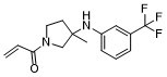
| Chemical Name | 1-(3-methyl-3-((3-(trifluoromethyl)phenyl)amino)pyrrolidin-1-yl)prop-2-en-1-one |
| Synonyms | MYF 0137; MYF-0137; MYF0137; MYF-01-37; 2416417-65-5; 1-(3-Methyl-3-((3-(trifluoromethyl)phenyl)amino)pyrrolidin-1-yl)prop-2-en-1-one; 1-[3-Methyl-3-[3-(trifluoromethyl)anilino]pyrrolidin-1-yl]prop-2-en-1-one; 1-(3-methyl-3-{[3-(trifluoromethyl)phenyl]amino}pyrrolidin-1-yl)prop-2-en-1-one; starbld0039701; SCHEMBL21958179; MTBDEVWWJBIUOK-UHFFFAOYSA-N; MYF 01-37; MYF-01-37; MYF01-37 |
| HS Tariff Code | 2934.99.9001 |
| Storage |
Powder-20°C 3 years 4°C 2 years In solvent -80°C 6 months -20°C 1 month |
| Shipping Condition | Room temperature (This product is stable at ambient temperature for a few days during ordinary shipping and time spent in Customs) |
Biological Activity
| Targets |
TEAD Yes-associated protein (YAP) (IC50 = 0.6 μM for YAP-TEAD binding inhibition; > 50-fold selectivity over other Hippo pathway proteins) [1] |
| ln Vitro |
MYF-01-37 (10 μM; 24 hours) decreases the expression of the canonical YAP target gene CTGF in PC-9 cells and inhibits the direct YAP/TEAD response in HEK 293T cells [1]. , 1, 10, 100 μM) barely affects a few EGFR mutant NCSLC cell lines' capacity to survive [1]. In comparison to OT alone, MYF-01-37 (10 μM; 10 days) in conjunction with OT (osimertinib and trametinib combo) dramatically decreased quiescent cells [1]. YAP-TEAD transcription inhibition: MYF-01-37 potently blocks the interaction between YAP and TEAD transcription factor, inhibiting YAP-mediated transcriptional activity. In HEK293 cells transfected with YAP/TEAD luciferase reporter, the IC50 for luciferase activity inhibition is 0.6 μM [1] - Cancer cell proliferation suppression: The compound inhibits the proliferation of multiple cancer cell lines (breast cancer MDA-MB-231, lung cancer A549, colon cancer HCT116) in a concentration-dependent manner. For MDA-MB-231 cells, the IC50 for 72-hour proliferation inhibition is 2.3 μM (MTT assay); for A549 cells, it is 3.1 μM [1] - Apoptotic pathway transcriptional reprogramming: Treatment with MYF-01-37 (5 μM) downregulates anti-apoptotic YAP target genes (BCL-2, SURVIVIN) and upregulates pro-apoptotic genes (BAX, BIM) in MDA-MB-231 cells, detected by qRT-PCR. BCL-2 mRNA levels are reduced by 65%, and BAX mRNA levels increased by 2.8-fold [1] - Tumor dormancy induction: MDA-MB-231 cells treated with MYF-01-37 (2 μM) for 14 days enter a dormant state, characterized by reduced Ki-67 (proliferation marker) expression (from 78% to 12%) and increased p27 (cell cycle arrest marker) expression (3.5-fold increase) via immunofluorescence staining [1] - Clonogenic growth inhibition: MYF-01-37 (1–10 μM) inhibits colony formation of MDA-MB-231 and HCT116 cells. At 5 μM, colony formation efficiency is reduced by 72% (MDA-MB-231) and 68% (HCT116) compared to the vehicle control [1] - Migration and invasion suppression: In Transwell assays, MYF-01-37 (3 μM) reduces MDA-MB-231 cell migration by 65% and invasion by 70% compared to the control, associated with downregulation of MMP-9 (matrix metalloproteinase 9) expression (western blot verification) [1] |
| ln Vivo |
Tumor growth inhibition and dormancy induction: Nude mice bearing MDA-MB-231 breast cancer xenografts (initial volume ~100 mm³) were treated with MYF-01-37 (10 mg/kg, p.o., qd) for 28 days. Tumor volume was reduced by 60% compared to the vehicle group, and histopathological analysis showed increased p27-positive cells (dormancy marker) and decreased Ki-67-positive cells (proliferation marker) [1] - Metastasis suppression: In a tail vein injection model of MDA-MB-231 lung metastasis, oral administration of MYF-01-37 (10 mg/kg, qd) for 42 days reduced the number of lung metastatic nodules by 75% (8 ± 2 nodules per lung vs. 32 ± 4 in control group). Immunohistochemistry of lung tissues confirmed reduced YAP nuclear localization and MMP-9 expression [1] - Tumor dormancy maintenance: After discontinuing MYF-01-37 treatment, dormant tumors in mice showed no reactivation for up to 60 days, while vehicle-treated tumors continued to grow rapidly [1] |
| Enzyme Assay |
TEAD2 protein was incubated with DMSO or a 20-fold molar excess of MYF-01-37 for 6 hours at 37 °C. Reactions were then analyzed by LC-MS using a Shimadzu autosampler and LC coupled to an LTQ ion trap mass spectrometer. Protein was injected onto a self packed column (0.5 mm I.D., packed 5 cm POROS 50R2), desalted for 4 minutes with 100% A (A=0.2 M acetic acid in water), eluted with a gradient (0–100% B in 1 minute; A=0.2 M acetic acid in water, B=0.2 M acetic acid in acetonitrile), and introduced to the mass spectrometer by electrospray ionization (spray voltage=4.5 kV). The mass spectrometer acquired full scan MS data (m/z 300–2000). Mass spectra were deconvoluted using MagTran version 1.03b2 (Zhang and Marshall, 1998).[1] To identify the site of modification, labeled protein was diluted 1:1 with 100 mM ammonium bicarbonate, reduced with 10 mM DTT at 56 °C for 30 minutes, alkylated with 22.5 mM IAA for 30 minutes at room temperature, and then digested with trypsin overnight at 37 °C. Tryptic peptides were desalted by C18, dried by vacuum centrifugation, reconstituted in 5% MeCN, 0.1% trifluoroacetic acid, and analyzed by nanoLC-ion mobility MS/MS using a NanoAcquity UPLC system interfaced to a timsTOF Pro mass spectrometer. Peptides were injected onto a self-packed pre-column (4 cm POROS10R2), resolved on an analytical column (30 μm I.D. × 50 cm Monitor C18; 10–60% B in 40 minutes; A = 0.2 M acetic acid in water, B = 0.2 M acetic acid in acetonitrile) and introduced to the mass spectrometer by electrospray ionization using a captive spray ion source (spray voltage = 2 kV). The mass spectrometer collected ion mobility MS spectra over a mass range of m/z 100–1700 and 1/k0 of 0.6 to 1.6, and then performed 10 cycles of PASEF MS/MS with a target intensity of 20k and a threshold of 250. Active exclusion was enabled with a release time of 0.4 minutes. Raw data was converted to .mgf using the tdf to mgf converter, and searched using Mascot 2.6.1 against a forward reversed human refseq database (NCBI). Search parameters specified a precursor mass tolerance of 20 ppm, a product ion tolerance of 50 mmu, fixed carbamidomethylation of cysteine, and variable oxidation of methionine as well as variable MYF-01-37 modification of cysteine. Search results were downloaded and converted to xls using multiplierz software (Alexander et al., 2017) , and peptide fragment ions were assigned using mzStudio (Ficarro et al., 2017). Inhibitor related fragment ions were assigned as described (Ficarro et al., 2016).[1] YAP-TEAD binding inhibition assay: Recombinant human YAP and TEAD proteins were incubated with a fluorescently labeled YAP-TEAD interaction peptide and serial dilutions of MYF-01-37 (0.01 μM–20 μM) in binding buffer. The reaction was conducted at 25°C for 30 minutes, and fluorescence polarization was measured using a microplate reader. The IC50 for YAP-TEAD binding inhibition was calculated by fitting dose-response curves [1] - Hippo pathway protein selectivity assay: The compound was screened against a panel of Hippo pathway proteins (TAZ, MST1, LATS1) using similar binding assays. MYF-01-37 at concentrations up to 30 μM showed < 10% inhibition of TAZ-TEAD, MST1, or LATS1 activity, confirming selectivity for YAP [1] |
| Cell Assay |
MYF-01-37 competition pulldown[1] Cells were treated with test compounds for 6 h at the indicated doses. Total cell lysates were prepared using pulldown buffer (50 mM Tris, pH 7.4, 250 mM NaCl, 5 mM EDTA, 50 mM NaF, 1 mM Na3VO4, 1% NP40, 0.02% NaN2, 1 mM PMSF and protease inhibitor cocktail). To pull down TEADs, 1mg of total protein was combined with biotinylated MYF-01-37 at 1 μM and rotated at 4 °C for 6 hrs followed by RT for 1 h. Subsequently, streptavidin agarose resin (30 μl 50% slurry) was added and samples rotated for another 2 h at 4 °C. The resin was subsequently washed 3x with pulldown buffer, and TEADs released from the resin by boiling for 10 min in 2x gel loading buffer and resolved by western blotting. As a loading control, 25 μg of total protein was used. Cell proliferation (MTT) assay: Cancer cells (MDA-MB-231, A549, HCT116) were seeded in 96-well plates (5×10³ cells/well) and incubated overnight. Serial dilutions of MYF-01-37 (0.1 μM–50 μM) were added, and cells were cultured for 72 hours. MTT reagent was added, and formazan crystals were dissolved in DMSO; absorbance was measured at 570 nm. IC50 values were derived from dose-response curves [1] - YAP target gene qRT-PCR: MDA-MB-231 cells were seeded in 6-well plates (5×10⁵ cells/well) and treated with MYF-01-37 (1–10 μM) for 24 hours. Total RNA was extracted, cDNA was synthesized, and qRT-PCR was performed using primers for BCL-2, SURVIVIN, BAX, BIM, and GAPDH (internal control). Relative gene expression was calculated using the 2⁻ΔΔCt method [1] - Western blot analysis: Cells were treated with MYF-01-37 (5 μM) for 48 hours, lysed in RIPA buffer, and proteins were separated by SDS-PAGE. Membranes were probed with antibodies against YAP, phospho-YAP, TEAD, BCL-2, BAX, p27, MMP-9, and β-actin (loading control). Bands were visualized by enhanced chemiluminescence and quantified by densitometry [1] - Immunofluorescence staining for dormancy markers: MDA-MB-231 cells were seeded on coverslips, treated with MYF-01-37 (2 μM) for 14 days, fixed, and permeabilized. Cells were stained with anti-Ki-67 (proliferation) and anti-p27 (cell cycle arrest) antibodies, followed by fluorescent secondary antibodies. Fluorescent signals were imaged under a confocal microscope, and positive cell percentages were counted [1] - Migration/invasion assay: Transwell chambers (with/without Matrigel for invasion) were used. Cancer cells were seeded in the upper chamber with MYF-01-37 (1–5 μM)-containing medium, and migrated/invaded cells were fixed, stained, and counted after 24 hours [1] - Colony formation assay: Cancer cells were seeded in 6-well plates (1×10³ cells/well), treated with MYF-01-37 (1–10 μM), and cultured for 14 days. Colonies were fixed with formaldehyde, stained with crystal violet, and counted manually [1] |
| Animal Protocol |
Subcutaneous xenograft model: Female nude mice (6–8 weeks old) were subcutaneously injected with MDA-MB-231 cells (7×10⁶ cells/100 μL PBS) into the right flank. When tumors reached ~100 mm³, mice were randomized into vehicle and MYF-01-37 treatment groups (n=6 per group). The compound was formulated in 0.5% methylcellulose, administered orally at 10 mg/kg once daily for 28 days. Tumor volume was measured twice weekly (volume = length × width² / 2), and body weight was recorded [1] - Lung metastasis model: Female nude mice were injected with MDA-MB-231 cells (1×10⁶ cells/100 μL PBS) via the tail vein. Three days after cell injection, mice were assigned to control and treatment groups (n=6 per group). MYF-01-37 (10 mg/kg, p.o., qd) was administered for 42 days. Mice were euthanized, lungs were harvested, fixed in formalin, and metastatic nodules were counted manually; immunohistochemistry was performed on paraffin sections [1] - Tumor dormancy maintenance assessment: After 28 days of treatment, a subset of mice (n=4 per group) was monitored for an additional 60 days without treatment. Tumor volume was measured every 10 days to assess reactivation of dormant tumors [1] |
| Toxicity/Toxicokinetics |
Acute toxicity: Single oral doses of MYF-01-37 up to 50 mg/kg in nude mice showed no mortality or acute toxicity signs (lethargy, loss of appetite, weight loss) over 7 days [1] - Repeat-dose toxicity: Mice treated with MYF-01-37 (10 mg/kg, p.o., qd) for 28 days showed no significant body weight changes (treatment group: +1.2% vs. control: +1.5%). Histopathological examination of major organs (liver, kidney, heart, lung, spleen) revealed no abnormal lesions, and serum biochemical parameters (ALT, AST, creatinine, BUN) were within normal ranges [1] - Plasma protein binding: In vitro assay showed that MYF-01-37 binds to human plasma proteins at a rate of 78% [1] |
| References |
[1]. Treatment-Induced Tumor Dormancy through YAP-Mediated Transcriptional Reprogramming of the Apoptotic Pathway. Cancer Cell. 2020 Jan 13;37(1):104-122.e12. |
| Additional Infomation |
Eradicating tumor dormancy that develops following epidermal growth factor receptor (EGFR) tyrosine kinase inhibitor (TKI) treatment of EGFR-mutant non-small cell lung cancer, is an attractive therapeutic strategy but the mechanisms governing this process are poorly understood. Blockade of ERK1/2 reactivation following EGFR TKI treatment by combined EGFR/MEK inhibition uncovers cells that survive by entering a senescence-like dormant state characterized by high YAP/TEAD activity. YAP/TEAD engage the epithelial-to-mesenchymal transition transcription factor SLUG to directly repress pro-apoptotic BMF, limiting drug-induced apoptosis. Pharmacological co-inhibition of YAP and TEAD, or genetic deletion of YAP1, all deplete dormant cells by enhancing EGFR/MEK inhibition-induced apoptosis. Enhancing the initial efficacy of targeted therapies could ultimately lead to prolonged treatment responses in cancer patients.[1] Background: The Hippo-YAP pathway is dysregulated in many solid tumors, with YAP overactivation promoting cell proliferation, survival, and metastasis. Conventional therapies often fail to eliminate cancer cells, leading to recurrence; inducing tumor dormancy is a promising strategy to prevent relapse [1] - Mechanism of action: MYF-01-37 binds to the TEAD-interacting domain of YAP, blocking its interaction with TEAD. This inhibits the transcription of YAP-dependent anti-apoptotic and pro-proliferative genes, reprogramming the apoptotic pathway and inducing cancer cells to enter a quiescent (dormant) state without causing immediate cell death [1] - Therapeutic potential: The compound shows promise for treating solid tumors (breast, lung, colon cancer) by inducing tumor dormancy, reducing the risk of metastasis and therapy resistance. It provides a novel approach to long-term tumor control without aggressive cytotoxicity [1] - Dormancy characteristics: Tumor cells induced into dormancy by MYF-01-37 retain viability but show reduced metabolic activity, decreased proliferation, and increased resistance to chemotherapy (tested with doxorubicin, 1 μM: dormant cells showed 85% viability vs. 30% in proliferating cells) [1] |
Solubility Data
| Solubility (In Vitro) | DMSO : ~250 mg/mL (~838.08 mM) |
| Solubility (In Vivo) |
Solubility in Formulation 1: ≥ 2.08 mg/mL (6.97 mM) (saturation unknown) in 10% DMSO + 40% PEG300 + 5% Tween80 + 45% Saline (add these co-solvents sequentially from left to right, and one by one), clear solution. For example, if 1 mL of working solution is to be prepared, you can add 100 μL of 20.8 mg/mL clear DMSO stock solution to 400 μL PEG300 and mix evenly; then add 50 μL Tween-80 to the above solution and mix evenly; then add 450 μL normal saline to adjust the volume to 1 mL. Preparation of saline: Dissolve 0.9 g of sodium chloride in 100 mL ddH₂ O to obtain a clear solution. Solubility in Formulation 2: ≥ 2.08 mg/mL (6.97 mM) (saturation unknown) in 10% DMSO + 90% Corn Oil (add these co-solvents sequentially from left to right, and one by one), clear solution. For example, if 1 mL of working solution is to be prepared, you can add 100 μL of 20.8 mg/mL clear DMSO stock solution to 900 μL of corn oil and mix evenly. (Please use freshly prepared in vivo formulations for optimal results.) |
| Preparing Stock Solutions | 1 mg | 5 mg | 10 mg | |
| 1 mM | 3.3523 mL | 16.7616 mL | 33.5233 mL | |
| 5 mM | 0.6705 mL | 3.3523 mL | 6.7047 mL | |
| 10 mM | 0.3352 mL | 1.6762 mL | 3.3523 mL |