MRS2578 (MRS-2578; MRS 2578) is a potent and selective P2Y6 receptor antagonist with potentially usefulness in controlling persistent storage symptoms in obstructed patients. It inhibits P2Y6 receptor with an IC50 of 37 nM and exhibits little activity against closely related P2Y1, P2Y2, P2Y4, and P2Y11 receptors. Activation of P2Y6 receptor amplifies mucosal adenosine triphosphate release underlying bladder overactivity in patients with benign prostatic hyperplasia. Therefore, selective P2Y6 receptor blockade as a novel therapeutic strategy is potentially useful to control persistent storage symptoms in obstructed patients.
Physicochemical Properties
| Molecular Formula | C20H20N6S4 | |
| Molecular Weight | 472.67 | |
| Exact Mass | 472.063 | |
| Elemental Analysis | C, 50.82; H, 4.27; N, 17.78; S, 27.13 | |
| CAS # | 711019-86-2 | |
| Related CAS # |
|
|
| PubChem CID | 16078986 | |
| Appearance | White to off-white solid powder | |
| Density | 1.3±0.1 g/cm3 | |
| Boiling Point | 652.7±65.0 °C at 760 mmHg | |
| Flash Point | 348.5±34.3 °C | |
| Vapour Pressure | 0.0±2.0 mmHg at 25°C | |
| Index of Refraction | 1.687 | |
| LogP | 5.1 | |
| Hydrogen Bond Donor Count | 4 | |
| Hydrogen Bond Acceptor Count | 6 | |
| Rotatable Bond Count | 9 | |
| Heavy Atom Count | 30 | |
| Complexity | 604 | |
| Defined Atom Stereocenter Count | 0 | |
| InChi Key | QOHNRGHTJPFMSL-UHFFFAOYSA-N | |
| InChi Code | InChI=1S/C20H20N6S4/c27-13-23-15-5-3-7-17(11-15)25-19(29)21-9-1-2-10-22-20(30)26-18-8-4-6-16(12-18)24-14-28/h3-8,11-12H,1-2,9-10H2,(H2,21,25,29)(H2,22,26,30) | |
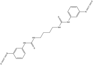
| Chemical Name | 1-(3-isothiocyanatophenyl)-3-[4-[(3-isothiocyanatophenyl)carbamothioylamino]butyl]thiourea | |
| Synonyms |
|
|
| HS Tariff Code | 2934.99.9001 | |
| Storage |
Powder-20°C 3 years 4°C 2 years In solvent -80°C 6 months -20°C 1 month Note: Please store this product in a sealed and protected environment, avoid exposure to moisture. |
|
| Shipping Condition | Room temperature (This product is stable at ambient temperature for a few days during ordinary shipping and time spent in Customs) |
Biological Activity
| Targets |
P2Y6 Receptor The target of MRS 2578 is the purinergic receptor P2Y6, with an IC50 of 10.8 nM for human P2Y6 receptor and 9.1 nM for rat P2Y6 receptor [1] |
| ln Vitro |
MRS2578 (1 μM) totally prevents 1321N1 astrocytoma cells from being protected by UDP during TNFα-induced apoptosis[1]. In HMEC-1 cells, MRS 2578 (10 μM) totally eliminates TNF-α-induced NF-κB reporter activity. In HMEC-1 cells, MRS 2578 (10 μM) dramatically lowers TNF-α-induced proinflammatory gene expression[2]. The physiological role of the P2Y(6) nucleotide receptor may involve cardiovascular, immune and digestive functions based on the receptor tissue distribution, and selective antagonists for this receptor are lacking. We have synthesized a series of symmetric aryl diisothiocyanate derivatives and examined their ability to inhibit phospholipase C (PLC) activity induced by activation of five subtypes of recombinant P2Y receptors. Several derivatives were more potent at inhibiting action of UDP at both human and rat P2Y(6) receptors expressed in 1321N1 human astrocytes than activation of human P2Y(1), P2Y(2), P2Y(4) and P2Y(11) receptors. The inhibition by diisothiocyanate derivatives of 1,2-diphenylethane (MRS2567) and 1,4-di-(phenylthioureido) butane (MRS2578) was concentration-dependent and insurmountable, with IC(50) values of 126+/-15 nM and 37+/-16 nM (human) and 101+/-27 nM and 98+/-11 nM (rat), respectively. A derivative of 1,4-phenylendiisothiocyanate (MRS2575) inhibited only human but not rat P2Y(6) receptor activity. MRS2567 and MRS2578 at 10microM did not affect the UTP (100nM)-induced responses of cells expressing P2Y(2) and P2Y(4) receptors, nor did they affect the 2-methylthio-ADP (30nM)-induced responses at the P2Y(1) receptor or the ATP (10microM)-induced responses at the P2Y(11) receptor. Other antagonists displayed mixed selectivities. The selective antagonists MRS2567, MRS2575 and MRS2578 (1microM) completely blocked the protection by UDP of cells undergoing TNFalpha-induced apoptosis. Thus, we have identified potent, insurmountable antagonists of P2Y(6) receptors that are selective within the family of PLC-coupled P2Y receptors. [1] Treatment with MRS2578, a selective P2Y6 receptor antagonist, suppressed mechanical stretch-induced Rho activation in a concentration-dependent manner, with an IC50 value of about 0.1 μM (Figure 5A and B). [2] P2Y6 receptor antagonist MRS 2578 dampens mediator-induced inflammation of vascular endothelia in vitro [3] After having shown that P2Y6 receptor transcript and protein expressions are selectively increased on inflammatory stimulation, we next studied functional consequences of endothelial P2Y6 signaling in vitro. As in the in vitro model, we transfected HMEC-1 cells with a NF-κB reporter plasmid containing the binding sites for p50/p65. Treatment of HMEC-1 cells with the P2Y6 agonist uridine diphosphate only resulted in a modest increase in NF-κB activity (1.64-fold ± 0.45[fold; P < .05; supplemental Figure 1). However, and consistent with previous studies implicating P2Y6 signaling in NF-κB activity,26,27 we observed a profound inhibition of basal NF-κB activity in the presence of the P2Y6 antagonist MRS 2578. This reduction of NF-κB activity was time (Figure 3A) and dose (Figure 3B) dependent. As such, these studies indicate that downstream targets of P2Y6 activation are necessary but not sufficient for NF-κB activation and speak for an indirect effect in enhancing vascular inflammation[3]. In cells transfected with human or rat P2Y6 receptors, MRS 2578 concentration-dependently inhibited UDP-induced P2Y6 receptor activation, blocked receptor-mediated intracellular Ca²⁺ elevation, and the inhibitory effect was irreversible [1] - Selectivity experiments showed that MRS 2578 had no significant inhibitory activity on other purinergic receptors (P2Y1, P2Y2, P2Y4, P2Y11, P2Y12, P2X1, P2X2, P2X4, P2X7). Even at a concentration of 10 μmol/L, the inhibition rate on these receptors was less than 20% [1] - In in vitro experiments of human umbilical vein endothelial cells (HUVECs), pretreatment with MRS 2578 significantly inhibited UDP-induced mRNA and protein expression of inflammatory factors (IL-8, MCP-1, ICAM-1), and simultaneously blocked the activation of the NF-κB signaling pathway [3] - In in vitro culture experiments of mouse airway epithelial cells, MRS 2578 inhibited IL-4-induced upregulation of P2Y6 receptor expression, reduced the secretion of chemokines (CCL11, CCL24), and thereby inhibited eosinophil chemotaxis [4] - In rat cardiac fibroblasts, MRS 2578 could block the activation of the P2Y6-Gα12/13 signaling pathway and inhibit cell proliferation and the synthesis of collagen (Col1a1, Col3a1) [2] |
| ln Vivo |
After transverse aortic constriction (TAC), MRS2578 (3 mg/kg; ip; for 3 days) dramatically reduces pressure overload-induced collagen deposition without influencing cardiomyocyte hypertrophy[4]. Inhibition of P2Y6 receptors attenuates pressure overload-induced cardiac fibrosis in vivo [2] Researchers next examined whether purinergic receptors actually participate in pressure overload-induced cardiac fibrosis in vivo. Treatment with MRS2578 after TAC significantly suppressed pressure overload-induced collagen deposition without affecting cardiomyocyte hypertrophy (Figure 6A–C). Treatment with MRS2578 significantly suppressed LV dysfunction induced by pressure overload (Figure 6D and E and Supplementary Table 3). Furthermore, the treatment with MRS2578 suppressed the increases in mRNA expressions of ANP, β-MHC, procollagen type I, periostin, and TGF-β2 by pressure overload (Figure 6F). We also found that MRS2578 inhibited pressure overload-induced Rho activation and TAC-induced increases in expression of periostin, mature TGF-βs, and ACE proteins (Figure 6G and H). Furthermore, we found that treatment with suramin also suppressed pressure overload-induced collagen deposition and LV dysfunction (Supplementary Figure 7 and Supplementary Table 4). These results suggest that inhibition of P2Y6 receptors actually attenuates pressure overload-induced cardiac fibrosis and LV dysfunction. Measurements and main results: We observed that the intratracheal application of a P2Y6R antagonist (MRS2578) and P2Y6R deficiency inhibited cardinal features of asthma, such as bronchoalveolar lavage eosinophilia, airway remodeling, Th2 cytokine production, and bronchial hyperresponsiveness in the ovalbumin-alum model. MRS2578 was also effective in reducing airway inflammation in a model using house dust mite extracts to induce allergic lung inflammation. Experiments with bone marrow chimeras revealed the importance of the P2Y6R expression on lung structural cells in airway inflammation. In accordance with this finding, we found a strong up-regulation of P2Y6 expression on airway epithelial cells of animals with experimental asthma. Concerning the underlying mechanism, we observed that MRS2578 inhibited the release of IL-6 and IL-8/KC by lung epithelial cells in vivo, whereas intrapulmonary application of the P2Y6R agonist uridine-5'-diphosphate increased the bronchoalveolar levels of IL-6 and KC. In addition, selective activation of P2Y6 receptors induced the release of IL-6 and KC/IL-8 by murine and human lung epithelial cells in vitro. Conclusions: P2Y6R expression on airway epithelial cells is up-regulated during acute and chronic allergic airway inflammation, and selective blocking of P2Y6R or P2Y6R deficiency on the structural cells reduces cardinal features of experimental asthma. Thus, blocking pulmonary P2Y6R might be a target for the treatment of allergic airway inflammation [4]. In a mouse model of pressure overload-induced cardiac fibrosis (induced by transverse aortic constriction), intraperitoneal injection of MRS 2578 (10 mg/kg, once daily for 4 weeks) significantly reduced collagen deposition in myocardial tissue, inhibited the expression of fibroblast activation marker (α-SMA), and improved cardiac diastolic function [2] - In a mouse model of vascular inflammation induced by carotid artery ligation, intraperitoneal injection of MRS 2578 (10 mg/kg, once daily for 14 days) reduced inflammatory cell infiltration (macrophages, neutrophils) in the vascular wall, decreased the expression of inflammatory factors (TNF-α, IL-6) and adhesion molecule (VCAM-1), and inhibited vascular intimal hyperplasia [3] - In a mouse model of allergic airway inflammation (ovalbumin-sensitized and challenged), intraperitoneal injection of MRS 2578 (5 mg/kg, once every 2 days for 14 days after sensitization) significantly reduced airway eosinophil infiltration, decreased the levels of Th2 cytokines (IL-4, IL-5, IL-13) in bronchoalveolar lavage fluid, and improved airway hyperresponsiveness and airway remodeling (reduced airway smooth muscle thickening and mucus secretion) [4] |
| Enzyme Assay |
Radioligand binding assays [1] P2Y1 receptor binding experiments were performed as previously described. Briefly, membranes (40 μg protein) from astrocytoma cells stably expressing human P2Y1 receptors were incubated with [3H]MRS2279 (8 nM) for 30 min at 4 °C in a total assay volume of 200 μl. For adenosine A1 receptor binding, an agonist radioligand [3H]R-PIA (2.0 nM) was incubated with membranes (40 μg protein/tube) from CHO cells stably expressing human adenosine A1 receptors for 60 min at 25 °C. Radiolabeled ligand concentrations used in all assays approximated the Kd values of the receptor. Binding reactions were terminated by filtration through Whatman GF/B glass-fiber filters under reduced pressure with a MT-24 cell harvester (Brandel), and radioactivity was determined with a 1414 liquid scintillation counter. P2Y6 receptor activity inhibition assay: Cells transfected with human or rat P2Y6 receptors were seeded in 96-well plates. After culturing to confluence, Ca²⁺ fluorescent probes were loaded. The cells were pretreated with MRS 2578 at different concentrations for 30 minutes, then stimulated with UDP. The change in intracellular Ca²⁺ fluorescence intensity was detected in real-time by a fluorescence microplate reader to calculate the IC50 value [1] - Receptor selectivity assay: Cells transfected with different purinergic receptors (P2Y1, P2Y2, etc.) were used. According to the above Ca²⁺ detection method, the effect of MRS 2578 at 10 μmol/L on the Ca²⁺ response mediated by each receptor was detected to evaluate its selectivity [1] |
| Cell Assay |
Evaluation of nuclear factor κB activity [3] We used reporter assays to assess nuclear factor κB (NF-κB) activity. To measure the transcriptional activity of NF-κB, endothelial cells were plated in 24-well plates at a density of 2.5 × 10⁴ cells/well and were allowed to adhere overnight. The monolayers were then transfected with 0.25 μg of either NF-κB promoter reporter or control pGL3 vector for 4 hours with the use of GeneJuice Transfection Reagent according to the manufacturer's instructions. Cells were exposed to the P2Y6 receptor antagonist MRS2578 or solvent (dimethyl sulfoxide [DMSO]) for 30 minutes. Subsequently, 10 ng/mL TNF-α were added for another 2 hours. At the end of the incubation period, cells were washed twice in ice-cold phosphate-buffered saline, and luciferase activity was measured with the use of the Luciferase Assay System. For normalization protein concentration was determined with the use of a BCA Protein Assay kit. Repression of inflammatory cytokine mRNA by MRS2578 [3] HMEC-1 cells were preincubated with 10μM MRS 2578 for 30 minutes. TNF-α (10 ng/mL) was added, and cells were lysed after indicated time points. mRNA levels of NF-κB–induced genes were determined with the use of the primer sets summarized in supplemental Table 1. Isolation of primary normal human bronchial epithelial cells. [4] The normal human bronchial epithelial cells were obtained from the bronchi of explanted lungs or their bronchial rings. The study was approved by the local ethics committee of Freiburg. Bronchi were longitudinally opened and mechanically dissected by using a disposable scalpel and subsequently washed in ice-cold Hanks’ balanced salt solution. The mucosa was minced in small pieces and digested in Dispase II in 80 ml PII solution, supplemented with 100 μl DNase and penicillin and streptomycin for 90 minutes in a water bath at 37°C. The crude solution was filtered using a cell strainer of 100 μm, centrifuged at 1,500 rpm, 5 minutes at 4°C and then resuspended in RPMI 1649 medium supplemented with penicillin and streptomycin for 15 minutes and placed in a Petri dish for 15 minutes. Nonadherent cells were carefully harvested and counted and then cultured (1 × 10~6 cells/well) in 6-well plates using Quantum 286 for Epithelial Cells medium. The cells were rested for 24 hours. Then the medium was changed and the cells were stimulated with the indicated concentrations of UDP and MRS2578. After 24 hours, cell culture supernatants were collected for cytokine measurements by ELISA. Human and mouse epithelial cell lines A549, BEAS-2B, and LA-4. [4] Human cell line cells (BEAS-2B) were cultured in RPMI 1640, supplemented with 10% fetal calf serum (FCS), 100 U/ml gentamicin, and 1% glutamine. LA-4 murine bronchial epithelial cells were cultured in F12K Nutrient Mixture supplemented with 15% FCS, 100 U/ml gentamicin, and 1% glutamine. A549 cells were grown in Eagle's minimum essential medium supplemented with 5% FCS, 100 U/ml gentamicin, and 1% glutamine. For each experiment, 1 × 10⁶ cells were seeded into 24-well plates and rested for 24 hours. Then medium was changed and the cells were stimulated with the indicated concentrations of UDP and MRS2578. After 24 hours, cell culture supernatants were collected for cytokine measurements by ELISA. Endothelial cell inflammatory response assay: HUVECs were seeded and cultured to 80% confluence, pretreated with MRS 2578 (1, 10, 100 nM) for 1 hour, then stimulated with UDP for 6 hours (for mRNA detection) or 24 hours (for protein detection); real-time quantitative PCR was used to detect the mRNA expression of IL-8, MCP-1, and ICAM-1, and Western blot was used to detect the phosphorylation level of NF-κB p65 [3] - Cardiac fibroblast proliferation and collagen synthesis assay: Rat cardiac fibroblasts were isolated, seeded, pretreated with MRS 2578 (100 nM) for 30 minutes, and stimulated with UDP for 48 hours; CCK-8 assay was used to detect cell proliferation activity, real-time quantitative PCR was used to detect the mRNA expression of Col1a1 and Col3a1, and immunofluorescence was used to detect α-SMA protein expression [2] - Airway epithelial cell chemokine secretion assay: Mouse airway epithelial cells were seeded, pretreated with MRS 2578 (10, 100 nM) for 1 hour, and stimulated with IL-4 for 24 hours; ELISA was used to detect the concentrations of CCL11 and CCL24 in cell culture supernatants, and real-time quantitative PCR was used to detect P2Y6 receptor mRNA expression [4] |
| Animal Protocol |
Animal/Disease Models: 6weeks old male C57BL/6J mice[4] Doses: 3 mg/kg Route of Administration: intraperitoneal (ip)injection; daily for 3 days after TAC Experimental Results: Dramatically suppressed pressure overload-induced collagen deposition. Animals and TAC surgery [2] Transgenic C57BL/6J mice expressing p115-RGS were tried to generate three times. We obtained only one line that was used in this study. Two lines of transgenic mice expressing CA-Gα13 were generated (lines 1 and 5). Heterozygote of line 5 was used in this study. Age-matched male WT C57BL/6J mice were used as control. TAC surgery was performed on 8- to 10-week-old male p115-Tg and WT C57BL/6J mice. A mini-osmotic pump (Alzet) filled with saline, MRS2578, or suramin was implanted intraperitoneally 3 days after TAC into 6-week-old male C57BL/6J mice. Details can be found in Supplementary methods at The EMBO Journal Online (http://embojournal.org). Murine endotoxinemia model [3] C57BL/6 mice or previously described P2Y6−/− mice on C57BL/6 background19 or corresponding littermate controls matched in age, sex, and weight were used. Mice were anesthetized, and 300 μg of LPS (Escherichia coli O26:B6) or vehicle were injected into the jugular vein. Where indicated, 100 μL of 10μM antagonist MRS2578 were given before and 1 hour after the application of LPS. All animal studies were approved by the local animal ethics committee and performed according to the respective guidelines. Ovalbumin/alum model of acute and chronic allergic airway inflammation. [4] Acute model: Female C57BL/6 mice, P2Y6R−/− and P2Y6R+/+ littermates on a C57Bl/6 background (n = 5 per group) were sham or ovalbumin (OVA) sensitized and challenged with OVA grade III, as previously published. The generation of P2Y6R−/− animals was previously described (12); animals were backcrossed to C57bl/6 background for at least eight generations. Briefly, mice were OVA or sham sensitized by intraperitoneal injection of OVA/alum or phosphate-buffered saline (PBS)/alum on Days 0 and 7 and were challenged with OVA aerosols on Days 17 to 19. Thirty minutes before each allergen challenge, animals were anesthetized with ketamine and xylazine and given an intratracheal injection of control vehicle or the receptor antagonist MRS2578 or the agonist UDP . Experiments were repeated three times. [4] Chronic model: Female C57BL/6 mice (6–9 wk, n = 8 per group) were sham or OVA sensitized by intraperitoneal injection on Day 0 and 7 and subsequently challenged with OVA aerosols three times weekly for 8 weeks. Treatment with MRS2578 was performed three times weekly for the last 2 weeks of OVA aerosol challenge, 30 minutes before each allergen challenge. Experiments were repeated three times. [4] Twenty-four hours after the last OVA exposure, in both acute and chronic OVA models, measurement of airway hyperresponsiveness, fluorescence-activated cell sorter analysis of the bronchoalveolar lavage fluid (BALF), and lung resection for histology and immunohistochemistry were performed as previously described. The levels of cytokines were measured in BALF and in restimulated mediastinal lymph nodes (MLN). For details about the airway hyperresponsiveness method, cytokine measurement, and histology and immunohistochemistry, see online supplement. House dust mite–induced allergic airway inflammation. [4] Female C57BL/6 mice (6–9 wk, n = 5 per group) were injected intratracheally with 100 μg Dermatophagoides pteronyssinus extract dissolved in 80 μl PBS on Day 0, Day 7, and Day 14. In the MRS2578-treated group, house dust mite extract was admixed with MRS2578 on Days 7 and 14. Animals were assessed for the classic features of asthma, such as airway hyperresponsiveness, inflammation, and remodeling, and cytokine levels in restimulated cells of MLN on day 17, as previously described. Experiments were repeated three times. For details, see online supplement. Mouse cardiac fibrosis experiment: 8-week-old C57BL/6 mice were subjected to transverse aortic constriction to establish a pressure overload model, while the sham-operated group only underwent thoracotomy without aortic constriction. From the first day after surgery, the model group was intraperitoneally injected with MRS 2578 (10 mg/kg), and the control group was injected with an equal volume of normal saline, once daily for 4 weeks. At the end of the experiment, cardiac function, myocardial collagen content, and related protein expression were detected [2] - Mouse vascular inflammation experiment: 8-week-old C57BL/6 mice were subjected to carotid artery ligation. From the first day after surgery, MRS 2578 (10 mg/kg) or normal saline was intraperitoneally injected once daily for 14 days. After the mice were sacrificed, the ligated carotid artery was isolated for tissue section staining to detect inflammatory cell infiltration and intimal hyperplasia, and real-time quantitative PCR was used to detect the expression of inflammatory factors in vascular tissue [3] - Mouse allergic airway inflammation experiment: 6-8-week-old BALB/c mice were sensitized by intraperitoneal injection of ovalbumin + aluminum adjuvant, and challenged by ovalbumin nebulization 14 days later to establish the model. From the first day after sensitization, the administration group was intraperitoneally injected with MRS 2578 (5 mg/kg) once every 2 days for 14 days. After the challenge, airway hyperresponsiveness, cell classification and cytokine levels in bronchoalveolar lavage fluid were detected, and the pathological changes of airway tissue were observed [4] |
| Toxicity/Toxicokinetics |
In in vitro experiments, MRS 2578 had no obvious cytotoxicity on P2Y6 receptor-transfected cells, HUVECs, cardiac fibroblasts, etc. at a concentration of 10 μmol/L, and the cell survival rate was higher than 90% [1][2][3] - In in vivo experiments, after intraperitoneal injection of MRS 2578 in mice (maximum dose 10 mg/kg for 4 weeks), no significant weight loss, behavioral abnormalities, or increases in liver and kidney function indicators (ALT, AST, BUN, Cr) were observed [2][3][4] |
| References |
[1]. Diisothiocyanate derivatives as potent, insurmountable antagonists of P2Y6 nucleotide receptors. Biochem Pharmacol, 2004, 67(9), 1763-1770. [2]. P2Y6 receptor-Galpha12/13 signalling in cardiomyocytes triggers pressure overload-induced cardiacfibrosis. EMBO J. 2008 Dec 3;27(23):3104-15. [3]. Selective induction of endothelial P2Y6 nucleotide receptor promotes vascular inflammation. Blood, 2011, 117(8), 2548-2555. [4]. Purinergic receptor type 6 contributes to airway inflammation and remodeling in experimental allergic airway inflammation. Am J Respir Crit Care Med, 2011, 184(2), 215-223. |
| Additional Infomation |
The present study demonstrated that MRS2567 and MRS2578 block agonist effects at both human and rat P2Y6 receptors, and MRS2575 selectively blocks effects at human P2Y6 but not rat P2Y6 receptors. This is the first report of selective antagonists for P2Y6 receptors, albeit insurmountable. Previous studies have shown that DIDS and H2DIDS block P2Y6 receptors at concentrations of 10–100 μM and are clearly less potent than MRS2567, MRS2578 and MRS2575. Compounds MRS2567 and MRS2578 can block UDP-stimulated activity at concentrations less than 1 μM (for human P2Y6 at IC50 values 126 ± 15 nM and 37 ± 16 nM, for rat P2Y6 at IC50 values 101 ± 27 nM and 98 ± 11 nM, respectively). Interestingly, MRS2575 is a selective antagonist for human P2Y6 receptors with an IC50 value of 155 ± 49 nM, while it has no effect on rat P2Y6 receptors. Since other compounds in this series inhibited other P2Y receptor subtypes, a panel of such diisothiocyanates may be useful in characterizing a given pharmacological response to extracellular nucleotides. For example, the following mixed selectivities were observed: MRS 2564 (P2Y6, P2Y11), MRS 2576 (P2Y1, P2Y2, P2Y4, P2Y6), and MRS 2577 (P2Y4, P2Y6).[1] Recently it has been reported that P2Y2R and P2X7R signaling on hematopoietic cells (such as eosinophils and DCs) contributes to the development of allergic airway inflammation. Functional expression of P2Y6R has also been described on DCs, eosinophils, mast cells, monocytes, and neutrophils, suggesting that P2Y6R might also be involved in the pathogenesis of allergic airway inflammation by affecting the function of these hematopoietic cells. We therefore investigated the effect of an intratracheal application of the P2Y6R-specific antagonist MRS2578 in different mouse models of asthma. Indeed, MRS2578 reduced the several features of asthma in the acute and chronic OVA-alum model as well as in a model wherein allergic airway inflammation had been induced by a house dust mite extract. Similarly, P2Y6R deficiency confirmed the importance of P2Y6R in the modulation of allergic inflammation, because P2Y6R-deficient animals sensitized and challenged with OVA presented decreased numbers of eosinophils, lymphocytes, neutrophils, and macrophages in BALF, as well as a decreased production of IL-4, IL-5, and IL-13 by restimulated cells of mediastinal lymph nodes.[4] MRS 2578 is a potent and highly selective irreversible antagonist of the P2Y6 receptor. Its chemical structure is a diisothiocyanate derivative, which blocks UDP-mediated signal transduction by binding to specific sites of the P2Y6 receptor [1] - By inhibiting P2Y6 receptor-related inflammatory responses and fibroblast activation, MRS 2578 exerts a protective effect in disease models such as cardiac fibrosis, vascular inflammation, and allergic airway inflammation, and is an important tool drug for studying the physiological functions of the P2Y6 receptor and the pathogenesis of related diseases [2][3][4] - Its mechanism of action is closely related to blocking the P2Y6-Gα12/13-NF-κB signaling pathway, which can inhibit the release of downstream inflammatory factors, cell proliferation, and the synthesis of fibrosis-related proteins [2][3] |
Solubility Data
| Solubility (In Vitro) |
|
|||
| Solubility (In Vivo) |
Solubility in Formulation 1: ≥ 2.5 mg/mL (5.29 mM) (saturation unknown) in 10% DMSO + 40% PEG300 + 5% Tween80 + 45% Saline (add these co-solvents sequentially from left to right, and one by one), clear solution. For example, if 1 mL of working solution is to be prepared, you can add 100 μL of 25.0 mg/mL clear DMSO stock solution to 400 μL PEG300 and mix evenly; then add 50 μL Tween-80 to the above solution and mix evenly; then add 450 μL normal saline to adjust the volume to 1 mL. Preparation of saline: Dissolve 0.9 g of sodium chloride in 100 mL ddH₂ O to obtain a clear solution. Solubility in Formulation 2: 2.5 mg/mL (5.29 mM) in 10% DMSO + 90% (20% SBE-β-CD in Saline) (add these co-solvents sequentially from left to right, and one by one), suspension solution; with ultrasonication. For example, if 1 mL of working solution is to be prepared, you can add 100 μL of 25.0 mg/mL clear DMSO stock solution to 900 μL of 20% SBE-β-CD physiological saline solution and mix evenly. Preparation of 20% SBE-β-CD in Saline (4°C,1 week): Dissolve 2 g SBE-β-CD in 10 mL saline to obtain a clear solution. Solubility in Formulation 3: ≥ 2.08 mg/mL (4.40 mM) (saturation unknown) in 10% DMSO + 90% Corn Oil (add these co-solvents sequentially from left to right, and one by one), clear solution. For example, if 1 mL of working solution is to be prepared, you can add 100 μL of 20.8 mg/mL clear DMSO stock solution to 900 μL of corn oil and mix evenly.. Solubility in Formulation 4: 30% propylene glycol, 5% Tween 80, 65% D5W: 30 mg/mL (Please use freshly prepared in vivo formulations for optimal results.) |
| Preparing Stock Solutions | 1 mg | 5 mg | 10 mg | |
| 1 mM | 2.1156 mL | 10.5782 mL | 21.1564 mL | |
| 5 mM | 0.4231 mL | 2.1156 mL | 4.2313 mL | |
| 10 mM | 0.2116 mL | 1.0578 mL | 2.1156 mL |