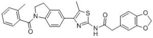
Physicochemical Properties
| Molecular Formula | C29H25N3O4S | |
| Molecular Weight | 511.60 | |
| Exact Mass | 511.156 | |
| Elemental Analysis | C, 68.08; H, 4.93; N, 8.21; O, 12.51; S, 6.27 | |
| CAS # | 846557-71-9 | |
| Related CAS # |
|
|
| PubChem CID | 1383822 | |
| Appearance | White to yellow solid powder | |
| Density | 1.4±0.1 g/cm3 | |
| Index of Refraction | 1.693 | |
| LogP | 5.47 | |
| Hydrogen Bond Donor Count | 1 | |
| Hydrogen Bond Acceptor Count | 6 | |
| Rotatable Bond Count | 5 | |
| Heavy Atom Count | 37 | |
| Complexity | 844 | |
| Defined Atom Stereocenter Count | 0 | |
| SMILES | O=C(N1CCC2C1=CC=C(C1=C(C)SC(NC(CC3C=C4C(OCO4)=CC=3)=O)=N1)C=2)C1C(C)=CC=CC=1 |
|
| InChi Key | LINHYWKZVCNAMQ-UHFFFAOYSA-N | |
| InChi Code | InChI=1S/C29H25N3O4S/c1-17-5-3-4-6-22(17)28(34)32-12-11-20-15-21(8-9-23(20)32)27-18(2)37-29(31-27)30-26(33)14-19-7-10-24-25(13-19)36-16-35-24/h3-10,13,15H,11-12,14,16H2,1-2H3,(H,30,31,33) | |
| Chemical Name | 2-(1,3-benzodioxol-5-yl)-N-[5-methyl-4-[1-(2-methylbenzoyl)-2,3-dihydroindol-5-yl]-1,3-thiazol-2-yl]acetamide | |
| Synonyms | ML385; ML 385; ML385; 846557-71-9; ML-385; 2-(benzo[d][1,3]dioxol-5-yl)-N-(5-methyl-4-(1-(2-methylbenzoyl)indolin-5-yl)thiazol-2-yl)acetamide; 2-Benzo[1,3]dioxol-5-yl-N-{5-methyl-4-[1-(2-methyl-benzoyl)-2,3-dihydro-1H-indol-5-yl]-thiazol-2-yl}-acetamide; SMR000173724; 2-(1,3-benzodioxol-5-yl)-N-[5-methyl-4-[1-(2-methylbenzoyl)-2,3-dihydroindol-5-yl]-1,3-thiazol-2-yl]acetamide; N-[4-[2,3-Dihydro-1-(2-methylbenzoyl)-1H-indol-5-yl]-5-methyl-2-thiazolyl]-1,3-benzodioxole-5-acetamide; ML-385 | |
| HS Tariff Code | 2934.99.9001 | |
| Storage |
Powder-20°C 3 years 4°C 2 years In solvent -80°C 6 months -20°C 1 month |
|
| Shipping Condition | Room temperature (This product is stable at ambient temperature for a few days during ordinary shipping and time spent in Customs) |
Biological Activity
| Targets |
NRF2 (IC50 = 1.9 μM)
Nuclear Factor Erythroid 2-Related Factor 2 (NRF2) (IC50 = 1.9 μM, ARE-luciferase reporter assay; IC50 = 3.2 μM, NRF2-dependent gene expression inhibition in H460 cells) [1] |
| ln Vitro |
ML385 exhibits anti-tumor activity in NSCLC (subcutaneous and orthotopic NSCLC models) both as a single agent and in combination with carboplatin. ML385 has a half-life (t1/2 = 2.82 h) after IP injection (30 mg/kg), according to the PK profile examined in CD-1 mice[1]. The pancreatic injury might be reduced by ML385[2]. 1. NRF2 pathway inhibition: ML385 dose-dependently inhibited NRF2 transcriptional activity in HEK293 cells transfected with an ARE-luciferase reporter construct, with an IC50 of 1.9 μM. In KEAP1-deficient NSCLC cell lines (H460, A549, H1299), ML385 (1-10 μM) significantly reduced mRNA and protein levels of NRF2 downstream target genes, including NQO1 (inhibition rate 50-70% at 5 μM), HO-1 (inhibition rate 45-65% at 5 μM), and GCLC (inhibition rate 40-60% at 5 μM), as detected by qRT-PCR and Western blot [1] 2. Antiproliferative activity: ML385 exhibited selective antiproliferative effects on KEAP1-deficient NSCLC cells, with IC50 values of 3.2 μM (H460), 4.5 μM (A549), and 5.1 μM (H1299) after 72 hours of treatment (MTT assay). No significant antiproliferative activity was observed in KEAP1-proficient NSCLC cells (H23, IC50 > 20 μM) or normal human bronchial epithelial cells (BEAS-2B, IC50 > 20 μM) [1] 3. Apoptosis induction: ML385 (5-10 μM) induced apoptosis in H460 and A549 cells, as evidenced by Annexin V-FITC/PI staining (apoptotic rate increased from 3-5% to 25-35% after 48 hours) and activation of caspase-3/7 (2.5-3.5 fold increase compared to control). Western blot showed increased cleavage of PARP and caspase-3 [1] 4. Clonogenic inhibition: ML385 (1-5 μM) dose-dependently inhibited colony formation of H460 cells, with colony formation rates reduced by 30% (1 μM), 55% (3 μM), and 80% (5 μM) compared to the control group [1] 5. Chemosensitization: Combination of ML385 (2 μM) with cisplatin (1-5 μM) synergistically enhanced antiproliferative activity in H460 cells (combination index < 0.8), increasing cisplatin-induced apoptosis by 2-fold compared to cisplatin alone [1] |
| ln Vivo |
ML385 in combination with DNA alkylating agent carboplatin leads to a significant reduction in tumor cell proliferation, as demonstrated by fewer Ki-67 positive cells. NRF2 protein levels and its downstream target genes are significantly decreased in tumor samples treated with ML385. \n\nML385 shows anti-tumor activity in NSCLC both as a single agent and in combination with carboplatin [1] \nTo determine whether ML385 has an appropriate pharmacokinetic (PK) profile for in vivo studies, we dosed CD-1 mice at 30 mg/kg IP. The PK profile showed that ML385 has a half-life (t1/2 = 2.82 h) after IP injection supporting its use in in vivo efficacy studies (Supplementary Figure 7). To determine whether the combination of ML385 and carboplatin observed in cell culture could be recapitulated in vivo, we performed subcutaneous xenograft experiments using A549 and H460 cells. Mice were dosed with ML385, carboplatin, or ML385 in combination with carboplatin for 3–4 weeks and the tumor volumes were measured biweekly. A549 and H460 tumors treated with ML385 in combination with carboplatin showed a significant reduction in tumor growth in both cell lines compared to vehicle. Although the treatment with a single agent (either ML385 or carboplatin) led to a reduction in tumor growth, the magnitude of these effects was variable between cell lines and did not reach statistical significance (Figure 6a–d, Supplementary Figure 8a–b). These results are consistent with prior findings that were obtained with NRF2 siRNA18 in combination with chemotherapeutic drugs. Tumor samples were analyzed for exposure to ML385 4–6 h post last treatment with ML385. We detected ML385 at intratumoral concentrations of ~1 μM in both single agent and combination treatment cohorts. Mice tolerated the combination treatment and the analysis of serum samples for liver and toxicity-related markers revealed no evident signs of toxicity (Supplementary Table 3). ML385 in combination with carboplatin led to a significant reduction in tumor cell proliferation, demonstrated by fewer Ki-67 positive cells (Figure 6e, Supplementary Figure 8c). The RT-PCR and immunoblot analyses of tumor samples were used to determine whether antitumor activity correlated with modulation of the pharmacodynamic markers of NRF2 signaling by ML385. Tumor samples treated with ML385 showed a significant reduction in NRF2 protein level and its downstream target genes (Figure 6f–g). Determination of platinum levels in A549 and H460 tumors treated with carboplatin alone or ML385 in combination with carboplatin by inductively-coupled plasma mass spectrometry (ICP-MS) revealed ~2-fold higher platinum levels in tumors treated with combination therapy (Figure 6h). Collectively, these results suggest that ML385 potentiates the cytotoxic activity of carboplatin partly by blocking the NRF2-dependent drug detoxification pathway leading to increased drug retention in the tumor. The anti-tumor activity of ML385 in combination with carboplatin was replicated in an independent investigator’s laboratory using H460 xenografts.\n \nML385 and carboplatin combination therapy blocks orthotopic human lung tumor growth [1] \nWe evaluated the therapeutic efficacy of ML385, alone and in combination with carboplatin in orthotopic models of human NSCLC that closely recapitulate clinical patterns of lung cancer progression. In our model, we sought to establish a single tumor within the left or right lung. Animals underwent micro-CT imaging, and the mice with approximately 3–7 mm nodules were randomly allocated into treatment groups. In the A549 lung cancer orthotopic model, mice treated with vehicle had 34% lung volume at 3 weeks compared to their pre-treatment volume. Single agent carboplatin or ML385-treated groups had 42% and 57% of their pretreatment lung volume at 3 weeks, respectively (Figure 7a–b). Although the differences in lung volume between the vehicle and the single agent (carboplatin and ML385)-treated groups did not reach a statistical significance, mice in the vehicle-treated group died immediately after 3 weeks, while those in the ML385 or carboplatin-treated group survived. The antitumor and anti-metastatic effect of carboplatin and ML385 combination treatment, as determined by tumor free lung volume, was significantly higher than vehicle or carboplatin monotherapy with 74% lung volume retention at 3 weeks post initiation of the combination treatment\n \nA micro-CT analysis of the lungs of H460 tumor-bearing mice showed that those treated with ML385 monotherapy had 64% of the pre-treatment lung volume at 2 weeks post-treatment. Treatment with carboplatin yielded 50% lung volume compared to pre-treatment volume, which was not significantly different from that noted in the ML385 group (Figure 7c–d). Again, when ML385 and carboplatin were combined, the antitumor effects were significantly enhanced over carboplatin monotherapy, with mice showing 73% of pre-treatment lung volume at 2 weeks post-treatment (Figure 7d; Supplementary Figure 10b). Overall, these data indicate that ML385 in combination with carboplatin has a substantial in vivo efficacy in orthotopic NSCLC models [1]. 1. Tumor growth inhibition in xenograft models: Nude mice (BALB/c nu/nu) were subcutaneously implanted with H460 cells (KEAP1-deficient). Oral administration of ML385 (25, 50 mg/kg/day) for 21 days dose-dependently inhibited tumor growth, with tumor growth inhibition (TGI) rates of 42% (25 mg/kg) and 68% (50 mg/kg) compared to the vehicle group. The 50 mg/kg group significantly reduced tumor weight from 1.2 g (vehicle) to 0.4 g [1] 2. NRF2 pathway modulation in vivo: Tumor tissues from ML385-treated mice (50 mg/kg) showed reduced NRF2 protein expression (by 60%) and downstream target genes NQO1 (by 55%) and HO-1 (by 50%) compared to vehicle-treated mice, as detected by Western blot and immunohistochemistry [1] 3. Safety profile: No significant changes in body weight (vehicle vs. 50 mg/kg group: 20.5 g vs. 19.8 g) or liver/kidney function parameters (ALT, AST, BUN, creatinine) were observed in ML385-treated mice. Histopathological analysis of major organs (liver, kidney, heart, lung) showed no obvious pathological damage [1] |
| Enzyme Assay |
Nickel pull-down streptavidin-HRP assay[1] Full-length NRF2 (1-605 AA), Neh1, the Cap-n-collar (CNC) bZip domain of NRF2 (434–561 AA) and ΔNeh1 fragments were cloned in a pET14B expression vector. The excess amount of purified histidine-tagged NRF2 proteins was bound to the pre-charged and pre-equilibrated Ni-NTA beads and was incubated for 30 min on ice. After incubation, the NRF2-bound NTA-resin was washed (3×) with PBS. Subsequently, biotin-labeled ML385 or control compounds were added at a concentration of 10 μM. After 1 h incubation on ice, beads with protein were washed (3×) with PBS. For the competition assay, ML385 and compound 3 were added at a concentration of 10 μM, incubated on ice, and washed (3×) with PBS. Next, 5 μg of horseradish peroxidase (HRP)-conjugated streptavidin was added to the tube, followed by a 30-min incubation on ice, followed by an 8× wash with PBS. Lastly, bound protein-drug complex was eluted with PBS containing 10 mM EDTA, mixed 1:1 with SuperSignal West PICO solution, and the HRP activity was measured using well-scan mode in a Flexistation-3. 1. ARE-luciferase reporter gene assay: HEK293 cells were co-transfected with an ARE-luciferase reporter plasmid and a Renilla luciferase plasmid (internal control) using transfection reagent. After 24 hours of transfection, cells were seeded in 96-well plates and treated with different concentrations of ML385 (0.1-20 μM) for 18 hours. Luciferase activity was measured using a dual-luciferase reporter assay system, and the ratio of firefly luciferase to Renilla luciferase activity was calculated to determine NRF2 transcriptional inhibition efficiency. The IC50 value was derived from dose-response curves [1] 2. NRF2 protein stability assay: H460 cells were treated with ML385 (5 μM) or vehicle, and cycloheximide (CHX, 100 μg/mL) was added at different time points (0, 1, 2, 4 hours) to inhibit protein synthesis. Cells were lysed, and NRF2 protein levels were detected by Western blot. The half-life of NRF2 protein was calculated, showing that ML385 reduced NRF2 protein stability (half-life from 3.5 hours to 1.2 hours) [1] |
| Cell Assay |
NQO1 enzyme activity measurement[1] Cells were treated with vehicle or ML385 for 72 h. The enzyme activity in the total protein lysate was determined as described previously. Total antioxidant capacity and GSH measurement[1] Cells were treated with vehicle or ML385 for 72 h. The total antioxidant capacity and glutathione levels were measured using antioxidant and glutathione assay kits, respectively. Caspase activity assay[1] Caspase activity was measured using the Caspase-Glo® 3/7 Assay kit as per the manufacturer’s instructions. The CellTiter-Blue assay was utilized to quantify cell density and to normalize caspase activity. Briefly, cells were treated with ML385 for 36 h. An equal amount of CellTiter-Blue reagent was added to the wells and the fluorescence was measured after 30 min. The CellTiter-Blue reagent was discarded and the Caspase-Glo (100 μL) reagent was added to the cells and incubated at 37°C for an additional 60–90 min. The resulting luminescence was recorded and the caspase activity was normalized to cell number. 1. Cell proliferation assay: KEAP1-deficient (H460, A549, H1299) and KEAP1-proficient (H23) NSCLC cells, as well as BEAS-2B cells, were seeded in 96-well plates at a density of 2×10^3 cells/well. After 24 hours of adherence, cells were treated with ML385 (0.1-20 μM) for 72 hours. MTT reagent was added, and after 4 hours of incubation, formazan crystals were dissolved in DMSO, and absorbance at 570 nm was measured to calculate cell viability and IC50 values [1] 2. Apoptosis assay: H460 cells were seeded in 6-well plates (5×10^5 cells/well) and treated with ML385 (5, 10 μM) for 48 hours. Cells were harvested, washed with PBS, stained with Annexin V-FITC and PI for 15 minutes in the dark, and analyzed by flow cytometry to quantify apoptotic cells. Caspase-3/7 activity was measured using a fluorescent assay kit, with fluorescence intensity detected at 485 nm excitation and 535 nm emission [1] 3. Clonogenic assay: H460 cells were seeded in 6-well plates (200 cells/well) and allowed to adhere for 24 hours. ML385 (1, 3, 5 μM) was added, and cells were cultured for 14 days. Colonies were fixed with methanol, stained with crystal violet, and counted. The colony formation rate was calculated as (number of colonies in treatment group / number of colonies in control group) × 100% [1] 4. qRT-PCR assay: H460 cells were treated with ML385 (5 μM) for 24 hours. Total RNA was extracted, reverse-transcribed into cDNA, and qRT-PCR was performed using specific primers for NQO1, HO-1, GCLC, and GAPDH (internal control). Relative gene expression was calculated using the 2^(-ΔΔCt) method [1] 5. Western blot assay: Cells or tumor tissues were lysed in RIPA buffer containing protease inhibitors. Total protein was separated by SDS-PAGE, transferred to PVDF membranes, and probed with antibodies against NRF2, NQO1, HO-1, GCLC, cleaved PARP, cleaved caspase-3, and GAPDH. After incubation with secondary antibodies, chemiluminescent signals were detected and quantified using imaging software [1] |
| Animal Protocol |
8-week-old C57B/6 male mice 30 mg/kg; 7 days Intraperitoneal injection Pharmacokinetic analysis of ML385 in CD-1 mice[1] For pharmacokinetic analysis, male CD-1 mice (n=3/time point) were administered a 30 mg/kg intraperitoneal (IP) dose of (vehicle: Solutol/Cremophor EL/polyethylene glycol 400/water [15/10/35/40,v/v/v/v]) of ML385. Blood samples were collected at pre-treatment, 0.083, 0.25, 0.5, 1, 2, 4, 8 and 24 h and plasma samples were harvested. Plasma concentration of ML385 was determined using a qualified LC-MS/MS. A simulation was conducted to predict the in vivo exposure after a multiple-dose treatment based on the single-dose study results. Determination of ML385 concentration in tumor samples[1] An UPLC-MS/MS method was developed to determine the concentration of ML385 in tumor samples. The details are included in the supplementary methods section. Establishment of tumor xenografts and treatment[1] Tumor xenografts were established as described previously18. A549 cells (5.0×10⁶) and H460 cells (1.0×10⁶) were injected subcutaneously into the flank of athymic nude mice and the tumor dimensions were measured by caliper at an interval of 3–5 days18. The tumor volumes were calculated using the following formula: [length (mm) × width (mm) × width (mm) × 0.5]. Once the tumor volumes were approximately 50–100 mm3, mice were randomly allocated into 4 groups: vehicle, ML385, carboplatin, and ML385 in combination with carboplatin. Vehicle, carboplatin (5 mg/kg daily Monday to Friday)18, ML385 (30 mg/kg daily Monday to Friday), or ML385 in combination with carboplatin were administered intraperitoneally for 3 weeks. At the end of treatment period, mice were sacrificed and the tumor, blood, lung, and liver samples were collected.[1] For the orthotopic lung tumor model, A549 (1.0×10⁶) and H460 cells (1.0×10⁶) were diluted 1:1 in matrigel (30 μL) and were injected directly into the lungs. Ten days post-cell implantation, mice were imaged. Mice with visible localized lung tumor were randomly divided into 4 groups: vehicle, ML385, carboplatin, and ML385+carboplatin. Vehicle, carboplatin, ML385, or ML385 in combination with carboplatin were administered intraperitoneally for 2 weeks using the same regimen as described above. High-resolution lung micro-computed tomography (CT) images were acquired in 512 projections (270 μA, 75 kVp), and the data were reconstructed using the ordered subsets-expectation maximization algorithm. Volume-rendered whole lung images were generated using Amira 5.3.0 software. For each mouse, pretreatment available lung volume was defined as 100% compared to post-treatment lung volumes. Treatment with a Nrf2/HO-1 Pathway Inhibitor[2] A Nrf2 inhibitor (ML385) or a HO-1 inhibitor (ZnPP) was used to inhibit the Nrf2/HO-1 antioxidant pathway in vivo. ML385 was dissolved in 100% DMSO to prepare a stock solution and then diluted it into 5% DMSO solution with PBS before being used. ZnPP was dissolved as follows: 2.5 mg ZnPP was dissolved in 0.33 ml NaOH (0.2 M) in a dark room, and 0.2 M HCl was added to adjust the pH to 7.0. Finally, saline was added to 5 ml (0.5 mg/ml).[2] ML385 (30 mg/kg) or ZnPP (5 mg/kg) pretreatment was administered intraperitoneally 1 h before administration of caerulein, and the mice in the control group were treated with vehicle. In the MAP model, high-dose ISL (200 mg/kg) was administered after the first caerulein injection immediately to identify the underlying molecular mechanisms of ISL on AP.[2] 1. Xenograft tumor model establishment: Female BALB/c nu/nu mice (6-8 weeks old, 18-22 g) were subcutaneously injected with 5×10^6 H460 cells suspended in Matrigel (1:1 ratio with PBS) into the right flank. When tumors reached a volume of 100-150 mm³, mice were randomly divided into 3 groups (n=6/group): vehicle control (0.5% DMSO + 5% Cremophor EL + 94.5% normal saline), ML385 25 mg/kg, and ML385 50 mg/kg [1] 2. Drug administration: ML385 was dissolved in DMSO, diluted with Cremophor EL and normal saline to the final concentration, and administered orally by gavage once daily for 21 days. The vehicle group received the same volume of solvent without the drug [1] 3. Tumor and body weight measurement: Tumor volume was measured every 3 days using a caliper, calculated as (length × width²)/2. Body weight was recorded daily. At the end of the experiment, mice were sacrificed by cervical dislocation, tumors were excised, weighed, and stored at -80℃ for Western blot and immunohistochemical analysis. Major organs (liver, kidney, heart, lung) were collected for histopathological examination [1] 4. Immunohistochemistry: Tumor tissues were fixed in 4% paraformaldehyde, embedded in paraffin, and sectioned into 5 μm slices. Slices were deparaffinized, rehydrated, and subjected to antigen retrieval. After blocking with BSA, slices were incubated with primary antibodies against NQO1 and HO-1 overnight at 4℃, followed by secondary antibody incubation. DAB staining was performed, and sections were counterstained with hematoxylin. Positive staining was visualized under a microscope and quantified [1] |
| ADME/Pharmacokinetics |
1. Oral bioavailability: In mice, oral administration of ML385 (50 mg/kg) resulted in an absolute bioavailability of 38% [1] 2. Plasma pharmacokinetics: After oral administration of ML385 (50 mg/kg) in mice, the peak plasma concentration (Cmax) was 2.3 μM (achieved at 1 hour), area under the curve (AUC0-24h) was 15.6 μM·h, and elimination half-life (t1/2) was 4.8 hours [1] 3. Tissue distribution: In mice, 2 hours after oral administration of ML385 (50 mg/kg), the highest drug concentrations were detected in the liver (6.8 μM) and tumor tissue (3.5 μM), followed by the kidney (2.9 μM) and lung (1.8 μM). Brain concentration was undetectable (<0.1 μM), indicating no significant blood-brain barrier penetration [1] |
| Toxicity/Toxicokinetics |
1. Acute toxicity: In mice, single oral administration of ML385 at doses up to 200 mg/kg did not cause significant mortality or obvious toxic symptoms (e.g., lethargy, diarrhea, weight loss) within 14 days of observation [1] 2. Chronic toxicity: Mice treated with ML385 (50 mg/kg/day, oral) for 21 days showed no significant changes in liver function (ALT, AST) or kidney function (BUN, creatinine) compared to the vehicle group. Histopathological analysis of major organs revealed no abnormal lesions [1] 3. Plasma protein binding: ML385 showed 89% plasma protein binding in mouse plasma, determined by equilibrium dialysis [1] |
| References |
[1]. Small molecule inhibitor of NRF2 selectively intervenes therapeutic resistance in KEAP1-deficient NSCLC tumors. ACS Chem Biol. 2016 Nov 18;11(11):3214-3225. [2]. Isoliquiritigenin Ameliorates Acute Pancreatitis in Mice via Inhibition of Oxidative Stress and Modulation of the Nrf2/HO-1 Pathway. Oxid Med Cell Longev. 2018 Apr 26:2018:7161592. [3]. Nrf2 protects against seawater drowning-induced acute lung injury via inhibiting ferroptosis. Respir Res . 2020 Sep 9;21(1):232. |
| Additional Infomation |
Loss of function mutations in Kelch Like ECH Associated Protein 1 (KEAP1) or gain-of-function mutations in nuclear factor erythroid 2-related factor 2 (NRF2) are common in non-small cell lung cancer (NSCLC) and is associated with therapeutic resistance. To discover novel NRF2 inhibitors for targeted therapy, we conducted a quantitative high-throughput screen using a diverse set of ~400,000 small molecules (Molecular Libraries Small Molecule Repository Library, MLSMR) at the National Center for Advancing Translational Sciences. We identified ML385 as a probe molecule that binds to NRF2 and inhibits its downstream target gene expression. Specifically, ML385 binds to the Neh1, the Cap ‘N’ Collar Basic Leucine Zipper (CNC-bZIP) domain of NRF2, and interferes with the binding of the V-Maf Avian Musculoaponeurotic Fibrosarcoma Oncogene Homolog G (MAFG)-NRF2 protein complex to regulatory DNA binding sequences. In clonogenic assays, when used in combination with platinum-based drugs such as doxorubicin or taxol, ML385 substantially enhances cytotoxicity in NSCLC cells compared to single agents alone. ML385 shows specificity and selectivity for NSCLC cells with KEAP1 mutation leading to gain of NRF2 function. In preclinical models of NSCLC with gain of NRF2 function, ML385 in combination with carboplatin showed significant anti-tumor activity. We demonstrate the discovery and validation of ML385 as a novel and specific NRF2 inhibitor and conclude that targeting NRF2 may represent a promising strategy for the treatment of advanced NSCLC.[1] \n\n\n\nOxidative stress plays a crucial role in the pathogenesis of acute pancreatitis (AP). Isoliquiritigenin (ISL) is a flavonoid monomer with confirmed antioxidant activity. However, the specific effects of ISL on AP have not been determined. In this study, we aimed to investigate the protective effect of ISL on AP using two mouse models. In the caerulein-induced mild acute pancreatitis (MAP) model, dynamic changes in oxidative stress injury of the pancreatic tissue were observed after AP onset. We found that ISL administration reduced serum amylase and lipase levels and alleviated the histopathological manifestations of pancreatic tissue in a dose-dependent manner. Meanwhile, ISL decreased the oxidative stress injury and increased the protein expression of the Nrf2/HO-1 pathway. In addition, after administering a Nrf2 inhibitor (ML385) or HO-1 inhibitor (zinc protoporphyrin) to block the Nrf2/HO-1 pathway, we failed to observe the protective effects of ISL on AP in mice. Furthermore, we found that ISL mitigated the severity of pancreatic tissue injury and pancreatitis-associated lung injury in a severe acute pancreatitis model induced by L-arginine. Taken together, our data for the first time confirmed the protective effects of ISL on AP in mice via inhibition of oxidative stress and modulation of the Nrf2/HO-1 pathway.[2] \n\nBackground: Ferroptosis is a new type of nonapoptotic cell death model that was closely related to reactive oxygen species (ROS) accumulation. Seawater drowning-induced acute lung injury (ALI) which is caused by severe oxidative stress injury, has been a major cause of accidental death worldwide. The latest evidences indicate nuclear factor (erythroid-derived 2)-like 2 (Nrf2) suppress ferroptosis and maintain cellular redox balance. Here, we test the hypothesis that activation of Nrf2 pathway attenuates seawater drowning-induced ALI via inhibiting ferroptosis.\n\nMethods: we performed studies using Nrf2-specific agonist (dimethyl fumarate), Nrf2 inhibitor (ML385), Nrf2-knockout mice and ferroptosis inhibitor (Ferrostatin-1) to investigate the potential roles of Nrf2 on seawater drowning-induced ALI and the underlying mechanisms.\n\nResults: Our data shows that Nrf2 activator dimethyl fumarate could increase cell viability, reduced the levels of intracellular ROS and lipid ROS, prevented glutathione depletion and lipid peroxide accumulation, increased FTH1 and GPX4 mRNA expression, and maintained mitochondrial membrane potential in MLE-12 cells. However, ML385 promoted cell death and lipid ROS production in MLE-12 cells. Furthermore, the lung injury became more aggravated in the Nrf2-knockout mice than that in WT mice after seawater drowning.\n\nConclusions: These results suggested that Nrf2 can inhibit ferroptosis and therefore alleviate ALI induced by seawater drowning. The effectiveness of ferroptosis inhibition by Nrf2 provides a novel therapeutic target for seawater drowning-induced ALI.[3] 1. ML385 is a selective small-molecule inhibitor of NRF2, which binds to NRF2 and promotes its degradation, thereby inhibiting the NRF2-ARE signaling pathway. It exhibits selective antiproliferative activity against KEAP1-deficient NSCLC cells, as KEAP1 deficiency leads to constitutive activation of NRF2, which contributes to therapeutic resistance [1] 2. The drug enhances the sensitivity of KEAP1-deficient NSCLC cells to chemotherapeutic agents (e.g., cisplatin) by inhibiting NRF2-mediated antioxidant defense and detoxification pathways, providing a potential therapeutic strategy for overcoming chemotherapy resistance in KEAP1-deficient NSCLC [1] 3. ML385 has favorable pharmacokinetic properties, including moderate oral bioavailability, effective tumor tissue penetration, and low toxicity in preclinical models, supporting its potential as a targeted therapy for KEAP1-deficient NSCLC [1] |
Solubility Data
| Solubility (In Vitro) |
|
|||
| Solubility (In Vivo) |
Solubility in Formulation 1: ≥ 2.5 mg/mL (4.89 mM) (saturation unknown) in 10% DMSO + 90% Corn Oil (add these co-solvents sequentially from left to right, and one by one), clear solution. For example, if 1 mL of working solution is to be prepared, you can add 100 μL of 25.0 mg/mL clear DMSO stock solution to 900 μL of corn oil and mix evenly. Solubility in Formulation 2: ≥ 2.08 mg/mL (4.07 mM) (saturation unknown) in 10% DMSO + 40% PEG300 + 5% Tween80 + 45% Saline (add these co-solvents sequentially from left to right, and one by one), clear solution. For example, if 1 mL of working solution is to be prepared, you can add 100 μL of 20.8 mg/mL clear DMSO stock solution to 400 μL PEG300 and mix evenly; then add 50 μL Tween-80 to the above solution and mix evenly; then add 450 μL normal saline to adjust the volume to 1 mL. Preparation of saline: Dissolve 0.9 g of sodium chloride in 100 mL ddH₂ O to obtain a clear solution. Solubility in Formulation 3: 6% DMSO+40% PEG 300+5%Tween80+ 49%ddH2O: 1.5mg/ml Solubility in Formulation 4: 10 mg/mL (19.55 mM) in 50% PEG300 50% Saline (add these co-solvents sequentially from left to right, and one by one), suspension solution; with ultrasonication. Preparation of saline: Dissolve 0.9 g of sodium chloride in 100 mL ddH₂ O to obtain a clear solution. Solubility in Formulation 5: 9.01 mg/mL (17.61 mM) in 0.5% CMC-Na/saline water (add these co-solvents sequentially from left to right, and one by one), clear solution; with ultrasonication. Preparation of saline: Dissolve 0.9 g of sodium chloride in 100 mL ddH₂ O to obtain a clear solution. Solubility in Formulation 6: 5 mg/mL (9.77 mM) in 15% Solutol HS 15 10% Cremophor EL 35% PEG 400 40% water (add these co-solvents sequentially from left to right, and one by one), suspension solution; with ultrasonication. (Please use freshly prepared in vivo formulations for optimal results.) |
| Preparing Stock Solutions | 1 mg | 5 mg | 10 mg | |
| 1 mM | 1.9547 mL | 9.7733 mL | 19.5465 mL | |
| 5 mM | 0.3909 mL | 1.9547 mL | 3.9093 mL | |
| 10 mM | 0.1955 mL | 0.9773 mL | 1.9547 mL |