ML347 (formerly known as ML-347; LDN193719; ML 347; LDN-193719) is a novel, highly potent and selective inhibitor of BMP (bone morphogenetic protein) receptor with potential anticancer activity. It inhibits ALK2 with an IC50 of 32 nM, and exhibits >300-fold selectivity over ALK3. ML347 was discovered as a selective inhibitor of BMP type-I receptor ALK2 versus ALK3 and is identified as a probe molecule. In the in vitro kinase assay, ML347 shows potent inhibitory activities against ALK1 and ALK2 with IC50 values of 46 and 32 nM, respectively. The IC50 value of it for ALK3 is more than 10μM, demonstrating that ML347 is 300-fold more potent against ALK2. Besides that, ML347 exerts no inhibition effect on other related kinases such as ALK6 and KDR.
Physicochemical Properties
| Molecular Formula | C22H16N4O | |
| Molecular Weight | 352.39 | |
| Exact Mass | 352.132 | |
| Elemental Analysis | C, 74.98; H, 4.58; N, 15.90; O, 4.54 | |
| CAS # | 1062368-49-3 | |
| Related CAS # |
|
|
| PubChem CID | 44577753 | |
| Appearance | Light yellow to yellow solid powder | |
| Density | 1.3±0.1 g/cm3 | |
| Melting Point | 209-210℃ | |
| Index of Refraction | 1.695 | |
| LogP | 2.63 | |
| Hydrogen Bond Donor Count | 0 | |
| Hydrogen Bond Acceptor Count | 4 | |
| Rotatable Bond Count | 3 | |
| Heavy Atom Count | 27 | |
| Complexity | 495 | |
| Defined Atom Stereocenter Count | 0 | |
| InChi Key | FVRYPYDPKSZGNS-UHFFFAOYSA-N | |
| InChi Code | InChI=1S/C22H16N4O/c1-27-17-9-7-15(8-10-17)16-12-24-22-20(13-25-26(22)14-16)18-4-2-6-21-19(18)5-3-11-23-21/h2-14H,1H3 | |
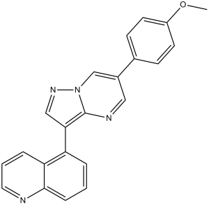
| Chemical Name | 5-[6-(4-methoxyphenyl)pyrazolo[1,5-a]pyrimidin-3-yl]quinoline | |
| Synonyms |
|
|
| HS Tariff Code | 2934.99.9001 | |
| Storage |
Powder-20°C 3 years 4°C 2 years In solvent -80°C 6 months -20°C 1 month |
|
| Shipping Condition | Room temperature (This product is stable at ambient temperature for a few days during ordinary shipping and time spent in Customs) |
Biological Activity
| Targets |
Bone morphogenetic protein (BMP) signaling cascade; ALK1 (IC50 = 46 nM); ACVR1 (IC50 = 32 nM); BMPR1A (IC50 = 10800 nM) ML347 is a selective inhibitor of bone morphogenetic protein (BMP) type I receptors, with preference for ALK2 over ALK3 (ALK2 IC50 = 1.9 nM; ALK3 IC50 = 140 nM) [1] ML347 shows weak or no inhibition of other ALK receptors (ALK1, ALK4-6: IC50 > 1 μM) and unrelated kinases (PKA, PKC, ERK1/2: IC50 > 10 μM) [1] |
|
| ln Vitro |
The more potent (and selective) compound, 7g (ML347), has IC50’s of 46 and 32 nM, respectively, against ALK1 and ALK2; however, the IC50 against ALK3 is 10,800 nM, >300-fold selective over ALK3. In addition, 7g is completely inactive against all the other kinases tested (with weak activity against ALK6, 9830 nM and KDR (VEGFR2) 19,700 nM). It is interesting to note that it appears to be a combination of the 5-quinoline and 4-methoxyphenyl which gives rise to the selectivity profile, as 13m still retains significant ALK3 activity (539 nM). Due to the potency of 7g against the BMP4 cell assay, ALK1 and ALK2 and the significant selectivity against the other kinases, 7g, has been declared a probe molecule in the MLPCN and redesignated ML347.[1] By suppressing ALK1/ALK2, ML347 can prevent the transduction of the TGF-β signal [2]. In recombinant ALK2/ALK3 kinase assays, ML347 dose-dependently inhibits kinase activity. At 10 nM, it inhibits ALK2 activity by 90% and ALK3 activity by 35%;at 100 nM, ALK3 inhibition reaches 68%. It blocks BMP-induced Smad1/5/8 phosphorylation in C2C12 myoblasts, with 75% reduction at 5 μM after 24 hours [1] - In mouse dental epithelial cells (mDECs), ML347 (10 μM) inhibits BMP2-induced cell proliferation by 52% after 48 hours (CCK-8 assay). It downregulates p-Smad1/5/8 protein expression by 65% and reduces the mRNA level of BMP target gene ID1 by 60% [2] - In normal mouse dental mesenchymal cells, ML347 shows no significant toxicity at concentrations up to 20 μM (cell viability > 85% vs. control) [2] |
|
| ln Vivo |
|
|
| Enzyme Assay |
In order to further the BMP community as to the utility of ML347, researchers evaluated this molecule in our Tier 1 in vitro pharmacokinetic assays (Table 4). These studies are useful in order to evaluate the metabolic stability and predicted clearance in a number of species in order to inform on possible dosing routes. Utilizing rapid equilibrium dialysis, the protein binding of ML347 was determined in human, rat and mouse plasma. The results were similar in all three species with ML347 displaying high plasma protein biding (Fu ~0.01-0.015). ML347 was also assessed for its intrinsic clearance in hepatic microsomes. This measure will help predict the in vivo clearance in the same three species (CLHEP). [1] These SAR studies culminated in the discovery of a highly selective ALK2 inhibitor, ML347, which shows >300-fold selectivity for ALK2 vs. ALK3. ML347 is potent in the BMP4 cell assay (152 nM) as well as the in vitro kinase assay for ALK1 (46 nM) and ALK2 (32 nM) and is devoid of activity in a number of related kinases. Further studies are planned for this selective inhibitor in a number of in vivo animal disease models, such as FOP, and results will be reported in due course. ALK2/ALK3 kinase activity assay: Purified recombinant human ALK2 or ALK3 was incubated with Smad1-derived substrate peptide and ML347 (0.1 nM-1 μM) in assay buffer (50 mM Tris-HCl, pH 7.5, 10 mM MgCl₂, 1 mM DTT, 0.1 mM ATP) at 30°C for 60 minutes. Phosphorylated substrate was detected by radiolabeled ATP counting, and IC50 values were calculated from dose-response curves [1] - Kinase selectivity assay: ML347 (10 μM) was screened against a panel of 40+ kinases (including ALK1, ALK4-6, PKA, PKC, ERK1/2) using respective substrate peptides and assay buffers. Kinase activity was quantified by colorimetric assay, with no significant off-target inhibition (>50% activity reduction) observed [1] |
|
| Cell Assay |
Immunofluorescence[2] Cell Types: Primary dental epithelial cells were cultured in Dulbecco's Modified Eagle Medium (DMEM)/ F12 supplemented with 20% fetal bovine serum and 1% penicillin/streptomycin. Tested Concentrations: 25 μM Incubation Duration: 2 hrs (hours) Experimental Results: Inhibited ALK1 /ALK2 then blocking Smad1/5 by TGF-β1. C2C12 cell BMP signaling assay: C2C12 myoblasts were seeded in 6-well plates at 2×10⁵ cells/well and pretreated with ML347 (0.5-10 μM) for 1 hour, then stimulated with BMP4 (10 ng/mL) for 24 hours. Western blot detected p-Smad1/5/8 and total Smad1 [1] - mDEC proliferation and BMP signaling assay: Mouse dental epithelial cells were seeded in 96-well plates (proliferation) or 6-well plates (signaling) at 3×10³ cells/well or 2×10⁵ cells/well respectively. Cells were pretreated with ML347 (1-20 μM) for 1 hour, then stimulated with BMP2 (5 ng/mL) for 24-48 hours. CCK-8 assay assessed proliferation; qPCR analyzed ID1 mRNA level; Western blot detected p-Smad1/5/8 [2] |
|
| Animal Protocol | This measure will help predict the in vivo clearance in the same three species (CLHEP). ML347 was unstable to oxidative metabolism – possibly due to the labile methoxy group – and therefore was predicted to display high clearance in human and mouse, and moderate-to-high clearance in the rat. Going forward, the intrinsic clearance is predicting high clearance after oral dosing, a more appropriate dosing paradigm might be intraperitoneal dosing for this compound. Further in vivo experiments, including PK, will be reported in due course.[1] | |
| References |
[1]. Synthesis and structure-activity relationships of a novel and selective bone morphogenetic protein receptor (BMP) inhibitor derived from the pyrazolo[1.5-a]pyrimidine scaffold of dorsomorphin: the discovery of ML347 as an ALK2 versus ALK3 selective MLPCN probe. Bioorg Med Chem Lett. 2013 Jun 1;23(11):3248-52. [2]. Dual roles of TGF-β signaling in the regulation of dental epithelial cell proliferation. J Mol Histol. 2021 Feb;52(1):77-86. |
|
| Additional Infomation |
The purpose of this study is to investigate the molecular mechanisms and biological function of TGF-β-activated Smad1/5 in dental epithelium. Immunohistochemistry was used to detect the expressions of TGF-β signaling-related gene in mice molar germ. Primary dental epithelial cells were cultured and treated with TGF-β1 at a concentration of 0.5 or 5 ng/mL. Small molecular inhibitors, SB431542 and ML347, was used to inhibite ALK5 and ALK1/2, respectively. Small interfering RNA was used to knock down Smad1/5 or Smad2/3. The proliferation rate of cells was evaluated by EdU assay. In the basal layer of dental epithelial bud TGF-β1 and p-Smad1/5 were highly expressed, and in the interior of the epithelial bud TGF-β1 was lowly expressed, whereas p-Smad2/3 was highly expressed. In primary cultured dental epithelial cells, low concentration of TGF-β1 activated Smad2/3 but not Smad1/5, while high concentration of TGF-β1 was able to activate both Smad2/3 and Smad1/5. SB431542 but not ML347 was able to block the phosphorylation of Smad2/3 by TGF-β1. Either SB431542 or ML347 was able to block the phosphorylation of Smad1/5 by TGF-β1. EdU staining showed that high concentration of TGF-β1 promoted dental epithelial cell proliferation, which was reversed by silencing Smad1/5, whereas low concentration of TGF-β1 inhibited cell proliferation, which was reversed by silencing Smad2/3. In conclusions, TGF-β exhibits dual roles in the regulation of dental epithelial cell proliferation through two pathways. On the one hand, TGF-β activates canonical Smad2/3 signaling through ALK5, inhibiting the proliferation of internal dental epithelial cells. On the other hand, TGF-β activates noncanonical Smad1/5 signaling through ALK1/2-ALK5, promoting the proliferation of basal cells in the dental epithelial bud.[2] A structure-activity relationship of the 3- and 6-positions of the pyrazolo[1,5-a]pyrimidine scaffold of the known BMP inhibitors dorsomorphin, 1, LDN-193189, 2, and DMH1, 3, led to the identification of a potent and selective compound for ALK2 versus ALK3. The potency contributions of several 3-position substituents were evaluated with subtle structural changes leading to significant changes in potency. From these studies, a novel 5-quinoline molecule was identified and designated an MLPCN probe molecule, ML347, which shows >300-fold selectivity for ALK2 and presents the community with a selective molecular probe for further biological evaluation.[1] ML347 is a potent, ALK2-selective small-molecule inhibitor derived from the pyrazolo[1.5-a]pyrimidine scaffold of dorsomorphin [1] - Its mechanism of action involves competitive binding to the ATP-binding pocket of ALK2 (with higher affinity than ALK3), inhibiting its kinase activity and blocking downstream BMP/Smad1/5/8 signaling pathway activation [1][2] - ML347 exhibits in vitro inhibitory activity against BMP signaling in myoblasts and dental epithelial cells, and suppresses BMP-induced proliferation of dental epithelial cells [1][2] - It serves as a MLPCN (Molecular Libraries Probe Production Centers Network) probe for studying ALK2-selective BMP signaling in biological processes and diseases [1] - In dental epithelial cells, it regulates proliferation by targeting BMP/Smad signaling, providing insights into BMP pathway functions in dental development [2] |
Solubility Data
| Solubility (In Vitro) |
|
|||
| Solubility (In Vivo) |
Note: Listed below are some common formulations that may be used to formulate products with low water solubility (e.g. < 1 mg/mL), you may test these formulations using a minute amount of products to avoid loss of samples. Injection Formulations (e.g. IP/IV/IM/SC) Injection Formulation 1: DMSO : Tween 80: Saline = 10 : 5 : 85 (i.e. 100 μL DMSO stock solution → 50 μL Tween 80 → 850 μL Saline) *Preparation of saline: Dissolve 0.9 g of sodium chloride in 100 mL ddH ₂ O to obtain a clear solution. Injection Formulation 2: DMSO : PEG300 :Tween 80 : Saline = 10 : 40 : 5 : 45 (i.e. 100 μL DMSO → 400 μLPEG300 → 50 μL Tween 80 → 450 μL Saline) Injection Formulation 3: DMSO : Corn oil = 10 : 90 (i.e. 100 μL DMSO → 900 μL Corn oil) Example: Take the Injection Formulation 3 (DMSO : Corn oil = 10 : 90) as an example, if 1 mL of 2.5 mg/mL working solution is to be prepared, you can take 100 μL 25 mg/mL DMSO stock solution and add to 900 μL corn oil, mix well to obtain a clear or suspension solution (2.5 mg/mL, ready for use in animals). Injection Formulation 4: DMSO : 20% SBE-β-CD in saline = 10 : 90 [i.e. 100 μL DMSO → 900 μL (20% SBE-β-CD in saline)] *Preparation of 20% SBE-β-CD in Saline (4°C,1 week): Dissolve 2 g SBE-β-CD in 10 mL saline to obtain a clear solution. Injection Formulation 5: 2-Hydroxypropyl-β-cyclodextrin : Saline = 50 : 50 (i.e. 500 μL 2-Hydroxypropyl-β-cyclodextrin → 500 μL Saline) Injection Formulation 6: DMSO : PEG300 : castor oil : Saline = 5 : 10 : 20 : 65 (i.e. 50 μL DMSO → 100 μLPEG300 → 200 μL castor oil → 650 μL Saline) Injection Formulation 7: Ethanol : Cremophor : Saline = 10: 10 : 80 (i.e. 100 μL Ethanol → 100 μL Cremophor → 800 μL Saline) Injection Formulation 8: Dissolve in Cremophor/Ethanol (50 : 50), then diluted by Saline Injection Formulation 9: EtOH : Corn oil = 10 : 90 (i.e. 100 μL EtOH → 900 μL Corn oil) Injection Formulation 10: EtOH : PEG300:Tween 80 : Saline = 10 : 40 : 5 : 45 (i.e. 100 μL EtOH → 400 μLPEG300 → 50 μL Tween 80 → 450 μL Saline) Oral Formulations Oral Formulation 1: Suspend in 0.5% CMC Na (carboxymethylcellulose sodium) Oral Formulation 2: Suspend in 0.5% Carboxymethyl cellulose Example: Take the Oral Formulation 1 (Suspend in 0.5% CMC Na) as an example, if 100 mL of 2.5 mg/mL working solution is to be prepared, you can first prepare 0.5% CMC Na solution by measuring 0.5 g CMC Na and dissolve it in 100 mL ddH2O to obtain a clear solution; then add 250 mg of the product to 100 mL 0.5% CMC Na solution, to make the suspension solution (2.5 mg/mL, ready for use in animals). Oral Formulation 3: Dissolved in PEG400 Oral Formulation 4: Suspend in 0.2% Carboxymethyl cellulose Oral Formulation 5: Dissolve in 0.25% Tween 80 and 0.5% Carboxymethyl cellulose Oral Formulation 6: Mixing with food powders Note: Please be aware that the above formulations are for reference only. InvivoChem strongly recommends customers to read literature methods/protocols carefully before determining which formulation you should use for in vivo studies, as different compounds have different solubility properties and have to be formulated differently. (Please use freshly prepared in vivo formulations for optimal results.) |
| Preparing Stock Solutions | 1 mg | 5 mg | 10 mg | |
| 1 mM | 2.8378 mL | 14.1888 mL | 28.3776 mL | |
| 5 mM | 0.5676 mL | 2.8378 mL | 5.6755 mL | |
| 10 mM | 0.2838 mL | 1.4189 mL | 2.8378 mL |