ML204 HCl is a novel, potent, and selective TRPC4 (Transient receptor potential canonical) channel inhibitor identified from high throughput fluorescent screen of 305,000 compounds of the Molecular Libraries Small Molecule Repository for inhibitors that blocked intracellular Ca(2+) rise in response to stimulation of mouse TRPC4β by μ-opioid receptors. ML204 inhibited TRPC4β-mediated intracellular Ca(2+) rise with an IC(50) value of 0.96 μm and exhibited 19-fold selectivity against muscarinic receptor-coupled TRPC6 channel activation. In whole-cell patch clamp recordings, ML204 blocked TRPC4β currents activated through either μ-opioid receptor stimulation or intracellular dialysis of guanosine 5'-3-O-(thio)triphosphate (GTPγS), suggesting a direct interaction of ML204 with TRPC4 channels rather than any interference with the signal transduction pathways. Selectivity studies showed no appreciable block by 10-20 μm ML204 of TRPV1, TRPV3, TRPA1, and TRPM8, as well as KCNQ2 and native voltage-gated sodium, potassium, and calcium channels in mouse dorsal root ganglion neurons. In isolated guinea pig ileal myocytes, ML204 blocked muscarinic cation currents activated by bath application of carbachol or intracellular infusion of GTPγS, demonstrating its effectiveness on native TRPC4 currents. Therefore, ML204 represents an excellent novel tool for investigation of TRPC4 channel function and may facilitate the development of therapeutics targeted to TRPC4.
Physicochemical Properties
| Molecular Formula | C15H19CLN2 | |
| Molecular Weight | 262.777762651443 | |
| Exact Mass | 262.123 | |
| CAS # | 2070015-10-8 | |
| Related CAS # | 5465-86-1;2070015-10-8 (HCl); | |
| PubChem CID | 49786978 | |
| Appearance | White to off-white solid powder | |
| Hydrogen Bond Donor Count | 1 | |
| Hydrogen Bond Acceptor Count | 2 | |
| Rotatable Bond Count | 1 | |
| Heavy Atom Count | 18 | |
| Complexity | 247 | |
| Defined Atom Stereocenter Count | 0 | |
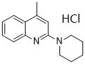
| SMILES | Cl.N1(C2C=C(C)C3C=CC=CC=3N=2)CCCCC1 |
|
| InChi Key | QCABWRXQHZUBPW-UHFFFAOYSA-N | |
| InChi Code | InChI=1S/C15H18N2.ClH/c1-12-11-15(17-9-5-2-6-10-17)16-14-8-4-3-7-13(12)14;/h3-4,7-8,11H,2,5-6,9-10H2,1H3;1H | |
| Chemical Name |
|
|
| Synonyms |
|
|
| HS Tariff Code | 2934.99.9001 | |
| Storage |
Powder-20°C 3 years 4°C 2 years In solvent -80°C 6 months -20°C 1 month Note: Please store this product in a sealed and protected environment, avoid exposure to moisture. |
|
| Shipping Condition | Room temperature (This product is stable at ambient temperature for a few days during ordinary shipping and time spent in Customs) |
Biological Activity
| Targets |
TRPC4/TRPC5 ML204 hydrochloride shows 19-fold selectivity against muscarinic receptor-coupled TRPC6 channel activation and inhibits TRPC4β-mediated intracellular Ca2+ increase with an IC50 value of 0.96 μM (HEK293 cells)[1]. ML204 hydrochloride inhibits TRPC4β activity that is triggered by endogenous M3-like muscarinic receptors stimulating Gq/11 or Gi/o activation by μ-opioid, 5HT1A serotonin, and M2 muscarinic receptors[1]. LPS-induced TRPC5 channel activity is inhibited by ML204 hydrochloride[3]. |
| ln Vitro |
ML204 hydrochloride shows 19-fold selectivity against muscarinic receptor-coupled TRPC6 channel activation and inhibits TRPC4β-mediated intracellular Ca2+ increase with an IC50 value of 0.96 μM (HEK293 cells)[1]. ML204 hydrochloride inhibits TRPC4β activity that is triggered by endogenous M3-like muscarinic receptors stimulating Gq/11 or Gi/o activation by μ-opioid, 5HT1A serotonin, and M2 muscarinic receptors[1]. LPS-induced TRPC5 channel activity is inhibited by ML204 hydrochloride[3]. In a fluorescence-based Ca²⁺ assay using HEK293 cells co-expressing mouse TRPC4β and μ-opioid receptors, ML204 inhibited DAMGO-induced intracellular Ca²⁺ rise with an IC₅₀ of 0.96 ± 0.26 µM. Complete inhibition was achieved at 20 µM. [1] In automated whole-cell patch clamp (QPatch16) recordings, ML204 blocked TRPC4β currents activated by 50 nM DAMGO with an IC₅₀ of ≈2.9–3.55 µM. [1] ML204 (3.33 µM) also blocked TRPC4β currents activated by intracellular dialysis of GTPγS (IC₅₀ ≈ 2.85 µM), indicating a direct effect on the channel independent of GPCR signaling. [1] In manual patch clamp recordings, 10 µM ML204 nearly completely blocked currents through guinea pig TRPC4β channels activated by 10 µM carbachol. [1] ML204 (10 µM) exhibited modest inhibition (≈38%) of TRPC6 currents activated via M₅ muscarinic receptors but showed no appreciable block of TRPC6 currents activated directly by 10 µM OAG. [1] In a membrane potential assay, ML204 blocked acetylcholine-induced TRPC6-mediated depolarization with an IC₅₀ of 18.4 µM. [1] ML204 (10 µM) significantly blocked native muscarinic receptor-activated cation currents (mIₐₐₜ) in freshly isolated guinea pig ileal smooth muscle cells, inhibiting carbachol (100 µM)-evoked currents by 86 ± 2% and GTPγS (200 µM)-induced currents by 65 ± 4%. [1] In broad profiling studies (Ricerca Lead Profiling Screen of 68 targets at 10 µM), ML204 showed >50% inhibition in only 7 out of 68 binding assays. [1] |
| ln Vivo |
In LPS-injected mice, ML204 hydrochloride (1 mg/kg; sc; twice a day; for 5 days) reduces peritoneal leukocyte counts and cytokines and causes mortality linked to worsened hypothermia[4]. Dual TRPC4/TRPC5 blockade by ML204 increased mortality and hypothermia in thioredoxin-treated LPS mice but preserved macrophage's ability to phagocytose. TRPC5 deletion did not alter body temperature but promoted additional accumulation of peritoneal leukocytes and inflammatory mediator release in thioredoxin-administered LPS mice. Thioredoxin diminished macrophage-mediated phagocytosis in wild-type but not TRPC5 knockout animals. TRPC5 ablation did not affect LPS-induced responses. However, ML204 caused mortality associated with exacerbated hypothermia and decreased peritoneal leukocyte numbers and cytokines in LPS-injected mice. These results suggest that bacterial thioredoxin effects under LPS stimuli are mediated by TRPC4 and TRPC5, shedding light on the additional mechanisms of bacterial virulence and on the pathophysiological roles of these receptors. Notably, ML204-perfused mice were protected from PS-induced FPE (Figure 7A). These results were quantified in a blinded fashion by counting the number of FPs over a measured length of GBM in TEM images (n = 90–105 images per group). By this analysis, ML204-perfused mice showed significant protection from the effects of PS (Figure 7B). These results are in line with our observations in Trpc5-KO mice.[3] Next, we tested the effect of ML204 in the LPS model. ML204 was injected (20 mg/kg/d i.p.) twice at 12-hour intervals after injection of LPS. Control PBS-injected mice had no observable structural changes and no albuminuria (Figure 7, C–E). LPS injection resulted in FPE (Figure 7D), although this was milder than the PS-induced FPE. Importantly, the ML204-treated mice were protected from LPS-induced FPE and albuminuria[3]. In a vascular perfusion model, co-perfusion of ML204 (10 µM) with protamine sulfate (PS) in wild-type mice preserved foot processes (FPs) and protected from PS-induced foot process effacement (FPE), similar to HBSS controls.[3] In an LPS-induced albuminuria model, treatment of wild-type mice with ML204 (20 mg/kg/day, intraperitoneal injection) mitigated LPS-induced foot process effacement (FPE).[3] Treatment with ML204 (20 mg/kg/day, i.p.) significantly reduced LPS-induced albuminuria in wild-type mice.[3] |
| Enzyme Assay |
ML204 has a 19-fold coupling to muscarinic receptor-coupled TRPC6 channel activation and inhibits TRPC4β-mediated intracellular Ca2+ rise with an IC50 value of 0.96 μM (HEK293 cells) [1]. The activation of Gq/11 by the M2 muscarinic receptor or endogenous M3-like muscarinic receptor, u-opioid, 5HT1A hematin, Gi/o, and TRPC4β is blocked by ML204[1]. ML204 inhibits TRPC5 channel activity that is triggered by LPS [3]. ML204 was identified as a novel TRPC4 channel inhibitor following a high throughput fluorescent screen of the MLSMR library and SAR analysis of active compounds. ML204 inhibited calcium influx through TRPC4 channels activated by μ-opioid receptor stimulation with an IC50 value of 0.96 μM and exhibited 19-fold selectivity against TRPC6 channels in similar fluorescent assays. ML204 blocked TRPC4 channels in an electrophysiological assay with an IC value of 2.6 μM and was also active in fluorescent and electrophysiological assays in which TRPC4 channels were activated by different mechanisms, indicating direct block of TRPC4 channels. Selectivity for block of TRPC4 channels was examined in fluorescent and electrophysiological experiments against closely related TRPC channels and more distantly related TRPV, TRPA and TRPM channels, and against non-TRP ion channels. ML204 afforded good selectivity (19-fold) against TRPC6 channels and more modest selectivity against TRPC3 and TRPC5 (9-fold) channels. Little or no block of TRPV, TRPA, TRPM or voltage-gated ion channels was observed. ML204 exhibited properties useful for a variety of in vitro investigations[2]. Rac1 activation assay.[3] Rac1 activation assays were done as previously described, with some modifications. Podocytes were treated with 300 μg/ml PS for 1 hour, followed by harvest and Rac1 pulldown experiments. In the ML204 experiments, cells were pretreated with 30 μM ML204 for 20 minutes before PS was applied. Activated Rac1 was analyzed with a commercial Rac1 activation assay kit using a GST-tagged fusion protein corresponding to the p21-binding domain (PBD; residues 67–150) of human PAK-1, according to the manufacturer’s instructions. After pulldown, the eluted active Rac1 was detected by immunoblotting using a mouse monoclonal Rac1 antibody. Total Rac1 and GADPH were measured in the cell lysates used for the pulldown studies and served as loading controls. Electrophysiology.[3] Patch-clamp electrophysiology was performed in the whole-cell configuration or on outside-out patches. Patch pipettes with resistances of 3–4 MΩ were pulled from borosilicate glass with a P-97 puller and filled with a solution containing 135 mM CH3SO3Cs, 10 mM CsCl, 3 mM MgATP, 0.2 mM NaGTP, 0.2 mM EGTA, 0.13 mM CaCl2, and 10 mM HEPES (pH 7.3) with CsOH. The bath solution contained 135 mM CH3SO3Na, 5 mM CsCl, 2 mM CaCl2, 1 mM MgCl2, 10 mM HEPES, and 10 mM glucose (pH 7.4) with NaOH. Angiotensin II (500 nM), LPS (100 μg/ml), and ML204 (10 μM) were applied to the bath solution. Whole-cell currents were recorded from –100 mV to +100 mV voltage ramps over 400 ms and a holding potential of –60 mV. For single-channel recordings in the outside-out configuration, we used a voltage step protocol from –100 mV to +100 mV delivered at 20-mV intervals and a holding potential of 0 mV. Average pipette resistance filled with pipette solution was 3–5 MΩ. Data were sampled at 10 kHz and filtered at 5 kHz. Single-channel data were further off-line filtered at 500 Hz before analysis. In single-channel traces, currents were idealized using a manually defined amplitude criterion to assign ion channel opening and closing transitions. Ensemble averages were expressed as Po (average current divided by unitary current amplitude and number of channels per patch) and plotted as histograms. All data were acquired at room temperature and analyzed using pClamp 10. |
| Cell Assay |
PS, LPS, and Cch treatment of cultured podocytes.[3] Differentiated cultured podocytes grown at >90% confluence were incubated with 1–30 μM ML204 for 20 minutes, and then exposed to 300 μg/ml PS, 100 μg/ml LPS, or 100 μM Cch as appropriate. For PS experiments, as soon as changes in cell morphology could be seen by light microscopy (70–90 min), cells were fixed with 4% paraformaldehyde in PBS for 15 minutes before permeabilization with 0.1% Triton X-100 for 10 minutes. For LPS and Cch experiments, cells were fixed as above at 24 hours after treatment. For immunostaining, podocytes were incubated with synaptopodin “NT” antibody and detected with Alexa Fluor 488–conjugated secondary antibody. Actin structures were labeled with Alexa Fluor 594–conjugated phalloidin as described previously. 3 independent trials were analyzed, with 3 dishes per condition in each trial and 10 images per dish with comparable cell density. A total of 1,600–2,000 cells was analyzed for the PS/ML204 experiment and 1,400–1,600 cells for the PS/KD experiments. A total of 1,000 cells was analyzed for the LPS experiment and 1,400 cells for the Cch experiment. The number of cells was counted by DAPI staining and analysis with an automated script in ImageJ, which was subsequently corrected manually. Affected cells were defined as either collapsed with a very bright, condensed actin staining (for PS experiments), or cells without clearly visible stress fibers (for LPS and Cch experiments), as previously described. Images were acquired with a Zeiss LSM510 upright confocal microscope. Images from an optical slice of 3–4 μm were acquired using Zeiss Pascal software. Statistical significance was evaluated by ANOVA and Dunnett’s multiple-comparison test. Primary High-Throughput Fluorescence Ca²⁺ Screen: A stable HEK293 cell line co-expressing mouse TRPC4β and μ-opioid receptors was used. Cells were seeded in 384-well plates, loaded with Fluo4-AM, and stimulated with DAMGO. Test compounds were added before agonist addition. Intracellular Ca²⁺ changes were monitored using a fluorescence plate reader. [1] Selectivity Screening via Fluorescence Ca²⁺ Assay: HEK293 cells stably expressing various TRP channels (TRPV1, TRPV3, TRPM8, TRPA1) were seeded in 96-well plates, loaded with Fluo4-AM, and exposed to respective channel agonists. Ca²⁺ influx was measured to assess compound effects. [1] Membrane Potential Assay for TRPC6: HEK293 cells stably expressing TRPC6 (with endogenous muscarinic receptors) were loaded with a membrane potential-sensitive dye. Cells were stimulated with acetylcholine, and membrane depolarization was measured in the presence or absence of test compounds. [1] Automated Patch Clamp (QPatch16): HEK293 cells stably expressing TRPC4β and μ-OR were prepared as a single-cell suspension. Whole-cell currents were recorded using a voltage ramp protocol. Agonists (DAMGO) and compounds were applied via perfusion. [1] Manual Whole-Cell Patch Clamp for TRPC Channels: HEK293 cells heterologously expressing specific TRPC channels (with or without co-expressed GPCRs) were voltage-clamped. Currents were elicited by voltage ramps. Drugs were applied via a gravity-driven perfusion system. [1] Electrophysiology on Native Neurons: Dorsal root ganglion neurons from mice were cultured. Voltage-gated Na⁺, K⁺, and Ca²⁺ channel currents were recorded using specific voltage step protocols in the presence or absence of ML204. [1] Electrophysiology on Native Smooth Muscle Cells: Ileal smooth muscle myocytes were freshly isolated from guinea pigs. Whole-cell recordings were performed to measure mIₐₐₜ induced by bath-applied carbachol or intracellular GTPγS infusion. ML204 was applied via perfusion. [1] |
| Animal Protocol |
Animal/Disease Models: Nonfasted male C57BL/6 (2 -3 months)[4] Doses: 1 mg/kg Route of Administration: subcutaneous (sc) injection, twice a day, for 5 days (prior to LPS injection) Experimental Results: Induces mortality associated with increased hypothermia in mice with LPS-induced systemic inflammatory response. LPS-induced albuminuria.[3] Induction of albuminuria in male WT and Trpc5-KO mice (20–25 g BW) by LPS injection was done as previously described, with some modifications. At 48 hours prior to injection, baseline urine was collected for 24 hours in metabolic cages. LPS (15 μg/g i.p., 1 mg/ml) was injected twice, at the 0- and 24-hour time points. PBS was injected i.p. twice, at 12 and 36 hours, to avoid dehydration. ML204 (20 mg/kg/d i.p.) was injected at 12 and 24 hours. Urine was collected for a 24-hour period beginning 24 hours after initial LPS injection using metabolic cages. To quantify the levels of albuminuria, 10 μl urine was analyzed by SDS-PAGE. Bovine serum albumin standards (0.25, 0.5, 1.0, 2.5, and 5.0 μg) were run on the same gel and used to identify and quantify urinary albumin bands. Coomassie signals were quantified using ImageJ. The resulting values — the product of area size and mean gray value of each albumin standard band — were used for construction of a standard curve and its associated mathematical function. Subsequently, the values of the sample bands were translated into albumin concentrations, which were extrapolated to the 24-hour total urine volume. Results were assessed by ANOVA and Bonferroni’s multiple-comparison test. For preparation of glomerular lysates for Western blotting, mice were treated as above and killed 36 hours after initial LPS injection. Mouse kidneys were perfused through the renal artery with Dynabeads for magnetic isolation of highly purified glomeruli. Protein extraction from isolated glomeruli, SDS-PAGE, and Western blotting was done as described previously, and proteins were detected with appropriate primary and secondary antibodies. PS model.[3] Adult WT (n = 15) and Trpc5-KO (n = 7) littermate mice were anesthetized with pentobarbital and placed on a heat pad set at 37°C, and their kidneys were perfused in situ through the renal artery at a pressure of approximately 240 mm Hg and an infusion rate of 9 ml/min as previously described (58), with some modifications. First, kidneys were flushed with HBSS or with HBSS plus 10 μM ML204 at 37°C for 2 minutes, followed by perfusion with 2 mg/ml PS in HBSS or with PS plus ML204 at 37°C for 15 minutes. All vascular perfusion solutions were kept at 37°C throughout the duration of the experiment. Pharmacological Treatments[4] C57BL/6, TRPC5+/+, and TRPC5−/− mice received a subcutaneous (s.c.) injection of phosphate-buffered saline (PBS) containing bacterial Trx (20 μg/150 μl/animal, twice a day; from E. coli) for 3 days prior to the induction of SIRS. In order to assess the role of TRPC4 and TRPC5 complexes in LPS-induced responses, C57BL/6 mice received ML204 [16, 21] (1 mg/kg, 150 μl/animal, twice a day) for 5 days and then LPS. In a separate set of experiments, C57BL/6 animals received ML204 (1 mg/kg, twice a day; in 6% dimethyl sulfoxide (DMSO) in PBS) for 2 days alone, and then, this drug was coinjected with bacterial Trx (20 μg/animal, twice a day) for another 3 days prior to LPS challenge. Vehicle-treated mice were used as controls. In the protamine sulfate (PS) perfusion model, adult wild-type mice were anesthetized, and kidneys were perfused in situ through the renal artery. Kidneys were first flushed with HBSS or HBSS containing 10 µM ML204 for 2 minutes, followed by perfusion with 2 mg/ml PS in HBSS or PS plus ML204 for 15 minutes. All solutions were kept at 37°C. Kidneys were then fixed for TEM analysis.[3] In the LPS-induced albuminuria model, male wild-type mice were injected intraperitoneally with LPS (15 µg/g) twice at 0 and 24 hours. ML204 (20 mg/kg/day, i.p.) was administered at 12 and 24 hours after the initial LPS injection. Control groups received PBS. Urine was collected in metabolic cages starting 24 hours post-LPS for albumin quantification. Kidneys were harvested 36 hours post-LPS for glomerular isolation and analysis.[3] |
| References |
[1]. Identification of ML204, a novel potent antagonist that selectively modulates native TRPC4/C5 ion channels. J Biol Chem. 2011 Sep 23;286(38):33436-46. [2]. Novel Chemical Inhibitor of TRPC4 Channels. Probe Reports from the NIH Molecular Libraries Program. [3]. Inhibition of the TRPC5 ion channel protects the kidney filter. J Clin Invest. 2013 Dec 2; 123(12): 5298–5309. [4]. Transient Receptor Potential Canonical Channels 4 and 5 Mediate Escherichia coli-Derived Thioredoxin Effects in Lipopolysaccharide-Injected Mice. Oxid Med Cell Longev. 2018 Jun 10;2018:4904696. |
| Additional Infomation |
Transient receptor potential canonical (TRPC) channels are Ca(2+)-permeable nonselective cation channels implicated in diverse physiological functions, including smooth muscle contractility and synaptic transmission. However, lack of potent selective pharmacological inhibitors for TRPC channels has limited delineation of the roles of these channels in physiological systems. Here we report the identification and characterization of ML204 as a novel, potent, and selective TRPC4 channel inhibitor. A high throughput fluorescent screen of 305,000 compounds of the Molecular Libraries Small Molecule Repository was performed for inhibitors that blocked intracellular Ca(2+) rise in response to stimulation of mouse TRPC4β by μ-opioid receptors. ML204 inhibited TRPC4β-mediated intracellular Ca(2+) rise with an IC(50) value of 0.96 μm and exhibited 19-fold selectivity against muscarinic receptor-coupled TRPC6 channel activation. In whole-cell patch clamp recordings, ML204 blocked TRPC4β currents activated through either μ-opioid receptor stimulation or intracellular dialysis of guanosine 5'-3-O-(thio)triphosphate (GTPγS), suggesting a direct interaction of ML204 with TRPC4 channels rather than any interference with the signal transduction pathways. Selectivity studies showed no appreciable block by 10-20 μm ML204 of TRPV1, TRPV3, TRPA1, and TRPM8, as well as KCNQ2 and native voltage-gated sodium, potassium, and calcium channels in mouse dorsal root ganglion neurons. In isolated guinea pig ileal myocytes, ML204 blocked muscarinic cation currents activated by bath application of carbachol or intracellular infusion of GTPγS, demonstrating its effectiveness on native TRPC4 currents. Therefore, ML204 represents an excellent novel tool for investigation of TRPC4 channel function and may facilitate the development of therapeutics targeted to TRPC4.[1] \n\nML204 was identified as a novel TRPC4 channel inhibitor following a high throughput fluorescent screen of the MLSMR library and SAR analysis of active compounds. ML204 inhibited calcium influx through TRPC4 channels activated by μ-opioid receptor stimulation with an IC50 value of 0.96 μM and exhibited 19-fold selectivity against TRPC6 channels in similar fluorescent assays. ML204 blocked TRPC4 channels in an electrophysiological assay with an IC value of 2.6 μM and was also active in fluorescent and electrophysiological assays in which TRPC4 channels were activated by different mechanisms, indicating direct block of TRPC4 channels. Selectivity for block of TRPC4 channels was examined in fluorescent and electrophysiological experiments against closely related TRPC channels and more distantly related TRPV, TRPA and TRPM channels, and against non-TRP ion channels. ML204 afforded good selectivity (19-fold) against TRPC6 channels and more modest selectivity against TRPC3 and TRPC5 (9-fold) channels. Little or no block of TRPV, TRPA, TRPM or voltage-gated ion channels was observed. ML204 exhibited properties useful for a variety of in vitro investigations.[2] \n\nAn intact kidney filter is vital to retention of essential proteins in the blood and removal of waste from the body. Damage to the filtration barrier results in albumin loss in the urine, a hallmark of cardiovascular disease and kidney failure. Here we found that the ion channel TRPC5 mediates filtration barrier injury. Using Trpc5-KO mice, a small-molecule inhibitor of TRPC5, Ca2+ imaging in isolated kidney glomeruli, and live imagining of podocyte actin dynamics, we determined that loss of TRPC5 or its inhibition abrogates podocyte cytoskeletal remodeling. Inhibition or loss of TRPC5 prevented activation of the small GTP-binding protein Rac1 and stabilized synaptopodin. Importantly, genetic deletion or pharmacologic inhibition of TRPC5 protected mice from albuminuria. These data reveal that the Ca2+-permeable channel TRPC5 is an important determinant of albuminuria and identify TRPC5 inhibition as a therapeutic strategy for the prevention or treatment of proteinuric kidney disease.[3] \n\nThioredoxin plays an essential role in bacterial antioxidant machinery and virulence; however, its regulatory actions in the host are less well understood. Reduced human Trx activates transient receptor potential canonical 5 (TRPC5) in inflammation, but there is no evidence of whether these receptors mediate bacterial thioredoxin effects in the host. Importantly, TRPC5 can form functional complexes with other subunits such as TRPC4. Herein, E. coli-derived thioredoxin induced mortality in lipopolysaccharide- (LPS-) injected mice, accompanied by reduction of leukocyte accumulation, regulation of cytokine release into the peritoneum, and impairment of peritoneal macrophage-mediated phagocytosis.[4] ML204 was identified from a high-throughput screen of 305,000 compounds from the Molecular Libraries Small Molecule Repository (MLSMR). [1] It is reported as the first potent and selective small-molecule blocker for TRPC4 channels. [1] The compound acts as a direct channel blocker rather than interfering with upstream GPCR signaling pathways. [1] Structure-activity relationship (SAR) studies indicated that small cycloalkyl amine substituents (e.g., piperidine, pyrrolidine) on the left-hand side and limited modification on the quinoline ring on the right-hand side are crucial for activity. [1] ML204 is proposed as an excellent pharmacological tool for investigating TRPC4/C5 channel function in native tissues and may facilitate therapeutic development targeting these channels. [1] |
Solubility Data
| Solubility (In Vitro) |
|
|||
| Solubility (In Vivo) |
Note: Listed below are some common formulations that may be used to formulate products with low water solubility (e.g. < 1 mg/mL), you may test these formulations using a minute amount of products to avoid loss of samples. Injection Formulations (e.g. IP/IV/IM/SC) Injection Formulation 1: DMSO : Tween 80: Saline = 10 : 5 : 85 (i.e. 100 μL DMSO stock solution → 50 μL Tween 80 → 850 μL Saline) *Preparation of saline: Dissolve 0.9 g of sodium chloride in 100 mL ddH ₂ O to obtain a clear solution. Injection Formulation 2: DMSO : PEG300 :Tween 80 : Saline = 10 : 40 : 5 : 45 (i.e. 100 μL DMSO → 400 μLPEG300 → 50 μL Tween 80 → 450 μL Saline) Injection Formulation 3: DMSO : Corn oil = 10 : 90 (i.e. 100 μL DMSO → 900 μL Corn oil) Example: Take the Injection Formulation 3 (DMSO : Corn oil = 10 : 90) as an example, if 1 mL of 2.5 mg/mL working solution is to be prepared, you can take 100 μL 25 mg/mL DMSO stock solution and add to 900 μL corn oil, mix well to obtain a clear or suspension solution (2.5 mg/mL, ready for use in animals). Injection Formulation 4: DMSO : 20% SBE-β-CD in saline = 10 : 90 [i.e. 100 μL DMSO → 900 μL (20% SBE-β-CD in saline)] *Preparation of 20% SBE-β-CD in Saline (4°C,1 week): Dissolve 2 g SBE-β-CD in 10 mL saline to obtain a clear solution. Injection Formulation 5: 2-Hydroxypropyl-β-cyclodextrin : Saline = 50 : 50 (i.e. 500 μL 2-Hydroxypropyl-β-cyclodextrin → 500 μL Saline) Injection Formulation 6: DMSO : PEG300 : castor oil : Saline = 5 : 10 : 20 : 65 (i.e. 50 μL DMSO → 100 μLPEG300 → 200 μL castor oil → 650 μL Saline) Injection Formulation 7: Ethanol : Cremophor : Saline = 10: 10 : 80 (i.e. 100 μL Ethanol → 100 μL Cremophor → 800 μL Saline) Injection Formulation 8: Dissolve in Cremophor/Ethanol (50 : 50), then diluted by Saline Injection Formulation 9: EtOH : Corn oil = 10 : 90 (i.e. 100 μL EtOH → 900 μL Corn oil) Injection Formulation 10: EtOH : PEG300:Tween 80 : Saline = 10 : 40 : 5 : 45 (i.e. 100 μL EtOH → 400 μLPEG300 → 50 μL Tween 80 → 450 μL Saline) Oral Formulations Oral Formulation 1: Suspend in 0.5% CMC Na (carboxymethylcellulose sodium) Oral Formulation 2: Suspend in 0.5% Carboxymethyl cellulose Example: Take the Oral Formulation 1 (Suspend in 0.5% CMC Na) as an example, if 100 mL of 2.5 mg/mL working solution is to be prepared, you can first prepare 0.5% CMC Na solution by measuring 0.5 g CMC Na and dissolve it in 100 mL ddH2O to obtain a clear solution; then add 250 mg of the product to 100 mL 0.5% CMC Na solution, to make the suspension solution (2.5 mg/mL, ready for use in animals). Oral Formulation 3: Dissolved in PEG400 Oral Formulation 4: Suspend in 0.2% Carboxymethyl cellulose Oral Formulation 5: Dissolve in 0.25% Tween 80 and 0.5% Carboxymethyl cellulose Oral Formulation 6: Mixing with food powders Note: Please be aware that the above formulations are for reference only. InvivoChem strongly recommends customers to read literature methods/protocols carefully before determining which formulation you should use for in vivo studies, as different compounds have different solubility properties and have to be formulated differently. (Please use freshly prepared in vivo formulations for optimal results.) |
| Preparing Stock Solutions | 1 mg | 5 mg | 10 mg | |
| 1 mM | 3.8055 mL | 19.0273 mL | 38.0546 mL | |
| 5 mM | 0.7611 mL | 3.8055 mL | 7.6109 mL | |
| 10 mM | 0.3805 mL | 1.9027 mL | 3.8055 mL |