Physicochemical Properties
| Molecular Formula | C21H29N5OS |
| Molecular Weight | 399.552862882614 |
| Exact Mass | 399.209 |
| CAS # | 2230047-87-5 |
| PubChem CID | 134817254 |
| Appearance | White to off-white solid powder |
| LogP | 3.9 |
| Hydrogen Bond Donor Count | 2 |
| Hydrogen Bond Acceptor Count | 4 |
| Rotatable Bond Count | 7 |
| Heavy Atom Count | 28 |
| Complexity | 511 |
| Defined Atom Stereocenter Count | 0 |
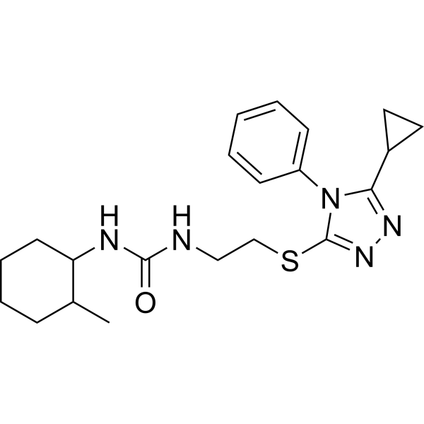
| SMILES | S(CCNC(NC1CCCCC1C)=O)C1=NN=C(C2CC2)N1C1C=CC=CC=1 |
| InChi Key | IDILCELZAIAPSY-UHFFFAOYSA-N |
| InChi Code | InChI=1S/C21H29N5OS/c1-15-7-5-6-10-18(15)23-20(27)22-13-14-28-21-25-24-19(16-11-12-16)26(21)17-8-3-2-4-9-17/h2-4,8-9,15-16,18H,5-7,10-14H2,1H3,(H2,22,23,27) |
| Chemical Name | 1-[2-[(5-cyclopropyl-4-phenyl-1,2,4-triazol-3-yl)sulfanyl]ethyl]-3-(2-methylcyclohexyl)urea |
| Synonyms | 2230047-87-5; 3-(2-((5-cyclopropyl-4-phenyl-4H-1,2,4-triazol-3-yl)sulfanyl)ethyl)-1-(2-methylcyclohexyl)urea; 3-{2-[(5-cyclopropyl-4-phenyl-4H-1,2,4-triazol-3-yl)sulfanyl]ethyl}-1-(2-methylcyclohexyl)urea; RefChem:490284; 845-220-5; MFN2 agonist-1; MFN2 agonist B-A l; orb1942962; . |
| HS Tariff Code | 2934.99.9001 |
| Storage |
Powder-20°C 3 years 4°C 2 years In solvent -80°C 6 months -20°C 1 month |
| Shipping Condition | Room temperature (This product is stable at ambient temperature for a few days during ordinary shipping and time spent in Customs) |
Biological Activity
| Targets |
- Mitofusin 2 (MFN2)
- MFN2 agonist-1 (B-A/l) is a direct allosteric activator of MFN2, targeting its GTPase domain and promoting mitochondrial fusion. It specifically binds to MFN2 residues Ser378 and adjacent regions, disrupting autoinhibitory interactions and enhancing GTP hydrolysis activity [1] |
| ln Vitro |
- Mitochondrial fusion restoration: 1. Patient-derived fibroblasts: Treatment of CMT2A patient fibroblasts with MFN2 agonist-1 (B-A/l) (10–20 μM) rescued mitochondrial fragmentation, increasing mitochondrial network connectivity by ~50% (mitochondrial aspect ratio >3.0) compared to untreated cells [1] 2. Neuronal cell models: In primary mouse dorsal root ganglion (DRG) neurons expressing mutant MFN2T105M, B-A/l (5–15 μM) restored mitochondrial motility, reducing static mitochondrial aggregates by ~60% and increasing anterograde transport velocity from 0.2 ± 0.05 μm/s (control) to 0.5 ± 0.1 μm/s [1] - Functional recovery: 1. ATP production: B-A/l (10 μM) increased ATP levels in MFN2-deficient HEK293 cells by ~40% (luciferin-luciferase assay), reversing mitochondrial dysfunction-induced bioenergetic deficits [1] 2. Mitochondrial membrane potential (ΔΨm): In CMT2A fibroblasts, B-A/l (15 μM) normalized ΔΨm (JC-1 staining), reducing the percentage of depolarized mitochondria from ~70% to ~30% [1] - Mechanistic insights: 1. Phosphorylation dynamics: B-A/l (10 μM) increased phosphorylation of MFN2 at Ser378 by ~2-fold (phospho-specific Western blot), mimicking PINK1 kinase activation and stabilizing MFN2 homodimers [1] 2. Protein-protein interactions: Co-immunoprecipitation showed B-A/l enhanced MFN2 binding to mitochondrial outer membrane proteins (e.g., Tom20), promoting mitochondrial tethering [1] |
| ln Vivo |
- CMT2A mouse model correction: 1. Motor function improvement: Mice expressing MFN2T105M treated with B-A/l (50 mg/kg, intraperitoneal, daily for 14 days) showed significant recovery in rotarod performance, increasing latency to fall from 8.2 ± 1.5 seconds (vehicle) to 15.6 ± 2.1 seconds [1] 2. Axonal mitochondrial transport: In sciatic nerve axons of treated mice, B-A/l restored mitochondrial anterograde transport frequency by ~70%, reducing axonal swellings and fragmented mitochondria [1] 3. Histological rescue: B-A/l treatment decreased muscle fiber atrophy (cross-sectional area increased from 180 ± 30 μm² to 250 ± 40 μm²) and preserved neuromuscular junctions (α-bungarotoxin staining) in CMT2A mice [1] |
| Enzyme Assay |
- MFN2 GTPase activity assay [1]: 1. Recombinant protein preparation: His-tagged human MFN2 (residues 1–650) was expressed in E. coli and purified using nickel affinity chromatography. GTPase activity was measured in buffer containing 50 mM Tris-HCl (pH 7.5), 10 mM MgCl₂, and 1 mM DTT. 2. Reaction setup: 50 μL reactions included MFN2 (2 μM), GTP (1 mM), and B-A/l (0.1–50 μM). Reactions were incubated at 37°C for 30 minutes, and GTP hydrolysis was quantified by malachite green assay. 3. Results: B-A/l increased MFN2 GTPase activity in a dose-dependent manner, with maximal activation (2.5-fold increase) at 10 μM. The EC₅₀ for activation was 3.2 μM [1] - Conformational change detection (FRET) [1]: 1. FRET pair labeling: Cysteine mutants of MFN2 (Cys102 and Cys589) were labeled with donor (Alexa Fluor 488) and acceptor (Alexa Fluor 594) dyes. 2. Binding assay: B-A/l (10 μM) induced a 20% decrease in FRET efficiency, indicating conformational relaxation of the MFN2 GTPase domain [1] |
| Cell Assay |
- Mitochondrial morphology analysis [1]: 1. Cell culture: CMT2A patient fibroblasts or MFN2T105M DRG neurons were seeded on glass coverslips and treated with B-A/l (0–20 μM) for 24 hours. 2. Staining: Cells were fixed with 4% paraformaldehyde, permeabilized with 0.1% Triton X-100, and stained with MitoTracker Red (50 nM) and DAPI (1 μg/mL). 3. Imaging: Confocal microscopy (63× oil immersion) was used to quantify mitochondrial aspect ratio (length/width) and connectivity. At 10 μM, B-A/l increased aspect ratio from 1.2 ± 0.2 to 3.5 ± 0.5 [1] - ATP production assay [1]: 1. Cell treatment: HEK293 cells transfected with siRNA against MFN2 were treated with B-A/l (0–20 μM) for 48 hours. 2. ATP measurement: Cell lysates were analyzed using an ATP Bioluminescence Assay Kit. B-A/l (10 μM) restored ATP levels to 85% of control (non-targeting siRNA) [1] |
| Animal Protocol |
- CMT2A mouse treatment [1]: 1. Animal model: MFN2T105M knock-in mice (8–10 weeks old, male) were randomized into treatment (n=8) and vehicle (n=8) groups. 2. Drug formulation: B-A/l was dissolved in DMSO (100 mM stock) and diluted in 0.9% saline containing 5% Tween 80 to a final concentration of 10 mg/mL. 3. Administration: Mice received intraperitoneal injections of B-A/l (50 mg/kg) or vehicle daily for 14 days. 4. Assessment: Rotarod performance, grip strength, and sciatic nerve morphology were evaluated weekly. Mice were euthanized on day 15 for tissue harvest [1] |
| ADME/Pharmacokinetics |
- Mouse pharmacokinetics [1]: 1. Plasma exposure: After a single intraperitoneal dose of B-A/l (50 mg/kg), peak plasma concentration (Cmax) was 25.3 μM at 1 hour, with a terminal half-life (t₁/₂) of 3.8 hours. 2. Tissue distribution: At 2 hours post-dose, B-A/l concentrations in sciatic nerve (18.7 μM) and spinal cord (12.5 μM) were ~75% and 50% of plasma levels, respectively. 3. Excretion: ~60% of the dose was excreted unchanged in urine within 24 hours, with minimal hepatic metabolism detected (<10% metabolites) [1] |
| Toxicity/Toxicokinetics |
- In vitro cytotoxicity: 1. Normal cell viability: B-A/l (up to 50 μM) showed no significant cytotoxicity in wild-type mouse embryonic fibroblasts (MTT assay, viability >90%) [1] 2. Genotoxicity: B-A/l (20 μM) did not induce micronuclei in human peripheral blood lymphocytes (cytokinesis-block micronucleus assay) [1] - In vivo safety: 1. General toxicity: Daily intraperitoneal administration of B-A/l (100 mg/kg) for 28 days caused no significant weight loss or histological abnormalities in liver, kidney, or heart tissues of mice [1] 2. Hematological parameters: Complete blood counts and serum chemistry (ALT, AST, creatinine) remained within normal ranges in treated mice [1] |
| References | [1]. MFN2 agonists reverse mitochondrial defects in preclinical models of Charcot-Marie-Tooth disease type 2A. Science. 2018 Apr 20;360(6386):336-341. |
| Additional Infomation |
- Mechanism of action: B-A/l disrupts the autoinhibitory interaction between MFN2's GTPase domain and helical bundle 2 (HB2), promoting homodimerization and mitochondrial tethering. This effect is dependent on phosphorylation of Ser378 by PINK1 kinase [1] - Therapeutic potential: B-A/l represents a first-in-class MFN2 agonist with preclinical efficacy in CMT2A, targeting mitochondrial dysdynamism. Its ability to cross the blood-brain barrier and accumulate in peripheral nerves makes it suitable for neurodegenerative disorders [1] - Development rationale: B-A/l was identified through high-throughput screening of a 50,000-compound library, prioritizing compounds that restored mitochondrial network integrity in MFN2-deficient cells [1] |
Solubility Data
| Solubility (In Vitro) | May dissolve in DMSO (in most cases), if not, try other solvents such as H2O, Ethanol, or DMF with a minute amount of products to avoid loss of samples |
| Solubility (In Vivo) |
Note: Listed below are some common formulations that may be used to formulate products with low water solubility (e.g. < 1 mg/mL), you may test these formulations using a minute amount of products to avoid loss of samples. Injection Formulations (e.g. IP/IV/IM/SC) Injection Formulation 1: DMSO : Tween 80: Saline = 10 : 5 : 85 (i.e. 100 μL DMSO stock solution → 50 μL Tween 80 → 850 μL Saline) *Preparation of saline: Dissolve 0.9 g of sodium chloride in 100 mL ddH ₂ O to obtain a clear solution. Injection Formulation 2: DMSO : PEG300 :Tween 80 : Saline = 10 : 40 : 5 : 45 (i.e. 100 μL DMSO → 400 μLPEG300 → 50 μL Tween 80 → 450 μL Saline) Injection Formulation 3: DMSO : Corn oil = 10 : 90 (i.e. 100 μL DMSO → 900 μL Corn oil) Example: Take the Injection Formulation 3 (DMSO : Corn oil = 10 : 90) as an example, if 1 mL of 2.5 mg/mL working solution is to be prepared, you can take 100 μL 25 mg/mL DMSO stock solution and add to 900 μL corn oil, mix well to obtain a clear or suspension solution (2.5 mg/mL, ready for use in animals). Injection Formulation 4: DMSO : 20% SBE-β-CD in saline = 10 : 90 [i.e. 100 μL DMSO → 900 μL (20% SBE-β-CD in saline)] *Preparation of 20% SBE-β-CD in Saline (4°C,1 week): Dissolve 2 g SBE-β-CD in 10 mL saline to obtain a clear solution. Injection Formulation 5: 2-Hydroxypropyl-β-cyclodextrin : Saline = 50 : 50 (i.e. 500 μL 2-Hydroxypropyl-β-cyclodextrin → 500 μL Saline) Injection Formulation 6: DMSO : PEG300 : castor oil : Saline = 5 : 10 : 20 : 65 (i.e. 50 μL DMSO → 100 μLPEG300 → 200 μL castor oil → 650 μL Saline) Injection Formulation 7: Ethanol : Cremophor : Saline = 10: 10 : 80 (i.e. 100 μL Ethanol → 100 μL Cremophor → 800 μL Saline) Injection Formulation 8: Dissolve in Cremophor/Ethanol (50 : 50), then diluted by Saline Injection Formulation 9: EtOH : Corn oil = 10 : 90 (i.e. 100 μL EtOH → 900 μL Corn oil) Injection Formulation 10: EtOH : PEG300:Tween 80 : Saline = 10 : 40 : 5 : 45 (i.e. 100 μL EtOH → 400 μLPEG300 → 50 μL Tween 80 → 450 μL Saline) Oral Formulations Oral Formulation 1: Suspend in 0.5% CMC Na (carboxymethylcellulose sodium) Oral Formulation 2: Suspend in 0.5% Carboxymethyl cellulose Example: Take the Oral Formulation 1 (Suspend in 0.5% CMC Na) as an example, if 100 mL of 2.5 mg/mL working solution is to be prepared, you can first prepare 0.5% CMC Na solution by measuring 0.5 g CMC Na and dissolve it in 100 mL ddH2O to obtain a clear solution; then add 250 mg of the product to 100 mL 0.5% CMC Na solution, to make the suspension solution (2.5 mg/mL, ready for use in animals). Oral Formulation 3: Dissolved in PEG400 Oral Formulation 4: Suspend in 0.2% Carboxymethyl cellulose Oral Formulation 5: Dissolve in 0.25% Tween 80 and 0.5% Carboxymethyl cellulose Oral Formulation 6: Mixing with food powders Note: Please be aware that the above formulations are for reference only. InvivoChem strongly recommends customers to read literature methods/protocols carefully before determining which formulation you should use for in vivo studies, as different compounds have different solubility properties and have to be formulated differently. (Please use freshly prepared in vivo formulations for optimal results.) |
| Preparing Stock Solutions | 1 mg | 5 mg | 10 mg | |
| 1 mM | 2.5028 mL | 12.5141 mL | 25.0282 mL | |
| 5 mM | 0.5006 mL | 2.5028 mL | 5.0056 mL | |
| 10 mM | 0.2503 mL | 1.2514 mL | 2.5028 mL |