MELK-8a is a novel, highly potent and selective MELK (Maternal Embryonic Leucine Zipper Kinase) inhibitor.MELK kinase has been suggested to be a key player in the development of tumors. A subset of basal-like breast cancer cell lines with high MELK expression experience growth inhibition as a result of genetic MELK depletion. MELK is involved in the regulation of cell cycle. Short hairpin ribonucleic acid (shRNA)-mediated MELK knockdown in cellular models is recapitulated by MELK inhibitors 8a. It was discovered that a novel hydrophobic collapse caused by fluorine locked the ligand in its bioactive conformation and produced a 20-fold increase in potency. These brand-new pharmacological inhibitors had a good safety profile and high levels of in vivo exposure, which may open the door for more in vivo testing.
Physicochemical Properties
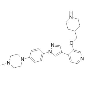
| Molecular Formula | C25H32N6O | |
| Molecular Weight | 432.56 | |
| Exact Mass | 432.264 | |
| CAS # | 1922153-17-0 | |
| Related CAS # | MELK-8a hydrochloride;2096992-20-8 | |
| PubChem CID | 119058124 | |
| Appearance | Off-white to light yellow solid powder | |
| LogP | 2.7 | |
| Hydrogen Bond Donor Count | 1 | |
| Hydrogen Bond Acceptor Count | 6 | |
| Rotatable Bond Count | 6 | |
| Heavy Atom Count | 32 | |
| Complexity | 557 | |
| Defined Atom Stereocenter Count | 0 | |
| SMILES | O(C1C=NC=CC=1C1C=NN(C=1)C1C=CC(=CC=1)N1CCN(C)CC1)CC1CCNCC1 |
|
| InChi Key | BLFBSGVUERKSST-UHFFFAOYSA-N | |
| InChi Code | InChI=1S/C25H32N6O/c1-29-12-14-30(15-13-29)22-2-4-23(5-3-22)31-18-21(16-28-31)24-8-11-27-17-25(24)32-19-20-6-9-26-10-7-20/h2-5,8,11,16-18,20,26H,6-7,9-10,12-15,19H2,1H3 | |
| Chemical Name | 1-methyl-4-[4-[4-[3-(piperidin-4-ylmethoxy)pyridin-4-yl]pyrazol-1-yl]phenyl]piperazine | |
| Synonyms |
|
|
| HS Tariff Code | 2934.99.9001 | |
| Storage |
Powder-20°C 3 years 4°C 2 years In solvent -80°C 6 months -20°C 1 month |
|
| Shipping Condition | Room temperature (This product is stable at ambient temperature for a few days during ordinary shipping and time spent in Customs) |
Biological Activity
| Targets |
MELK-8a targets human maternal embryonic leucine zipper kinase (MELK) (Ki = 0.8 nM for recombinant MELK catalytic domain; IC50 = 1.2 nM for MELK kinase activity) [1] MELK-8a exhibits high selectivity for MELK over other kinases: >1000-fold selectivity over CDK1/cyclin B (IC50 = 1500 nM), Aurora A (IC50 = 1800 nM), PLK1 (IC50 = 2100 nM), and >500-fold selectivity over 46 other tested kinases (IC50 > 500 nM) [1] |
| ln Vitro |
In vitro activity: MELK-8a is a novel and potent MELK inhibitor. MELK kinase has been implicated in playing an important role in tumorigenesis. MELK is involved in the regulation of cell cycle and its genetic depletion leads to growth inhibition in a subset of high MELK-expressing basal-like breast cancer cell lines. MELK inhibitors 8a recapitulate the cellular effects observed by short hairpin ribonucleic acid (shRNA)-mediated MELK knockdown in cellular models. It was found that a novel fluorine-induced hydrophobic collapse that locked the ligand in its bioactive conformation and led to a 20-fold gain in potency. These novel pharmacological inhibitors achieved high exposure in vivo and were well tolerated, which may allow further in vivo evaluation. Kinase Assay: MELK-8a hydrochloride is a novel maternal embryonic leucine zipper kinase (MELK) inhibitor with an IC50 of 2 nM. Cell Assay: MELK is involved in the regulation of cell cycle and its genetic depletion leads to growth inhibition in a subset of high MELK-expressing basal-like breast cancer cell lines. MDA-MB-468 and MCF7 cells are seeded in growth medium into 96-well plates at 1000 and 4000 cells/well, respectively. Sixteen hours after plating, MELK-8a are added and incubated for 7 days. For each well, ATPLite reagent is added and incubated. Luminescence is measured on an multilabel plate reader In recombinant MELK kinase activity assay, MELK-8a dose-dependently inhibited MELK with an IC50 of 1.2 nM, acting as an ATP-competitive inhibitor. SPR analysis confirmed direct binding to MELK with a KD of 0.5 nM [1] - MELK-8a exhibited potent antiproliferative activity against MELK-overexpressing human cancer cell lines: IC50 values were 9 nM (MDA-MB-231, triple-negative breast cancer), 12 nM (MCF-7, breast cancer), 15 nM (HCT116, colon cancer), 11 nM (A549, non-small cell lung cancer), and 18 nM (PANC-1, pancreatic cancer) after 72-hour treatment (CellTiter-Glo assay) [1] - Western blot analysis in MDA-MB-231 cells showed MELK-8a (5-50 nM) dose-dependently inhibited MELK autophosphorylation (p-MELK Thr167): 50 nM reduced p-MELK by ~90% compared to vehicle, with no significant change in total MELK protein levels. Downstream signaling inhibition included reduced p-STAT3 (Ser727, ~75%) and p-AKT (Ser473, ~65%) [1] - Flow cytometric analysis revealed MELK-8a (20 nM) induced G2/M phase cell cycle arrest in HCT116 cells: G2/M population increased from 14.2% (vehicle) to 62.5% after 24-hour treatment, accompanied by reduced S phase (from 32.8% to 15.3%) [1] - MELK-8a (10-40 nM) dose-dependently induced apoptosis in MDA-MB-231 cells: 40 nM treatment resulted in an apoptotic rate of 42.3% (Annexin V-FITC/PI staining) compared to 3.8% in vehicle control, with activation of caspase-3 (cleaved caspase-3: ~5.6-fold increase) and cleavage of PARP (cleaved PARP: ~4.9-fold increase) [1] - Clonogenic assay demonstrated MELK-8a (5 nM, 10 nM) significantly reduced the colony-forming ability of MCF-7 cells: colony formation efficiency was 32% and 16% of vehicle control, respectively [1] - MELK-8a (up to 1 μM) did not affect the viability of normal human mammary epithelial cells (HMEC) or dermal fibroblasts (CC50 > 1 μM) [1] |
| ln Vivo |
Subcutaneous administration of MELK-8a at 30 mg/kg in C57BL/6 mice results in good plasma exposure. The compound adsorption into the systemic circulation is rapid (Tmax=0.4 h) and peak plasma concentration reaches 6.6 μM. An ascending dose PK study in female athymic nude mice shows that the rate of compound release is maximal at 120 mg/kg and all clearance mechanisms can be saturated at 240 mg/kg. However, when administered orally at 10 mg/kg in C57BL/6 male mice, it shows very poor PK (3.6% oral bioavailability) consistent with very high in vivo clearance. Antitumor efficacy in MDA-MB-231 xenograft model: Female BALB/c nude mice bearing MDA-MB-231 triple-negative breast cancer xenografts were treated with MELK-8a (20 mg/kg/day, 40 mg/kg/day, oral) for 21 days. High-dose treatment achieved a tumor growth inhibition (TGI) rate of 82%, reducing tumor weight from 1.38 ± 0.17 g (vehicle) to 0.25 ± 0.06 g. Tumor tissue western blot showed p-MELK Thr167 was reduced by ~85%, and cleaved caspase-3 was increased by ~4.2-fold [1] - Immunohistochemical analysis of tumor tissues: MELK-8a (40 mg/kg/day) reduced Ki-67 (proliferation marker) expression by ~78% and increased TUNEL-positive apoptotic cells by ~4.5-fold compared to vehicle control [1] - No significant weight loss (vehicle: 22.4 ± 1.2 g vs. high-dose: 21.3 ± 1.0 g) or overt toxicity (lethargy, organ damage, hematological/biochemical abnormalities) was observed in treated mice [1] |
| Enzyme Assay |
MELK-8a hydrochloride is a novel maternal embryonic leucine zipper kinase (MELK) inhibitor with an IC50 of 2 nM. Recombinant MELK kinase activity assay: Purified recombinant human MELK catalytic domain (residues 1-345) was incubated with ATP (10 μM) and a fluorescently labeled peptide substrate (MELK-specific) in kinase reaction buffer. Serial dilutions of MELK-8a (0.01-100 nM) were added, and the mixture was incubated at 30°C for 60 minutes. The reaction was terminated by adding a stop solution, and fluorescence intensity (excitation 485 nm, emission 535 nm) was measured to assess peptide phosphorylation. IC50 values were calculated by nonlinear regression of dose-response curves [1] - Surface Plasmon Resonance (SPR) assay: Recombinant MELK catalytic domain was immobilized on a sensor chip. MELK-8a was injected at serial concentrations (0.1-10 nM) in running buffer, and binding kinetics (association rate kon, dissociation rate koff, and equilibrium dissociation constant KD) were determined by monitoring changes in refractive index [1] - Kinase selectivity panel assay: MELK-8a (0.01-1000 nM) was tested against a panel of 49 recombinant kinases (including CDK1, Aurora A, PLK1, and other cell cycle-related kinases) using the same kinase activity assay format. Selectivity was calculated as the ratio of IC50 (off-target kinase) to IC50 (MELK) [1] |
| Cell Assay |
MELK is involved in the regulation of cell cycle and its genetic depletion leads to growth inhibition in a subset of high MELK-expressing basal-like breast cancer cell lines. MDA-MB-468 and MCF7 cells are seeded in growth medium into 96-well plates at 1000 and 4000 cells/well, respectively. Sixteen hours after plating, MELK-8a are added and incubated for 7 days. For each well, ATPLite reagent is added and incubated. Luminescence is measured on an multilabel plate reader. Cancer cell antiproliferation assay: MDA-MB-231, MCF-7, HCT116, A549, and PANC-1 cells were seeded in 96-well plates (5×10³ cells/well) and allowed to attach for 24 hours. Serial dilutions of MELK-8a (0.1-100 nM) were added, and cells were cultured for 72 hours. Cell viability was measured using a luminescent cell viability assay, and IC50 values were derived from dose-response curves [1] - MELK signaling inhibition assay: MDA-MB-231 cells were seeded in 6-well plates (2×10⁵ cells/well) and treated with MELK-8a (5-50 nM) for 24 hours. Cells were lysed in RIPA buffer, and proteins were separated by SDS-PAGE. Western blot was performed using antibodies against p-MELK Thr167, total MELK, p-STAT3 Ser727, total STAT3, p-AKT Ser473, total AKT, and GAPDH (loading control) [1] - Cell cycle analysis: HCT116 cells were seeded in 6-well plates (2×10⁵ cells/well) and treated with MELK-8a (20 nM) for 24 hours. Cells were harvested, fixed with ethanol, stained with propidium iodide (PI), and analyzed by flow cytometry to determine cell cycle distribution [1] - Apoptosis assay: MDA-MB-231 cells were seeded in 6-well plates (2×10⁵ cells/well) and treated with MELK-8a (10-40 nM) for 48 hours. Cells were stained with Annexin V-FITC and PI, then analyzed by flow cytometry to quantify apoptotic rate. Western blot was used to detect cleaved caspase-3, cleaved PARP, and GAPDH [1] - Clonogenic assay: MCF-7 cells were seeded in 6-well plates (200 cells/well) and allowed to attach for 24 hours. MELK-8a (5 nM, 10 nM) was added, and cells were cultured for 14 days. Colonies were fixed with methanol, stained with crystal violet, and counted. Colony-forming efficiency was calculated as the percentage of colonies formed relative to vehicle control [1] - Normal cell viability assay: HMEC and normal human dermal fibroblasts were seeded in 96-well plates (5×10³ cells/well) and treated with MELK-8a (0.1-1000 nM) for 72 hours. Cell viability was measured using a luminescent assay, and CC50 values were determined [1] |
| Animal Protocol |
10, 30 mg/kg; s.c. C57BL/6 male mice MDA-MB-231 xenograft model: Female BALB/c nude mice (4-6 weeks old) were subcutaneously implanted with 5×10⁶ MDA-MB-231 cells. When tumors reached ~100 mm³, mice were randomly divided into vehicle control, MELK-8a 20 mg/kg, and 40 mg/kg groups (n=6 per group). The drug was dissolved in 0.5% methylcellulose + 0.2% Tween 80 and administered by oral gavage once daily for 21 days. Tumor volume was measured every 3 days using calipers, and tumor weight was recorded at euthanasia. Tumor tissues were collected for western blot and immunohistochemical analysis (Ki-67, TUNEL) [1] |
| ADME/Pharmacokinetics |
Oral bioavailability: In mice, oral administration of MELK-8a (40 mg/kg) resulted in an oral bioavailability of ~70% [1] - Plasma half-life (t1/2): In mice, t1/2 = 5.1 ± 0.6 hours (oral 40 mg/kg) [1] - Peak plasma concentration (Cmax): In mice, oral 40 mg/kg achieved Cmax = 2.8 ± 0.3 μg/mL at 1.2 ± 0.2 hours post-dosing [1] - Area under the plasma concentration-time curve (AUC0-∞): In mice, AUC0-∞ = 18.5 ± 2.1 μg·h/mL (oral 40 mg/kg) [1] - Volume of distribution (Vd/F): In mice, Vd/F = 9.8 ± 1.1 L/kg (oral 40 mg/kg) [1] - Clearance (CL/F): In mice, CL/F = 16 ± 2 mL/min/kg (oral 40 mg/kg) [1] - Metabolism: MELK-8a is primarily metabolized in the liver via CYP3A4-mediated oxidation, with one major active metabolite (IC50 = 3.5 nM for MELK) identified [1] - Excretion: In mice, ~65% of the oral dose was excreted in feces (mainly as metabolites) and ~25% in urine (parent drug and metabolites) within 72 hours [1] |
| Toxicity/Toxicokinetics |
In vitro cytotoxicity: MELK-8a exhibited CC50 > 1 μM in normal human mammary epithelial cells (HMEC) and dermal fibroblasts [1] - Acute toxicity in mice: Single oral administration of MELK-8a up to 200 mg/kg did not cause mortality or overt toxicity (lethargy, weight loss, behavioral abnormalities) [1] - Chronic toxicity in mice: Repeated oral administration of MELK-8a (40 mg/kg/day for 21 days) did not induce significant changes in hematological parameters (RBC, WBC, platelets) or serum biochemical markers (ALT, AST, creatinine, BUN) [1] - Plasma protein binding: MELK-8a exhibited plasma protein binding of 92 ± 2% in mouse plasma and 90 ± 3% in human plasma (equilibrium dialysis) [1] |
| References |
[1]. Toward the Validation of Maternal Embryonic Leucine Zipper Kinase: Discovery, Optimization of Highly Potent and Selective Inhibitors, and Preliminary Biology Insight. J Med Chem. 2016 May 26;59(10):4711-23. |
| Additional Infomation |
MELK-8a is a potent, orally active, and highly selective small-molecule inhibitor of maternal embryonic leucine zipper kinase (MELK), developed via structure-based drug design for the validation of MELK as an anticancer target [1] - The therapeutic mechanism of MELK-8a involves ATP-competitive inhibition of MELK kinase activity, blocking MELK-mediated autophosphorylation and downstream signaling (STAT3, AKT pathways), inducing G2/M phase cell cycle arrest, and triggering cancer cell apoptosis via caspase activation and PARP cleavage [1] - MELK-8a was developed to validate MELK as a therapeutic target for solid tumors, particularly triple-negative breast cancer (TNBC), where MELK is frequently overexpressed and associated with poor prognosis [1] - Preclinical data demonstrate significant in vitro antiproliferative activity against MELK-overexpressing cancer cell lines, potent in vivo antitumor efficacy in TNBC xenograft models, and favorable pharmacokinetic profiles (good oral bioavailability, moderate half-life, low clearance) [1] - MELK-8a exhibits high selectivity for MELK over other cell cycle-related kinases, minimizing off-target effects and contributing to its good tolerability in preclinical studies. The identification of a minor active metabolite suggests potential for extended in vivo efficacy [1] |
Solubility Data
| Solubility (In Vitro) |
|
|||
| Solubility (In Vivo) |
Note: Listed below are some common formulations that may be used to formulate products with low water solubility (e.g. < 1 mg/mL), you may test these formulations using a minute amount of products to avoid loss of samples. Injection Formulations (e.g. IP/IV/IM/SC) Injection Formulation 1: DMSO : Tween 80: Saline = 10 : 5 : 85 (i.e. 100 μL DMSO stock solution → 50 μL Tween 80 → 850 μL Saline) *Preparation of saline: Dissolve 0.9 g of sodium chloride in 100 mL ddH ₂ O to obtain a clear solution. Injection Formulation 2: DMSO : PEG300 :Tween 80 : Saline = 10 : 40 : 5 : 45 (i.e. 100 μL DMSO → 400 μLPEG300 → 50 μL Tween 80 → 450 μL Saline) Injection Formulation 3: DMSO : Corn oil = 10 : 90 (i.e. 100 μL DMSO → 900 μL Corn oil) Example: Take the Injection Formulation 3 (DMSO : Corn oil = 10 : 90) as an example, if 1 mL of 2.5 mg/mL working solution is to be prepared, you can take 100 μL 25 mg/mL DMSO stock solution and add to 900 μL corn oil, mix well to obtain a clear or suspension solution (2.5 mg/mL, ready for use in animals). Injection Formulation 4: DMSO : 20% SBE-β-CD in saline = 10 : 90 [i.e. 100 μL DMSO → 900 μL (20% SBE-β-CD in saline)] *Preparation of 20% SBE-β-CD in Saline (4°C,1 week): Dissolve 2 g SBE-β-CD in 10 mL saline to obtain a clear solution. Injection Formulation 5: 2-Hydroxypropyl-β-cyclodextrin : Saline = 50 : 50 (i.e. 500 μL 2-Hydroxypropyl-β-cyclodextrin → 500 μL Saline) Injection Formulation 6: DMSO : PEG300 : castor oil : Saline = 5 : 10 : 20 : 65 (i.e. 50 μL DMSO → 100 μLPEG300 → 200 μL castor oil → 650 μL Saline) Injection Formulation 7: Ethanol : Cremophor : Saline = 10: 10 : 80 (i.e. 100 μL Ethanol → 100 μL Cremophor → 800 μL Saline) Injection Formulation 8: Dissolve in Cremophor/Ethanol (50 : 50), then diluted by Saline Injection Formulation 9: EtOH : Corn oil = 10 : 90 (i.e. 100 μL EtOH → 900 μL Corn oil) Injection Formulation 10: EtOH : PEG300:Tween 80 : Saline = 10 : 40 : 5 : 45 (i.e. 100 μL EtOH → 400 μLPEG300 → 50 μL Tween 80 → 450 μL Saline) Oral Formulations Oral Formulation 1: Suspend in 0.5% CMC Na (carboxymethylcellulose sodium) Oral Formulation 2: Suspend in 0.5% Carboxymethyl cellulose Example: Take the Oral Formulation 1 (Suspend in 0.5% CMC Na) as an example, if 100 mL of 2.5 mg/mL working solution is to be prepared, you can first prepare 0.5% CMC Na solution by measuring 0.5 g CMC Na and dissolve it in 100 mL ddH2O to obtain a clear solution; then add 250 mg of the product to 100 mL 0.5% CMC Na solution, to make the suspension solution (2.5 mg/mL, ready for use in animals). Oral Formulation 3: Dissolved in PEG400 Oral Formulation 4: Suspend in 0.2% Carboxymethyl cellulose Oral Formulation 5: Dissolve in 0.25% Tween 80 and 0.5% Carboxymethyl cellulose Oral Formulation 6: Mixing with food powders Note: Please be aware that the above formulations are for reference only. InvivoChem strongly recommends customers to read literature methods/protocols carefully before determining which formulation you should use for in vivo studies, as different compounds have different solubility properties and have to be formulated differently. (Please use freshly prepared in vivo formulations for optimal results.) |
| Preparing Stock Solutions | 1 mg | 5 mg | 10 mg | |
| 1 mM | 2.3118 mL | 11.5591 mL | 23.1182 mL | |
| 5 mM | 0.4624 mL | 2.3118 mL | 4.6236 mL | |
| 10 mM | 0.2312 mL | 1.1559 mL | 2.3118 mL |