MCU-i4 is a negative modulator of of the MCU (mitochondrial calcium uniporter), acting by inhibiting IP3-dependent mitochondrial Ca2+-uptake and maintaining the gatekeeping role of their target. MCU-i4 and MCU-i11 represent leading molecules for the development of MICU1-targeting drugs.
Physicochemical Properties
| Molecular Formula | C23H27N3O2 |
| Molecular Weight | 377.4794 |
| Exact Mass | 377.21 |
| Elemental Analysis | C, 73.18; H, 7.21; N, 11.13; O, 8.48 |
| CAS # | 371924-24-2 |
| PubChem CID | 1568449 |
| Appearance | White to yellow solid powder |
| LogP | 5.7 |
| Hydrogen Bond Donor Count | 1 |
| Hydrogen Bond Acceptor Count | 5 |
| Rotatable Bond Count | 8 |
| Heavy Atom Count | 28 |
| Complexity | 488 |
| Defined Atom Stereocenter Count | 0 |
| InChi Key | RIWXBJCWHVMATR-UHFFFAOYSA-N |
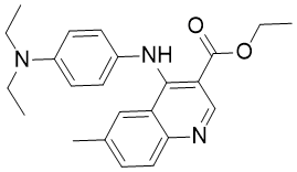
| InChi Code | InChI=1S/C23H27N3O2/c1-5-26(6-2)18-11-9-17(10-12-18)25-22-19-14-16(4)8-13-21(19)24-15-20(22)23(27)28-7-3/h8-15H,5-7H2,1-4H3,(H,24,25) |
| Chemical Name | Ethyl 4-((4-(diethylamino)phenyl)amino)-6-methylquinoline-3-carboxylate |
| Synonyms | MCUi4 MCU-i4 MCU i4 |
| HS Tariff Code | 2934.99.9001 |
| Storage |
Powder-20°C 3 years 4°C 2 years In solvent -80°C 6 months -20°C 1 month Note: This product requires protection from light (avoid light exposure) during transportation and storage. |
| Shipping Condition | Room temperature (This product is stable at ambient temperature for a few days during ordinary shipping and time spent in Customs) |
Biological Activity
| Targets |
Mitochondrial Calcium Uniporter Regulator 1 (MICU1) with an IC50 value of 2.3 μM for inhibiting MICU1-mediated mitochondrial calcium uptake [1] |
| ln Vitro |
MCU-i4 regulates mitochondrial Ca2+ to the intestine for 1 minute, i.e. exerts negative regulation, indicating that the main effect of these compounds is to suddenly and directly regulate MCU complex activity [1]. MCU-i4 has a negative impact on Δψ. MCU-i4 Cellular Differentiation Assay [1] In HEK293T cells overexpressing MICU1, MCU-i4 dose-dependently blocked mitochondrial calcium (Ca²⁺) uptake induced by histamine, with an IC50 of 2.3 μM. This effect was confirmed by mitochondrial Ca²⁺ imaging using a fluorescent probe, showing reduced Ca²⁺ influx into mitochondria compared to vehicle controls [1] - MCU-i4 selectively inhibited MICU1-dependent Ca²⁺ uptake without affecting MICU2/MICU3-mediated mitochondrial Ca²⁺ transport or cytosolic Ca²⁺ homeostasis. In MICU1-knockout HEK293T cells, the compound showed no significant effect on mitochondrial Ca²⁺ levels [1] - In cystic fibrosis (CF) bronchial epithelial cells (CFBE41o⁻), MCU-i4 (5 μM) reduced excessive mitochondrial Ca²⁺ accumulation and restored mitochondrial membrane potential. It also decreased the production of reactive oxygen species (ROS) by 45% and inhibited NF-κB activation, as detected by western blot and luciferase reporter assays [1] |
| ln Vivo |
In a murine model of cystic fibrosis (CFTRΔF508 mice), intraperitoneal administration of MCU-i4 (10 mg/kg/day for 14 days) improved lung function, as evidenced by increased forced expiratory volume and reduced airway resistance. Histological analysis showed decreased airway inflammation, with fewer neutrophils and macrophages infiltrating the lung parenchyma [1] - MCU-i4 treatment (5 mg/kg, intraperitoneal) in CF mice reduced lung tissue ROS levels by 52% and downregulated pro-inflammatory cytokine mRNA expression (IL-6, TNF-α, IL-1β) measured by qPCR. Mitochondrial Ca²⁺ levels in lung epithelial cells were normalized to wild-type levels [1] |
| Enzyme Assay |
MICU1 binding assay: Recombinant human MICU1 protein was immobilized on a sensor chip, and MCU-i4 was injected at concentrations ranging from 0.1 to 50 μM. Binding affinity was measured by surface plasmon resonance (SPR) technology, with equilibrium dissociation constant (KD) calculated from sensorgrams. The assay was performed at 25°C in running buffer containing 10 mM HEPES and 150 mM NaCl [1] - Mitochondrial Ca²⁺ uptake assay: Isolated mouse liver mitochondria were incubated with MCU-i4 for 30 minutes, then exposed to Ca²⁺ (10 μM) and a Ca²⁺-sensitive fluorescent dye. Fluorescence intensity (excitation 506 nm, emission 531 nm) was recorded in real time to quantify Ca²⁺ uptake rates. Results were normalized to mitochondrial protein concentration [1] |
| Cell Assay |
Cell Differentiation Assay [1] Cell Types: Growth medium of 90% confluent C2C12 myoblasts. Vitality has no effect [1]. Tested Concentrations: 10μM. Incubation Duration: 24 hrs (hours). Experimental Results: Myotube width decreases. Mitochondrial Ca²⁺ imaging assay: HEK293T or CFBE41o⁻ cells were loaded with a mitochondrial-targeted Ca²⁺ fluorescent probe for 30 minutes, then pretreated with MCU-i4 (0.1–10 μM) for 1 hour. Cells were stimulated with histamine (100 μM) or ATP (10 μM), and fluorescent signals were captured by confocal microscopy. Ca²⁺ influx was analyzed using ImageJ software to calculate peak fluorescence intensity [1] - ROS detection assay: CFBE41o⁻ cells were seeded in 96-well plates (2×10⁴ cells/well) and treated with MCU-i4 for 24 hours. ROS levels were measured by adding a ROS-sensitive fluorescent dye, and fluorescence intensity was recorded at 485 nm excitation and 520 nm emission. Data were normalized to cell viability (MTT assay) [1] - Western blot assay: Cells treated with MCU-i4 were lysed, and protein extracts were separated by SDS-PAGE. Membranes were probed with antibodies against phospho-NF-κB, total NF-κB, and β-actin. Immunoreactive bands were quantified by densitometry, with results normalized to β-actin expression [1] |
| Animal Protocol |
CFTRΔF508 mouse model for lung function assessment: 8-week-old CFTRΔF508 mice were randomly divided into treatment and vehicle groups. MCU-i4 was dissolved in DMSO:corn oil (1:9) and administered intraperitoneally at 10 mg/kg/day for 14 days. Vehicle controls received the same volume of DMSO:corn oil. Lung function parameters were measured using a small-animal spirometer on day 15 [1] - Inflammation and ROS analysis in CF mice: CFTRΔF508 mice were given MCU-i4 (5 mg/kg) or vehicle via intraperitoneal injection every 24 hours for 7 days. On day 8, mice were euthanized, and lung tissues were collected for ROS detection, qPCR analysis of cytokines, and histological examination. Mitochondria were isolated from lung tissue to measure Ca²⁺ levels [1] |
| References |
[1]. A High-Throughput Screening Identifies MICU1 Targeting Compounds. Cell Rep. 2020 Feb 18;30(7):2321-2331.e6. [2]. Update on Calcium Signaling in Cystic Fibrosis Lung Disease. Front Pharmacol. 2021 Mar 11;12:581645. |
| Additional Infomation |
MCU-i4 is a small-molecule inhibitor identified through high-throughput screening targeting MICU1, a key regulator of the mitochondrial calcium uniporter (MCU) complex [1] - The compound exerts its biological effects by binding to the N-terminal domain of MICU1, preventing MICU1 from activating the MCU channel and thereby inhibiting excessive mitochondrial Ca²⁺ uptake [1] - MCU-i4 shows potential therapeutic value for cystic fibrosis and other diseases associated with abnormal mitochondrial Ca²⁺ signaling and inflammation [1] |
Solubility Data
| Solubility (In Vitro) | DMSO : ~12.5 mg/mL (~33.11 mM) |
| Solubility (In Vivo) |
Solubility in Formulation 1: 1.25 mg/mL (3.31 mM) in 10% DMSO + 40% PEG300 + 5% Tween80 + 45% Saline (add these co-solvents sequentially from left to right, and one by one), suspension solution; with sonication. For example, if 1 mL of working solution is to be prepared, you can add 100 μL of 12.5 mg/mL clear DMSO stock solution to 400 μL PEG300 and mix evenly; then add 50 μL Tween-80 to the above solution and mix evenly; then add 450 μL normal saline to adjust the volume to 1 mL. Preparation of saline: Dissolve 0.9 g of sodium chloride in 100 mL ddH₂ O to obtain a clear solution. Solubility in Formulation 2: ≥ 1.25 mg/mL (3.31 mM) (saturation unknown) in 10% DMSO + 90% (20% SBE-β-CD in Saline) (add these co-solvents sequentially from left to right, and one by one), clear solution. For example, if 1 mL of working solution is to be prepared, you can add 100 μL of 12.5 mg/mL clear DMSO stock solution to 900 μL of 20% SBE-β-CD physiological saline solution and mix evenly. Preparation of 20% SBE-β-CD in Saline (4°C,1 week): Dissolve 2 g SBE-β-CD in 10 mL saline to obtain a clear solution. (Please use freshly prepared in vivo formulations for optimal results.) |
| Preparing Stock Solutions | 1 mg | 5 mg | 10 mg | |
| 1 mM | 2.6491 mL | 13.2457 mL | 26.4915 mL | |
| 5 mM | 0.5298 mL | 2.6491 mL | 5.2983 mL | |
| 10 mM | 0.2649 mL | 1.3246 mL | 2.6491 mL |