Physicochemical Properties
| Molecular Formula | C28H22N4O6 |
| Molecular Weight | 510.5 |
| Exact Mass | 510.153 |
| CAS # | 2315-97-1 |
| PubChem CID | 65099 |
| Appearance | Yellow to khaki solid powder |
| Melting Point | 250ºC |
| LogP | 6.183 |
| Hydrogen Bond Donor Count | 0 |
| Hydrogen Bond Acceptor Count | 6 |
| Rotatable Bond Count | 1 |
| Heavy Atom Count | 38 |
| Complexity | 522 |
| Defined Atom Stereocenter Count | 0 |
| InChi Key | KNJDBYZZKAZQNG-UHFFFAOYSA-N |
| InChi Code | InChI=1S/C28H22N2.2NO3/c1-29-23-15-7-3-11-19(23)27(20-12-4-8-16-24(20)29)28-21-13-5-9-17-25(21)30(2)26-18-10-6-14-22(26)28;2*2-1(3)4/h3-18H,1-2H3;;/q+2;2*-1 |
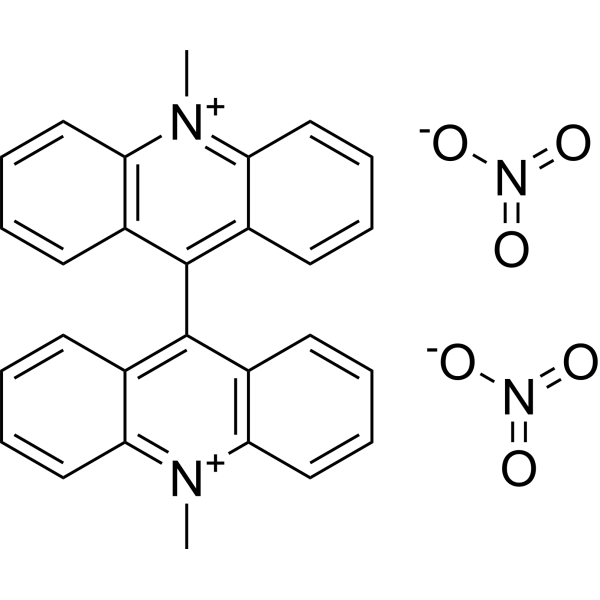
| Chemical Name | 10-methyl-9-(10-methylacridin-10-ium-9-yl)acridin-10-ium;dinitrate |
| HS Tariff Code | 2934.99.9001 |
| Storage |
Powder-20°C 3 years 4°C 2 years In solvent -80°C 6 months -20°C 1 month Note: Please store this product in a sealed and protected environment (e.g. under nitrogen), avoid exposure to moisture and light. |
| Shipping Condition | Room temperature (This product is stable at ambient temperature for a few days during ordinary shipping and time spent in Customs) |
Biological Activity
| Targets |
Lucigenin targets superoxide anion radical (O₂⁻) as a chemilumigenic probe[1] |
| ln Vitro |
1.1 Making the stock solution: To get 10 mM lucigenin, dissolve 1 milligram of lucigenin in 0.1919 mL of DMSO. Note: To prevent frequent freezing and thawing, it is advised to store the stock solution at -20°C or -80°C away from light. 1.2 Lucigenin working solution: To create a 5–10 μM Lucigenin working solution, dilute the original solution with serum-free cell solution or PBS. Note: Please modify the Lucigenin working solution concentration based on the actual circumstances. 2.1 Preparing suspended cells for staining: Centrifuge at 1000 g for 3–5 minutes at 4°C; discard supernatant. Use PBS to wash twice, for five minutes each time. Adherent cells: To create a single cell suspension, add islet dissociated cells and discard the cell culture media. Discard the supernatant after centrifuging at 1000g for three to five minutes at 4°C. Use PBS to wash twice, for five minutes each time. 2.2 After adding 1 milliliter of the Lucigenin working solution, season for 15 minutes. 2.3 Discard the supernatant after centrifuging at 400 g for three to four minutes at 4°C. 2.4 Use PBS twice, giving each wash five minutes. 2.5 Re-suspend the cells in PBS or serum-free cells and use a fluorescent microscope to observe. - Enzymatic system superoxide anion detection: Lucigenin produced dose-dependent chemiluminescence in the xanthine oxidase-xanthine system, with a linear response to O₂⁻ concentrations ranging from 0.1 to 10 nmol/L. The chemiluminescence signal was significantly inhibited (≥ 90%) by superoxide dismutase (SOD), confirming specificity for O₂⁻ [1] - Cellular system superoxide anion detection: Lucigenin detected O₂⁻ production in phorbol 12-myristate 13-acetate (PMA)-stimulated RAW 264.7 macrophages. The chemiluminescence signal peaked at 15-20 minutes after stimulation and was reduced by 85% in the presence of SOD. Unstimulated macrophages showed minimal chemiluminescence, indicating low background noise [1] - Selectivity verification: Lucigenin did not produce significant chemiluminescence with other reactive oxygen species (ROS) including hydrogen peroxide (H₂O₂), hydroxyl radical (·OH), and singlet oxygen (¹O₂), confirming high selectivity for O₂⁻ [1] |
| Enzyme Assay |
- Xanthine oxidase-xanthine system chemiluminescence assay: Reaction mixtures were prepared by mixing xanthine (final concentration 0.1 mM), xanthine oxidase (0.01 U/mL), and Lucigenin (final concentration 5 μM) in phosphate-buffered saline (pH 7.4). For selectivity testing, SOD (100 U/mL) was added to parallel mixtures. Chemiluminescence intensity was measured continuously for 30 minutes using a chemiluminometer. The linear range of O₂⁻ detection was determined by varying xanthine oxidase concentration to adjust O₂⁻ production [1] - Reactive oxygen species (ROS) selectivity assay: Separate reaction mixtures containing Lucigenin (5 μM) and individual ROS generators (H₂O₂: 1 mM; ·OH: generated by Fenton reaction; ¹O₂: generated by hypochlorite-hydrogen peroxide system) were prepared. Chemiluminescence was measured for 30 minutes to evaluate cross-reactivity with non-target ROS [1] |
| Cell Assay |
- Macrophage superoxide anion detection assay: RAW 264.7 macrophages were seeded into 96-well plates at a density of 5×10⁴ cells/well and incubated overnight. The medium was replaced with serum-free medium containing Lucigenin (5 μM), and the cells were pre-incubated for 30 minutes. PMA (final concentration 100 nM) was added to stimulate O₂⁻ production, while unstimulated cells served as the negative control. For specificity verification, SOD (100 U/mL) was added to parallel wells. Chemiluminescence intensity was measured every 5 minutes for 60 minutes using a microplate chemiluminometer [1] |
| References |
[1]. Validation of lucigenin (bis-N-methylacridinium) as a chemilumigenic probe for detecting superoxide anion radical production by enzymatic and cellular systems. J Biol Chem. 1998 Jan 23;273(4):2015-23. |
| Additional Infomation |
See also: Lucigenin (annotation moved to). - Chemical nature: Lucigenin (bis-N-methylacridinium) is a cationic organic compound belonging to the acridinium salt family, characterized by its ability to undergo chemiluminescent reactions with superoxide anion radicals [1] - Mechanism of action: Lucigenin reacts with O₂⁻ via a one-electron reduction to form a radical cation, which subsequently undergoes decomposition to emit light (maximum emission wavelength ~480 nm), enabling quantitative detection of O₂⁻ [1] - Probe advantages: Lucigenin exhibits high sensitivity for O₂⁻ (detection limit ~0.05 nmol/L), low background chemiluminescence, and compatibility with both enzymatic and cellular systems, making it a reliable tool for O₂⁻ research [1] - Critical validation: The specificity of Lucigenin for O₂⁻ is dependent on SOD inhibition—only O₂⁻-mediated chemiluminescence is abolished by SOD, distinguishing it from other ROS probes [1] |
Solubility Data
| Solubility (In Vitro) | DMSO : ~62.5 mg/mL (~122.43 mM) |
| Solubility (In Vivo) |
Solubility in Formulation 1: ≥ 2.08 mg/mL (4.07 mM) (saturation unknown) in 10% DMSO + 40% PEG300 + 5% Tween80 + 45% Saline (add these co-solvents sequentially from left to right, and one by one), clear solution. For example, if 1 mL of working solution is to be prepared, you can add 100 μL of 20.8 mg/mL clear DMSO stock solution to 400 μL PEG300 and mix evenly; then add 50 μL Tween-80 to the above solution and mix evenly; then add 450 μL normal saline to adjust the volume to 1 mL. Preparation of saline: Dissolve 0.9 g of sodium chloride in 100 mL ddH₂ O to obtain a clear solution. Solubility in Formulation 2: ≥ 2.08 mg/mL (4.07 mM) (saturation unknown) in 10% DMSO + 90% (20% SBE-β-CD in Saline) (add these co-solvents sequentially from left to right, and one by one), clear solution. For example, if 1 mL of working solution is to be prepared, you can add 100 μL of 20.8 mg/mL clear DMSO stock solution to 900 μL of 20% SBE-β-CD physiological saline solution and mix evenly. Preparation of 20% SBE-β-CD in Saline (4°C,1 week): Dissolve 2 g SBE-β-CD in 10 mL saline to obtain a clear solution. (Please use freshly prepared in vivo formulations for optimal results.) |
| Preparing Stock Solutions | 1 mg | 5 mg | 10 mg | |
| 1 mM | 1.9589 mL | 9.7943 mL | 19.5886 mL | |
| 5 mM | 0.3918 mL | 1.9589 mL | 3.9177 mL | |
| 10 mM | 0.1959 mL | 0.9794 mL | 1.9589 mL |