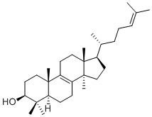
Lanosterol, a naturally-occurring triterpenoid derivative, is a key imtermediate in the biosynthesis of Cholesterol. Lanosterol is metabolized to Cholesterol and Cucurbitacins. Lanosterol is a naturally-occurring sterol and biosynthetic precursor of several animal, fungal, and protozoan steroids, including cholesterol and ergosterol. Defects in the processing of lanosterol contribute to a wide variety of disorders, including the formation of cataracts. Similarly, certain fungicides act by blocking lanosterol processing by fungi.
Physicochemical Properties
| Molecular Formula | C30H50O |
| Molecular Weight | 426.7174 |
| Exact Mass | 426.386 |
| CAS # | 79-63-0 |
| PubChem CID | 246983 |
| Appearance | White to off-white solid powder |
| Density | 1.0±0.1 g/cm3 |
| Boiling Point | 498.9±44.0 °C at 760 mmHg |
| Melting Point | 137 °C |
| Flash Point | 221.1±20.7 °C |
| Vapour Pressure | 0.0±2.9 mmHg at 25°C |
| Index of Refraction | 1.530 |
| LogP | 11 |
| Hydrogen Bond Donor Count | 1 |
| Hydrogen Bond Acceptor Count | 1 |
| Rotatable Bond Count | 4 |
| Heavy Atom Count | 31 |
| Complexity | 767 |
| Defined Atom Stereocenter Count | 7 |
| SMILES | C[C@H](CCC=C(C)C)[C@H]1CC[C@@]2([C@@]1(CCC3=C2CC[C@@H]4[C@@]3(CC[C@@H](C4(C)C)O)C)C)C |
| InChi Key | CAHGCLMLTWQZNJ-BQNIITSRSA-N |
| InChi Code | InChI=1S/C30H50O/c1-20(2)10-9-11-21(3)22-14-18-30(8)24-12-13-25-27(4,5)26(31)16-17-28(25,6)23(24)15-19-29(22,30)7/h10,21-22,25-26,31H,9,11-19H2,1-8H3/t21-,22-,25+,26+,28-,29-,30+/m1/s1 |
| Chemical Name | (3S,5R,10S,13R,14R,17R)-4,4,10,13,14-pentamethyl-17-((R)-6-methylhept-5-en-2-yl)-2,3,4,5,6,7,10,11,12,13,14,15,16,17-tetradecahydro-1H-cyclopenta[a]phenanthren-3-ol |
| Synonyms | NSC 60677; NSC-60677; NSC60677; |
| HS Tariff Code | 2934.99.9001 |
| Storage |
Powder-20°C 3 years 4°C 2 years In solvent -80°C 6 months -20°C 1 month |
| Shipping Condition | Room temperature (This product is stable at ambient temperature for a few days during ordinary shipping and time spent in Customs) |
Biological Activity
| Targets |
Co-chaperone CHIP (carboxyl terminus of Hsp70-interacting protein) – lanosterol increases its expression and stabilizes its protein levels, enhancing its cytoprotective function in protein quality control systems. Autophagy-lysosomal pathway components (LC3, Lamp2) – lanosterol upregulates their expression and enhances autophagic flux. Ubiquitin-proteasome system (UPS) – lanosterol enhances proteasomal degradation of misfolded proteins. Heat shock factor-1 (HSF-1) and heat shock response – indirectly influenced via chaperone induction.[1] |
| ln Vitro |
Through enhanced autophagy and co-chaperone CHIP expression, lanosterol reduces aberrant proteotoxic aggregation and lessens its cytotoxicity[1]. The oocyte is instructed to restart meiosis by lanosterol, which triggers a physiological signal in the cholesterol biosynthesis pathway and is a precursor of meiosis-activating sterols [2]. Lanosterol treatment (10 µM for 24 h) in Cos-7 and A549 cells upregulated autophagy markers LC3 and Lamp2, reduced p62/SQSTM1 levels, and increased co-chaperone CHIP expression at mRNA and protein levels in a dose- and time-dependent manner.[1] Lanosterol reduced cytoplasmic aggregates of mutant misfolded proteins such as GFP-Δ9CAT, mutant SOD1G37R, mutant α-synuclein (S87A), expanded polyglutamine proteins (ataxin-3(84Q) and huntingtin (HDQ74)), and heat-denatured luciferase in various cell models.[1] Lanosterol treatment enhanced the degradation of heat-denatured luciferase, which was inhibited by proteasome inhibitor MG132 and autophagy inhibitor bafilomycin, indicating involvement of both UPS and autophagy pathways.[1] Lanosterol treatment stabilized CHIP protein levels, slowing its degradation in cycloheximide chase assays.[1] Lanosterol did not induce morphological changes or apoptosis in Cos-7 cells at concentrations up to 10 µM for 24 h.[1] Lanosterol protected cells from stress-induced cytotoxicity caused by MG132, bafilomycin, tunicamycin, and β-mercaptoethanol, as measured by MTT assay.[1] |
| Enzyme Assay |
Molecular docking analysis was performed to predict the interaction between lanosterol and the TPR domain of CHIP. The structure of CHIP TPR domain (PDB ID: 3Q49) and lanosterol (PDB ID: 1W6K) were used. Docking was performed using Autodock Vina with a search space box of 46 Å × 54 Å × 50 Å. The best binding mode showed a binding affinity of −9.2 kcal/mol, with hydrophobic interactions involving Leu72, Lys73, Leu69, Phe99, and Phe132, and a hydrogen bond between Glu107 and lanosterol.[1] |
| Cell Assay |
Cell viability assay: Cos-7 cells were seeded in 96-well plates and treated with lanosterol (10 µM), MG132 (10 µM), bafilomycin (50 nM), tunicamycin (10 µg/ml), or β-mercaptoethanol (5 mM) for specified durations. Cell viability was assessed using MTT assay.[1] Immunofluorescence and aggregate counting: Cos-7 cells transfected with various misfolded protein constructs (e.g., GFP-Δ9CAT, SOD1G37R, α-synuclein S87A, ataxin-3 Q84, HDQ74) were treated with lanosterol (10 µM) with or without chloroquine (20 µM). Cells were fixed, permeabilized, stained with primary antibodies (e.g., anti-ubiquitin, anti-CHIP, anti-LC3) and fluorescent secondary antibodies, and visualized by fluorescence microscopy. Aggregates were manually counted.[1] Western blot analysis: Cells treated with lanosterol were lysed, proteins separated by SDS-PAGE, transferred to nitrocellulose membranes, and probed with antibodies against CHIP, LC3, Lamp2, p62, ubiquitin, luciferase, SOD1, α-synuclein, and β-actin.[1] RT-PCR and qRT-PCR: Total RNA was extracted from lanosterol-treated A549 cells using TRIzol. CHIP mRNA levels were analyzed by semiquantitative RT-PCR and quantitative real-time PCR using SYBR Green.[1] Luciferase degradation assay: Cells transfected with luciferase plasmid were treated with lanosterol, exposed to heat shock (43°C for 30 min), recovered at 37°C for 2 h, and luciferase activity was measured using a dual-luciferase reporter assay kit.[1] Cycloheximide chase assay: Cos-7 cells treated with lanosterol were incubated with cycloheximide (15 µg/ml) for various time points to assess CHIP protein stability.[1] Dot-blot analysis: Cell extracts from lanosterol-treated cells expressing misfolded proteins were applied to nitrocellulose membranes and probed with antibodies against synuclein, SOD1, GFP, and β-actin to assess protein solubility.[1] |
| Toxicity/Toxicokinetics |
Lanosterol at 10 µM for 24 h did not induce morphological changes, apoptosis, or cytotoxicity in Cos-7 cells.[1] Lanosterol provided cytoprotection against stress-induced cell death caused by proteasomal inhibition, autophagy inhibition, ER stress, and oxidative stress.[1] |
| References |
[1]. Lanosterol Suppresses the Aggregation and Cytotoxicity of Misfolded Proteins Linked with Neurodegenerative Diseases. Mol Neurobiol. 2018;55(2):1169-1182. [2]. Lanosterol influences cytoplasmic maturation of pig oocytes in vitro and improves preimplantation development of cloned embryos. Theriogenology. 2016;85(4):575-584. |
| Additional Infomation |
Lanosterol is a tetracyclic triterpenoid that is lanosta-8,24-diene substituted by a beta-hydroxy group at the 3beta position. It is the compound from which all steroids are derived. It has a role as a bacterial metabolite, a plant metabolite, a human metabolite, a Saccharomyces cerevisiae metabolite and a mouse metabolite. It is a 3beta-sterol, a tetracyclic triterpenoid and a 14alpha-methyl steroid. It derives from a hydride of a lanostane. Lanosterol has been reported in Camellia sinensis, Dictyuchus monosporus, and other organisms with data available. Lanosterin is a metabolite found in or produced by Saccharomyces cerevisiae. A triterpene that derives from the chair-boat-chair-boat folding of 2,3-oxidosqualene. It is metabolized to CHOLESTEROL and CUCURBITACINS. Lanosterol is an intermediate in cholesterol synthesis and has been shown to reduce protein aggregation in cataracts by dissolving preformed aggregates.[1] Lanosterol enhances cellular protein quality control systems by upregulating co-chaperone CHIP and autophagy, thereby reducing cytotoxicity caused by misfolded proteins in models of neurodegenerative diseases.[1] Lanosterol may represent a therapeutic strategy for protein misfolding disorders by promoting clearance of toxic protein aggregates via both ubiquitin-proteasome and autophagy-lysosomal pathways.[1] |
Solubility Data
| Solubility (In Vitro) |
Ethanol : ~10 mg/mL (~23.43 mM) DMSO : ~5 mg/mL (~11.72 mM) |
| Solubility (In Vivo) |
Solubility in Formulation 1: ≥ 1 mg/mL (2.34 mM) (saturation unknown) in 10% EtOH + 40% PEG300 + 5% Tween80 + 45% Saline (add these co-solvents sequentially from left to right, and one by one), clear solution. For example, if 1 mL of working solution is to be prepared, you can add 100 μL of 10.0 mg/mL clear EtOH stock solution to 400 μL of PEG300 and mix evenly; then add 50 μL of Tween-80 to the above solution and mix evenly; then add 450 μL of normal saline to adjust the volume to 1 mL. Preparation of saline: Dissolve 0.9 g of sodium chloride in 100 mL ddH₂ O to obtain a clear solution. Solubility in Formulation 2: ≥ 1 mg/mL (2.34 mM) (saturation unknown) in 10% EtOH + 90% Corn Oil (add these co-solvents sequentially from left to right, and one by one), clear solution. For example, if 1 mL of working solution is to be prepared, you can add 100 μL of 10.0 mg/mL clear EtOH stock solution to 900 μL of corn oil and mix well. (Please use freshly prepared in vivo formulations for optimal results.) |
| Preparing Stock Solutions | 1 mg | 5 mg | 10 mg | |
| 1 mM | 2.3435 mL | 11.7173 mL | 23.4346 mL | |
| 5 mM | 0.4687 mL | 2.3435 mL | 4.6869 mL | |
| 10 mM | 0.2343 mL | 1.1717 mL | 2.3435 mL |