LSZ-102 is a potent, orally bioactive, selective ERα antagonist that has the potential to treat estrogen receptor positive breast cancer. It also functions as an ERα degrader, with an IC50 of 0.2 nM. About 74% of cases of breast cancer are estrogen receptor alpha (ERα) positive, and in these cases, ERα is the main factor promoting cell division. Aromatase inhibitors, selective estrogen receptor modulators, and selective estrogen receptor destroyers (SERDs) have long been staples in the treatment of ERα positive breast cancer. LSZ102 is a substance that is presently undergoing clinical trials to treat breast cancer that is ERα positive.
Physicochemical Properties
| Molecular Formula | C25H17F3O4S | |
| Molecular Weight | 470.460296392441 | |
| Exact Mass | 470.08 | |
| Elemental Analysis | C, 63.83; H, 3.64; F, 12.11; O, 13.60; S, 6.81 | |
| CAS # | 2135600-76-7 | |
| Related CAS # |
|
|
| PubChem CID | 118574930 | |
| Appearance | White to light yellow solid powder | |
| LogP | 6.6 | |
| Hydrogen Bond Donor Count | 2 | |
| Hydrogen Bond Acceptor Count | 8 | |
| Rotatable Bond Count | 6 | |
| Heavy Atom Count | 33 | |
| Complexity | 712 | |
| Defined Atom Stereocenter Count | 0 | |
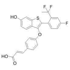
| SMILES | CC(C1=C(C=CC(=C1)F)C2=C(C3=C(S2)C=C(C=C3)O)OC4=CC=C(C=C4)/C=C/C(=O)O)(F)F |
|
| InChi Key | SJXNPGGVGZXKKI-NYYWCZLTSA-N | |
| InChi Code | InChI=1S/C25H17F3O4S/c1-25(27,28)20-12-15(26)5-9-18(20)24-23(19-10-6-16(29)13-21(19)33-24)32-17-7-2-14(3-8-17)4-11-22(30)31/h2-13,29H,1H3,(H,30,31)/b11-4+ | |
| Chemical Name | (E)-3-[4-[[2-[2-(1,1-difluoroethyl)-4-fluorophenyl]-6-hydroxy-1-benzothiophen-3-yl]oxy]phenyl]prop-2-enoic acid | |
| Synonyms |
|
|
| HS Tariff Code | 2934.99.9001 | |
| Storage |
Powder-20°C 3 years 4°C 2 years In solvent -80°C 6 months -20°C 1 month Note: Please store this product in a sealed and protected environment (e.g. under nitrogen), avoid exposure to moisture. |
|
| Shipping Condition | Room temperature (This product is stable at ambient temperature for a few days during ordinary shipping and time spent in Customs) |
Biological Activity
| Targets |
estrogen receptor
Estrogen Receptor α (ERα) (Ki = 0.08 nM for human ERα; IC₅₀ = 0.2 nM for ERα binding inhibition; IC₅₀ = 0.5 nM for ERα degradation in MCF-7 cells); Estrogen Receptor β (ERβ) (Ki = 8.5 nM for human ERβ; 106-fold selectivity over ERα) [1] |
| ln Vitro |
LSZ-102 estrogen receptor degrader that is being tested in Phase I/Ib trials for the treatment of ERα positive breast cancer. Its IC50 is 0.2 nM. MCF-7 cells treated with a 10 μM solution of LSZ-102 for 24 hours show significant degradation of ERα. When MCF-7 cells are incubated with LSZ-102 at a half inhibitory concentration of 1.7 nM, a strong inhibition of cell proliferation is seen. Results using charcoal-stripped serum treated with E2 show that LSZ-102 effectively inhibits the estrogen-induced activation of the ERE-luciferase reporter with an IC50 of 0.3 nM[1]. ERα binding and selective degradation: LSZ102 is a potent, orally bioavailable selective estrogen receptor degrader (SERD) with high affinity for human ERα (Ki = 0.08 nM) and weak affinity for ERβ (Ki = 8.5 nM), achieving 106-fold selectivity for ERα. It competitively inhibits [³H]-estradiol binding to ERα with an IC₅₀ of 0.2 nM. In ER+ breast cancer cell lines, it dose-dependently induces ERα proteasomal degradation: 92% degradation at 1 nM in MCF-7 cells (IC₅₀ = 0.5 nM), 88% degradation at 1 nM in T47D cells (IC₅₀ = 0.7 nM), and 85% degradation at 1 nM in ZR-75-1 cells (IC₅₀ = 0.9 nM). ERβ degradation is not observed at concentrations up to 10 μM [1] - Antiproliferative activity: LSZ102 potently inhibits proliferation of ER+ HER2- breast cancer cell lines, including endocrine-sensitive (MCF-7: IC₅₀ = 0.6 μM; T47D: IC₅₀ = 0.9 μM; ZR-75-1: IC₅₀ = 1.1 μM) and endocrine-resistant subtypes (MCF-7/LTED, tamoxifen-resistant: IC₅₀ = 1.8 μM; MCF-7/AI, aromatase inhibitor-resistant: IC₅₀ = 2.1 μM). It shows no antiproliferative effect on ER- breast cancer cells (MDA-MB-231: IC₅₀ > 50 μM) or normal mammary epithelial cells (MCF-10A: IC₅₀ > 50 μM) [1] - ER signaling pathway inhibition: In MCF-7 cells, LSZ102 (1 nM) suppresses ER-mediated transcriptional activity (ERE-luciferase assay, IC₅₀ = 0.4 nM) and downregulates expression of ER target genes (GREB1, PGR, TFF1, CCND1) at the mRNA (65–80% reduction) and protein levels (55–70% reduction) via qPCR and western blot. It also inhibits ERα nuclear localization, reducing nuclear ERα levels by 75% at 1 nM [1] - Apoptosis induction: In MCF-7 cells, LSZ102 (1–10 μM) induces apoptosis, as evidenced by increased cleaved caspase-3 (2.8-fold at 5 μM), cleaved PARP (3.2-fold at 5 μM), and Annexin V-positive cells (35% at 5 μM, 62% at 10 μM) after 48 hours of treatment [1] - Metabolic stability: In human liver microsomes, LSZ102 exhibits excellent metabolic stability with a half-life (t₁/₂) of 150 minutes and intrinsic clearance (CLint) of 7 μL/min/mg protein. In rat liver microsomes: t₁/₂ = 165 minutes; in dog liver microsomes: t₁/₂ = 180 minutes [1] |
| ln Vivo |
When mice are administered LSZ-102 once daily at a dose of 20 mg/kg, their tumor growth is significantly inhibited compared to the control group that receives vehicle alone. This leads to tumor stasis (mean change in tumor volume of LSZ-102 vs control=%ΔT/ΔC of 2.4% on day 48, p<0.05). Male Sprague-Dawley rats given a 3 mg/kg solution of LSZ-102 exhibit a dose-normalized exposure of 620 nM•h and 33% bioavailability[1]. Human ER+ breast cancer xenograft models (nude mice): Oral administration of LSZ102 (3, 10, 30 mg/kg, once daily for 21 days) dose-dependently inhibits tumor growth: - MCF-7 (endocrine-sensitive): Tumor growth inhibition (TGI) of 48%, 72%, and 85% compared to vehicle; 30 mg/kg reduces tumor weight by 82% and ERα protein levels in tumors by 88% [1] - MCF-7/LTED (tamoxifen-resistant): 30 mg/kg daily results in 78% TGI and 75% reduction in tumor weight; ER target gene (GREB1, PGR) expression in tumors is reduced by 70–75% [1] - ZR-75-1: 30 mg/kg daily achieves 80% TGI and reduces intratumoral Ki-67 (proliferation marker) positive cells by 65% [1] - Pharmacodynamic validation: In MCF-7 xenografts, oral LSZ102 (30 mg/kg) leads to sustained ERα degradation (≥80% reduction for 24 hours post-dosing) and suppression of ER target gene expression, correlating with tumor growth inhibition [1] |
| Enzyme Assay |
Reduction in growth factors In 96-well plates, MCF-7 ERE-luc cells are used and seeded (10 000 cells/well) in CSS medium. Cells are incubated for a full night before being treated with LSZ-102 for 24 hours while being exposed to 0.1 nM estradiol. After that, cells are lysed, and the Bright-Glo assay is used to measure the amount of luciferase activity[1]. ERα/ERβ radioligand binding assay: Recombinant human ERα or ERβ ligand-binding domains are suspended in binding buffer (Tris-HCl, EDTA, glycerol, DTT, 0.1% BSA). LSZ102 is serially diluted (0.001–1000 nM) and mixed with ER protein and [³H]-estradiol (0.5 nM). The mixture is incubated at 4°C for 18 hours, then filtered through glass fiber filters pre-soaked in binding buffer. Filters are washed with cold buffer, and radioactivity is measured by liquid scintillation counting. Ki and IC₅₀ values are calculated using nonlinear regression analysis of displacement curves [1] - ERE-luciferase reporter assay: MCF-7 cells stably transfected with an ERE-luciferase plasmid are seeded in 96-well plates (1×10⁴ cells/well) and incubated overnight. LSZ102 (0.001–100 nM) is preincubated with cells for 1 hour, followed by stimulation with 10 nM estradiol for 24 hours. Cells are lysed, and luciferase activity is measured using a luminometer. IC₅₀ values are derived from dose-response curves of normalized luciferase activity [1] |
| Cell Assay |
ERα degradation western blot assay: ER+ breast cancer cells (MCF-7, T47D, ZR-75-1) are seeded in 6-well plates (2×10⁵ cells/well) and incubated overnight. Cells are treated with LSZ102 (0.1–10 nM) for 24 hours, then lysed in RIPA buffer with protease/proteasome inhibitors. Proteins are separated by SDS-PAGE, transferred to PVDF membranes, and probed with antibodies against ERα and GAPDH (loading control). Band intensity is quantified by densitometry, and degradation efficiency is calculated relative to vehicle-treated cells [1] - Cell proliferation assay (MTT): ER+/- breast cancer cells or normal mammary epithelial cells are seeded in 96-well plates (5×10³ cells/well) and incubated overnight. LSZ102 (0.01–100 μM) is added, and cells are incubated for 72 hours. MTT reagent is added, and absorbance at 570 nm is measured. IC₅₀ values are calculated using nonlinear regression analysis of cell viability curves [1] - Apoptosis assay (Annexin V-FITC/PI): MCF-7 cells are seeded in 6-well plates (2×10⁵ cells/well) and treated with LSZ102 (1–10 μM) for 48 hours. Cells are harvested, stained with Annexin V-FITC and propidium iodide (PI), and analyzed by flow cytometry. The percentage of apoptotic cells (Annexin V-positive/PI-negative + Annexin V-positive/PI-positive) is calculated [1] - ER target gene expression assay (qPCR): MCF-7 cells are treated with LSZ102 (1 nM) for 24 hours. Total RNA is extracted, reverse-transcribed to cDNA, and qPCR is performed using primers for GREB1, PGR, TFF1, CCND1, and GAPDH (housekeeping gene). Relative mRNA levels are calculated using the 2⁻ΔΔCt method [1] |
| Animal Protocol |
In tumor xenograft studies, female athymic nude mice are employed. Injecting 200 μL of MCF-7 cells subcutaneously into the right axillary mammary fat pad area of each animal. There are twice-weekly measurements of tumor volume and body weight. Mice are randomly divided into groups once tumors have an average volume of less than 200 mm3. LSZ-102 (20 mg/kg) or tamoxifen (60 mg/kg) given orally to animals five days a week are the three options[1]. Human ER+ breast cancer xenograft studies: Female nude mice (6–8 weeks old, n=8 per group) are subcutaneously inoculated with 5×10⁶ ER+ breast cancer cells (MCF-7, MCF-7/LTED, or ZR-75-1) into the right flank. When tumors reach 100–150 mm³, LSZ102 is dissolved in 0.5% methylcellulose and administered orally at doses of 3, 10, or 30 mg/kg once daily for 21 days. Vehicle group receives 0.5% methylcellulose. Tumor volume (V = length × width² / 2) and body weight are measured every 3 days. At the end of treatment, mice are euthanized; tumors are excised to measure weight, analyze ERα protein levels (western blot), ER target gene expression (qPCR), and Ki-67 immunohistochemistry [1] - Rat and dog pharmacokinetic studies: Male Sprague-Dawley rats (200–250 g, n=5 per time point) and beagle dogs (8–10 kg, n=4 per time point) are administered LSZ102 via oral gavage (10 mg/kg) or intravenous injection (5 mg/kg). Blood samples are collected at 0.25, 0.5, 1, 2, 4, 8, 12, 24, 48 hours post-dosing. Plasma is separated, and drug concentrations are measured by LC-MS/MS. Pharmacokinetic parameters are calculated using non-compartmental analysis [1] - Tissue distribution study (rats): Male Sprague-Dawley rats (200–250 g, n=3 per time point) are administered LSZ102 via oral gavage (10 mg/kg). At 2, 6, 12, 24 hours post-dosing, rats are euthanized; major organs (liver, lung, spleen, kidney, tumor, brain) are harvested, homogenized, and drug concentrations are measured by LC-MS/MS. Tissue-to-plasma ratios are calculated [1] |
| ADME/Pharmacokinetics |
In rats: Oral administration (10 mg/kg) results in peak plasma concentration (Cₘₐₓ) = 3.2 μg/mL, time to Cₘₐₓ (Tₘₐₓ) = 1.0 hour, terminal half-life (t₁/₂) = 12.5 hours, volume of distribution (Vd) = 3.8 L/kg, and oral bioavailability = 78%. Intravenous administration (5 mg/kg) shows clearance (CL) = 0.28 L/h/kg [1] - In dogs: Oral administration (10 mg/kg) results in Cₘₐₓ = 3.7 μg/mL, Tₘₐₓ = 1.2 hours, t₁/₂ = 15.8 hours, Vd = 3.5 L/kg, and oral bioavailability = 83%. Intravenous administration (5 mg/kg) shows CL = 0.22 L/h/kg [1] - Tissue distribution (rats, 2 hours post-oral 10 mg/kg): Preferential distribution to tumor tissue (tissue-to-plasma ratio = 3.2), liver (2.9), spleen (2.7), lung (2.5), kidney (2.3), and mammary gland (2.1); low brain penetration (tissue-to-plasma ratio = 0.3) [1] - Excretion (rats): 72 hours after intravenous administration (5 mg/kg), 70% of the dose is excreted in feces (38% as parent drug, 32% as metabolites) and 20% in urine (12% as parent drug, 8% as metabolites) [1] - Metabolism: Major metabolic pathways in humans include oxidation (CYP3A4-mediated) and glucuronidation; no toxic metabolites are detected in liver microsome studies [1] |
| Toxicity/Toxicokinetics |
Plasma protein binding: LSZ102 has a plasma protein binding rate of 98% in human plasma, 96% in rat plasma, and 97% in dog plasma (measured by ultrafiltration) [1] - Acute toxicity: In rats and dogs, oral LD₅₀ > 400 mg/kg. No overt toxicity (weight loss, convulsions, respiratory depression, mortality) is observed at doses up to 200 mg/kg in a 7-day acute study [1] - Subchronic toxicity (28-day repeated oral dosing in rats): Doses of 10, 30, 100 mg/kg/day do not cause significant changes in body weight, food intake, hematological parameters (RBC, WBC, platelets), or liver/kidney function (ALT, AST, creatinine, BUN). No histopathological abnormalities are found in major organs (liver, kidney, heart, lung, spleen, mammary gland) [1] - Drug-drug interaction: In vitro studies show no inhibition of cytochrome P450 enzymes (CYP1A2, CYP2C9, CYP2C19, CYP2D6, CYP3A4) at concentrations up to 10 μM; weak induction of CYP3A4 (1.2-fold at 10 μM) is observed but considered clinically irrelevant [1] |
| References |
[1]. Discovery of LSZ102, a Potent, Orally Bioavailable Selective Estrogen Receptor Degrader (SERD) for the Treatment of Estrogen Receptor Positive Breast Cancer. J Med Chem. 2018 Apr 12;61(7):2837-2864. |
| Additional Infomation |
LSZ-102 is under investigation in clinical trial NCT02734615 (Phase I/Ib Trial of LSZ102 Single Agent or LSZ102 + LEE011 or LSZ102 + BYL719 in ER+ Breast Cancers). Selective Estrogen Receptor Degrader LSZ102 is an selective estrogen receptor (ER) degrader (SERD), with potential antineoplastic activity. Upon administration of LSZ102, this agent binds to the ER and induces the degradation of the receptor. This prevents ER activation and ER-mediated signaling, and inhibits the growth and survival of ER-expressing cancer cells. LSZ102 is a potent, highly selective, orally bioavailable selective estrogen receptor degrader (SERD) developed for the treatment of ER+ breast cancer, including endocrine-resistant subtypes [1] - Mechanism of action: Binds selectively to the ligand-binding domain of ERα with high affinity, inducing conformational changes that promote ERα ubiquitination (via recruitment of E3 ubiquitin ligases) and subsequent proteasomal degradation. This irreversibly blocks ER-mediated transcriptional activation, inhibits ER+ breast cancer cell proliferation, and induces apoptosis [1] - Therapeutic potential: Preclinical data support its utility in ER+ HER2- breast cancer, including tumors resistant to tamoxifen, aromatase inhibitors, or other SERDs. Its high oral bioavailability, long half-life, and tumor-targeted distribution address unmet needs in metastatic or recurrent ER+ breast cancer [1] - Druggability advantages: Excellent metabolic stability, low clearance, long half-life (12.5–15.8 hours) supporting once-daily oral administration; high ERα selectivity minimizes off-target effects on ERβ-expressing tissues (e.g., ovaries, uterus); low toxicity in preclinical studies supports chronic use [1] |
Solubility Data
| Solubility (In Vitro) |
|
|||
| Solubility (In Vivo) |
Solubility in Formulation 1: ≥ 1.67 mg/mL (3.55 mM) (saturation unknown) in 10% DMSO + 40% PEG300 + 5% Tween80 + 45% Saline (add these co-solvents sequentially from left to right, and one by one), clear solution. For example, if 1 mL of working solution is to be prepared, you can add 100 μL of 16.7 mg/mL clear DMSO stock solution to 400 μL PEG300 and mix evenly; then add 50 μL Tween-80 to the above solution and mix evenly; then add 450 μL normal saline to adjust the volume to 1 mL. Preparation of saline: Dissolve 0.9 g of sodium chloride in 100 mL ddH₂ O to obtain a clear solution. Solubility in Formulation 2: ≥ 1.67 mg/mL (3.55 mM) (saturation unknown) in 10% DMSO + 90% (20% SBE-β-CD in Saline) (add these co-solvents sequentially from left to right, and one by one), clear solution. For example, if 1 mL of working solution is to be prepared, you can add 100 μL of 16.7 mg/mL clear DMSO stock solution to 900 μL of 20% SBE-β-CD physiological saline solution and mix evenly. Preparation of 20% SBE-β-CD in Saline (4°C,1 week): Dissolve 2 g SBE-β-CD in 10 mL saline to obtain a clear solution. Solubility in Formulation 3: ≥ 1.67 mg/mL (3.55 mM) (saturation unknown) in 10% DMSO + 90% Corn Oil (add these co-solvents sequentially from left to right, and one by one), clear solution. For example, if 1 mL of working solution is to be prepared, you can add 100 μL of 16.7 mg/mL clear DMSO stock solution to 900 μL of corn oil and mix evenly. (Please use freshly prepared in vivo formulations for optimal results.) |
| Preparing Stock Solutions | 1 mg | 5 mg | 10 mg | |
| 1 mM | 2.1256 mL | 10.6279 mL | 21.2558 mL | |
| 5 mM | 0.4251 mL | 2.1256 mL | 4.2512 mL | |
| 10 mM | 0.2126 mL | 1.0628 mL | 2.1256 mL |