LDN193189 4HCl (LDN-193189; DM-3189; LDN 193189; DM 3189), the tetrahydrochloride salt of LDN193189, is a highly potent and selective inhibitor of the BMP (bone morphogenetic protein) signaling pathway with potential anticancer activity. LDN-193189 also exhibits inhibitory effects on associated vascular inflammation, osteogenic activity, and calcification. It inhibits the transcriptional activity of the BMP type I receptor kinases such as ALK2 (activin receptor-like kinase-2) and ALK3 with IC50 of 5 nM and 30 nM in C2C12 cells, respectively.
Physicochemical Properties
| Molecular Formula | C₂₅H₂₆CL₄N₆ | |
| Molecular Weight | 552.33 | |
| Exact Mass | 442.167 | |
| CAS # | 1062368-62-0 | |
| Related CAS # |
|
|
| PubChem CID | 54613581 | |
| Appearance | Typically exists as light yellow to yellow solids at room temperature | |
| LogP | 5.216 | |
| Hydrogen Bond Donor Count | 2 | |
| Hydrogen Bond Acceptor Count | 5 | |
| Rotatable Bond Count | 3 | |
| Heavy Atom Count | 32 | |
| Complexity | 587 | |
| Defined Atom Stereocenter Count | 0 | |
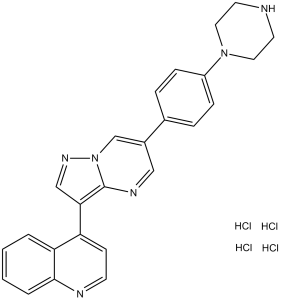
| SMILES | Cl[H].N1(C2C([H])=C([H])C(C3C([H])=NC4=C(C([H])=NN4C=3[H])C3=C([H])C([H])=NC4=C([H])C([H])=C([H])C([H])=C34)=C([H])C=2[H])C([H])([H])C([H])([H])N([H])C([H])([H])C1([H])[H] |
|
| InChi Key | PCCDKTWDGDFRME-UHFFFAOYSA-N | |
| InChi Code | InChI=1S/C25H22N6.ClH/c1-2-4-24-22(3-1)21(9-10-27-24)23-16-29-31-17-19(15-28-25(23)31)18-5-7-20(8-6-18)30-13-11-26-12-14-30;/h1-10,15-17,26H,11-14H2;1H | |
| Chemical Name | 4-[6-(4-piperazin-1-ylphenyl)pyrazolo[1,5-a]pyrimidin-3-yl]quinoline;hydrochloride | |
| Synonyms |
|
|
| HS Tariff Code | 2934.99.9001 | |
| Storage |
Powder-20°C 3 years 4°C 2 years In solvent -80°C 6 months -20°C 1 month |
|
| Shipping Condition | Room temperature (This product is stable at ambient temperature for a few days during ordinary shipping and time spent in Customs) |
Biological Activity
| Targets |
ACVR1 (IC50 = 5 nM); BMPR1A (IC50 = 30 nM); ALK2 (IC50 = 5 nM), ALK3 (IC50 = 30 nM)[1] LDN-193189 4HCl is a potent inhibitor of bone morphogenetic protein (BMP) type I receptors (ALK2, ALK3, ALK6) and transforming growth factor-beta type I receptor (TGFβR-I/ALK5) (ALK2 IC50 = 0.8 nM; ALK3 IC50 = 5.2 nM; ALK6 IC50 = 1.5 nM; ALK5 IC50 = 10.8 nM) [1][2] LDN-193189 4HCl shows weak inhibition of other ALK receptors (ALK1, ALK4: IC50 > 1 μM) and no significant inhibition of unrelated kinases (PKA, PKC: IC50 > 10 μM) [2] |
| ln Vitro |
In vitro activity: LDN193189 potently inhibits BMP4-mediated Smad1, Smad5 and Smad8 activation with IC50 of 5 nM, and efficiently inhibits transcriptional activity of the BMP type I receptors ALK2 and ALK3 with IC50 of 5 nM and 30 nM, respectively. Furthermore, LDN193189 also shows the inhibitory effect on the transcriptional activity induced by either constitutively active ALK2R206H or ALK2Q207D mutant proteins. A recent study shows that LDN-193189 blocks the production of reactive oxygen species induced by oxidized LDL during atherogenesis in human aortic endothelial cells. Kinase Assay: LDN193189 is a selective BMP type I receptor inhibitor, which efficiently inhibits ALK2 and ALK3 (IC50=5 nM and 30 nM, respectively), with weaker effects on ALK4, ALK5 and ALK7 (IC50≥500 nM). Cell Assay: Mouse PASMCs grown are transiently transfected to 50% confluence in six-well plates with 0.3 μg Id1promoter luciferase reporter construct (BRE-Luc) in combination with 0.6 μg of plasmid expressing constitutively active forms of BMP type I receptors (caALK2, caALK3 or caALK6). For both reporter plasmids, 0.2 μg of pRL-TKRenilla luciferase are used to control for transfection efficiency. PASMCs are incubated with LDN193189 (2 nM-32 μM) or vehicle starting 1 h after transfection. Cell extracts are harvested and quantified relative promoter activity by the ratio of firefly to Renilla luciferase activity with the dual luciferase assay kit. In human osteosarcoma cells (U2OS), LDN-193189 4HCl (5 μM) inhibits BMP2-induced Smad1/5/8 phosphorylation by 85% after 24 hours, downregulates BMP target genes (ID1, ID2) by 70-75% at mRNA level, and reduces cell proliferation by 62% (MTT assay) after 72 hours [1][3] - In human non-small cell lung cancer (NSCLC) cells (A549), LDN-193189 4HCl (2 μM) blocks TGF-β1-induced epithelial-mesenchymal transition (EMT), upregulates E-cadherin by 2.3-fold, downregulates vimentin by 68% at protein level, and inhibits cell migration by 70% (wound-healing assay) after 48 hours [3][4] - In mouse embryonic fibroblasts (MEFs), LDN-193189 4HCl (1 μM) inhibits both BMP4-induced Smad1/5/8 phosphorylation (80% reduction) and TGF-β1-induced Smad2 phosphorylation (75% reduction) after 12 hours, suppressing downstream target gene expression (PAI-1, CTGF) by 65-70% [2] - In normal human bronchial epithelial cells (HBECs) and osteoblasts (hFOB1.19), LDN-193189 4HCl shows low toxicity at concentrations up to 20 μM (cell viability > 85% vs. control) [3][4] |
| ln Vivo |
In conditional caALK2-transgenic mice with Ad.Cre on on postnatal day 7 (P7), LDN-193189 (3 mg/kg i.p) leads to mild calcifications surrounding the left tibia and fibula first visible at P13, and prevents radiographic lesions at P15 without causing weight loss or growth retardation, spontaneous fractures, decreased bone density or behavioral abnormalities. LDN193189 dorsalizes zebrafish embryos by inhibiting signaling pathways induced by bone morphogenetic protein (BMP)6 without effect on vascular development. In PCa-118b tumor-bearing mice, LDN-193189 treatment attenuates tumor growth and reduces bone formation in the tumors. In LDL receptor-deficient (LDLR-/-) mice, LDN-193189 potently inhibits development of atheroma. Moreover, LDN-193189 also exhibits the inhibitory effects on associated vascular inflammation, osteogenic activity, and calcification. In nude mice bearing subcutaneous A549 lung cancer xenografts, intraperitoneal administration of LDN-193189 4HCl (10 mg/kg/day for 21 days) inhibits tumor growth by 68% compared to vehicle controls. Tumor tissues show downregulated p-Smad1/5/8 (72% reduction) and vimentin (65% reduction), with increased E-cadherin expression [3] - In a mouse model of heterotopic ossification, oral administration of LDN-193189 4HCl (30 mg/kg/day for 14 days) reduces ectopic bone formation by 75% (micro-CT analysis) and decreases collagen deposition in affected tissues (Masson’s trichrome staining) [1] - In BALB/c mice with Lewis lung cancer lung metastasis, intravenous injection of LDN-193189 4HCl (5 mg/kg/3 days for 4 weeks) reduces lung metastatic nodules by 70% compared to vehicle, suppressing EMT in metastatic foci [4] |
| Enzyme Assay |
Alkaline phosphatase activity[1] We seeded C2C12 cells into 96-well plates at 2,000 cells per well in DMEM supplemented with 2% FBS. We treated the wells in quadruplicate with BMP ligands and LDN-193189 or vehicle. We collected the cells after 6 d in culture in 50 μl Tris-buffered saline and 1% Triton X-100. We added the lysates to p-nitro-phenylphosphate reagent in 96-well plates for 1 h and then evaluated alkaline phosphatase activity (absorbance at 405 nm). We measured cell viability and quantity by Cell Titer Aqueous One (absorbance at 490 nm), using replicate wells treated identically to those used for alkaline phosphatase measurements. Id1 and plasminogen activator inhibitor-1 promoter luciferase reporter assays[1] We transiently transfected mouse PASMCs grown to 50% confluence in six-well plates with 0.3 μg Id1 promoter luciferase reporter construct (BRE-Luc30, kindly provided by P. ten Dijke) in combination with 0.6 μg of plasmid expressing constitutively active forms of BMP type I receptors (caALK2, caALK3 or caALK631, kindly provided by K. Miyazono), using Fugene6. To assess activin and TGF-β type I receptor function, we transiently transfected PASMCs with 0.3 μg PAI1 (plasminogen activator inhibitor-1) promoter luciferase reporter construct (CAGA-Luc32, provided by P. ten Dijke) in combination with 0.6 μg of plasmid expressing constitutively active forms of type I receptors (caALK4, caALK5 and caALK733,). For both reporter plasmids, we used 0.2 μg of pRL-TK Renilla luciferase to control for transfection efficiency. We incubated PASMCs with LDN-193189 (2 nM–32 μM) or vehicle starting 1 h after transfection. We harvested cell extracts and quantified relative promoter activity by the ratio of firefly to Renilla luciferase activity with the dual luciferase assay kit. BMP/TGF-β type I receptor kinase activity assay: Purified recombinant human ALK2, ALK3, ALK6, or ALK5 was incubated with respective Smad-derived substrate peptides (Smad1 for BMP receptors, Smad2 for ALK5) and LDN-193189 4HCl (0.1 nM-100 nM) in assay buffer (50 mM Tris-HCl, pH 7.5, 10 mM MgCl₂, 1 mM DTT, 0.1 mM ATP) at 30°C for 60 minutes. Phosphorylated substrate was detected by radiolabeled ATP counting, and IC50 values were calculated from dose-response curves [1][2] - Kinase selectivity assay: LDN-193189 4HCl (10 μM) was screened against a panel of 40+ kinases (including ALK1, ALK4, PKA, PKC, ERK1/2) using respective substrate peptides and assay buffers. Kinase activity was quantified by colorimetric assay, with no significant off-target inhibition (>50% activity reduction) observed [2] |
| Cell Assay |
Cell culture [1] We isolated PASMCs from wild-type and CAG-Z-EGFP-caALK2–transgenic mice as previously described and cultured them in RPMI medium supplemented with 10% FBS. We induced recombination of PASMCs expressing conditional caALK2 in vitro by infecting with Ad.Cre (multiplicity of infection of 50) or Ad.GFP as a control and then culturing for 3 d and passaging. We cultured C2C12 myofibroblast cells (American Type Culture Collection) in DMEM supplemented with glutamine and 10% FBS. We preincubated cells with pharmacological inhibitors for 10 min and then exposed them to BMP4, TGF-β or platelet-derived growth factor-BB ligands for 30 minutes at 37 °C.[1] Immunoblot analysis of Smad1, Smad5 and Smad8 phosphorylation[1] We mechanically homogenized cell extracts in SDS-lysis buffer (62.5 mM Tris-HCl (pH 6.8), 2% SDS, 10% glycerol, 50 mM dithiothreitol and 0.01% bromophenol blue), separated the proteins by SDS-PAGE, immunoblotted with polyclonal antibodies specific for phosphorylated Smad1, Smad5 and Smad8, phosphorylated Smad2 or rabbit monoclonal antibodies specific for Smad1 or Smad2 , and visualized the immunoreactive proteins with ECL Plus.[1] Osteosarcoma cell proliferation and BMP signaling assay: U2OS cells were seeded in 96-well plates (proliferation) or 6-well plates (signaling) at 3×10³ cells/well or 2×10⁵ cells/well respectively. Cells were pretreated with LDN-193189 4HCl (0.1-10 μM) for 1 hour, then stimulated with BMP2 (10 ng/mL) for 24-72 hours. MTT assay assessed proliferation; Western blot detected p-Smad1/5/8 and total Smad1; qPCR analyzed ID1/ID2 mRNA levels [1][3] - NSCLC cell EMT and migration assay: A549 cells were seeded in 6-well plates (EMT) or wound-healing chambers (migration) at 2×10⁵ cells/well or 5×10⁴ cells/chamber respectively. Cells were pretreated with LDN-193189 4HCl (0.5-5 μM) for 1 hour, then stimulated with TGF-β1 (5 ng/mL) for 48 hours. Western blot detected E-cadherin and vimentin; wound-healing assay evaluated migration; immunofluorescence visualized EMT marker localization [3][4] - MEF dual signaling inhibition assay: MEFs were seeded in 6-well plates at 2×10⁵ cells/well, pretreated with LDN-193189 4HCl (0.1-5 μM) for 1 hour, then stimulated with BMP4 (10 ng/mL) or TGF-β1 (5 ng/mL) for 12 hours. Western blot detected p-Smad1/5/8, total Smad1, p-Smad2, and total Smad2; qPCR analyzed PAI-1/CTGF mRNA levels [2] |
| Animal Protocol |
Dissolved in DMSO and then diluted in water; 3 mg/kg; i.p. injection Ad.Cre on P7 is injected into conditional caALK2–transgenic and wild-type mice Conditionally-expressed, constitutively-active ALK2–transgenic mice[1] The construction of mice expressing a single conditionally expressed allele of the gene encoding constitutively-active ALK2Q207D (CAG-Z-EGFP-caALK2) on a C57BL/6 background was previously described14. We obtained CAGGS-CreER mice, which express a tamoxifen-inducible Cre recombinase ubiquitously under the control of the cytomegalovirus immediate-early enhancer and the chicken β-actin promoter/enhancer20, from the Jackson Laboratory. Mice[3] In the first experiment, SCID mice are implanted with MDA-PCa-118b tumors. After 7 days when tumors reached measurable sizes, mice are injected with LDN-193189 (3 mg/kg) or with vehicle intraperitoneally twice a day. Tumor sizes and body weights are measured weekly. Mice are injected with calcein at three days and one day prior to sacrifice. Blood is collected and tumors are weighed. A portion of the tumors are fixed in formaldehyde for micro-computed tomography, using EVS CT, or further decalcified for bone histomorphometric analysis, using OsteoMeasure Analysis System, or flash frozen for RNA preparation. Osteocalcin in the mouse serum is determined by ELISA. In the second experiment, PCa-118b tumors are first digested with Accumax, and the isolated cells are plated overnight, resuspended in Matrigel in 1:1 ratio, and injected into SCID mice (1×10⁶ cells/mouse) subcutaneously. Mice are treated with LDN-193189 five days post-injection. Rats[4] Male Sprague-Dawley (SD) rats, 8 weeks of age, weighing 200-220 g, are randomly assigned to one of seven experiment groups (n=6 per group). Rats are housed with free access to food and water under a natural 12/12 h day/night cycle. The Monocrotaline is administered (60 mg/kg) to rats by subcutaneous injection into the back region. The animal’s lungs are harvested at 28th day of the study after hemodynamic assessment. The Sildenafil group received daily intragastric administration of Sildenafil after the administration of MCT (60 mg/kg). The LDN-193189 group received daily intragastric administration of Sildenafil (50 mg/kg) and intraperitoneal injection of LDN-193189 (10 mg/kg). In other groups, the same volume saline is given. Mouse heterotopic ossification model: C57BL/6 mice were induced with heterotopic ossification via muscle injury. One day post-induction, LDN-193189 4HCl was suspended in 0.5% carboxymethylcellulose sodium and administered orally at 30 mg/kg/day for 14 days. Vehicle group received carboxymethylcellulose sodium. Mice were euthanized, and injured muscle tissues were collected for micro-CT analysis and Masson’s trichrome staining [1] - Nude mouse A549 xenograft model: 6-8 weeks old nude mice were subcutaneously inoculated with A549 cells (5×10⁶ cells/mouse). When tumors reached ~100 mm³, mice were randomly divided into vehicle and LDN-193189 4HCl groups. The drug was dissolved in saline and administered intraperitoneally at 10 mg/kg/day for 21 days. Tumor volume was measured every 3 days; tumor tissues were collected for Western blot (p-Smad1/5/8, vimentin, E-cadherin) [3] - BALB/c mouse lung metastasis model: BALB/c mice were intravenously injected with Lewis lung cancer cells (1×10⁶ cells/mouse). One day post-injection, LDN-193189 4HCl was dissolved in saline and administered intravenously at 5 mg/kg every 3 days for 4 weeks. Vehicle group received saline. Lungs were harvested to count metastatic nodules; immunofluorescence detected EMT markers in metastatic tissues [4] |
| Toxicity/Toxicokinetics |
In vitro, LDN-193189 4HCl shows low toxicity to normal human cells (HBECs IC50 > 20 μM; hFOB1.19 IC50 > 25 μM) [3][4] - In in vivo studies, administration of LDN-193189 4HCl at tested doses (5-30 mg/kg/day, oral/ip/iv) causes no significant body weight loss (<5% vs. baseline) or overt lethality in mice [1][3][4] - No significant changes in liver function (ALT, AST) or renal function (creatinine, BUN) were observed in LDN-193189 4HCl-treated mice compared to vehicle controls [3][4] - Plasma protein binding rate of LDN-193189 4HCl is 92-95% in mice (in vitro plasma binding assay) [2] |
| References |
[1]. Nat Med.2008 Dec;14(12):1363-9. [2]. Br J Pharmacol.2010 Sep;161(1):140-9. [3]. Cancer Res, 2011, 71(15), 5194-5203. [4]. Int J Clin Exp Pathol. 2014 Jul 15;7(8):4674-84. |
| Additional Infomation |
Fibrodysplasia ossificans progressiva (FOP) is a congenital disorder of progressive and widespread postnatal ossification of soft tissues and is without known effective treatments. Affected individuals harbor conserved mutations in the ACVR1 gene that are thought to cause constitutive activation of the bone morphogenetic protein (BMP) type I receptor, activin receptor-like kinase-2 (ALK2). Here we show that intramuscular expression in the mouse of an inducible transgene encoding constitutively active ALK2 (caALK2), resulting from a glutamine to aspartic acid change at amino acid position 207, leads to ectopic endochondral bone formation, joint fusion and functional impairment, thus phenocopying key aspects of human FOP. A selective inhibitor of BMP type I receptor kinases, LDN-193189 (ref. 6), inhibits activation of the BMP signaling effectors SMAD1, SMAD5 and SMAD8 in tissues expressing caALK2 induced by adenovirus specifying Cre (Ad.Cre). This treatment resulted in a reduction in ectopic ossification and functional impairment. In contrast to localized induction of caALK2 by Ad.Cre (which entails inflammation), global postnatal expression of caALK2 (induced without the use of Ad.Cre and thus without inflammation) does not lead to ectopic ossification. However, if in this context an inflammatory stimulus was provided with a control adenovirus, ectopic bone formation was induced. Like LDN-193189, corticosteroid inhibits ossification in Ad.Cre-injected mutant mice, suggesting caALK2 expression and an inflammatory milieu are both required for the development of ectopic ossification in this model. These results support the role of dysregulated ALK2 kinase activity in the pathogenesis of FOP and suggest that small molecule inhibition of BMP type I receptor activity may be useful in treating FOP and heterotopic ossification syndromes associated with excessive BMP signaling.[4] LDN-193189 4HCl is a potent, multi-target inhibitor of BMP type I receptors (ALK2/3/6) and TGFβR-I (ALK5), with high selectivity for the TGF-β/BMP signaling pathway [1][2] - Its mechanism of action involves competitive binding to the ATP-binding pockets of target receptors, inhibiting their kinase activity and blocking downstream Smad-dependent signaling (Smad1/5/8 for BMPs, Smad2/3 for TGF-β) [1][2][3][4] - LDN-193189 4HCl exhibits in vitro anti-tumor (osteosarcoma, NSCLC) activity, EMT inhibitory effects, and dual signaling inhibition (BMP/TGF-β) in fibroblasts [1][3][4] - In vivo, it suppresses heterotopic ossification, tumor growth, and lung metastasis, supporting its potential in treating BMP/TGF-β-driven diseases (e.g., ectopic ossification, cancer) [1][3][4] - It is widely used as a tool compound to study the cross-talk between BMP and TGF-β signaling pathways in development, cancer, and fibroproliferative diseases [1][2][3][4] |
Solubility Data
| Solubility (In Vitro) |
|
|||
| Solubility (In Vivo) |
Note: Listed below are some common formulations that may be used to formulate products with low water solubility (e.g. < 1 mg/mL), you may test these formulations using a minute amount of products to avoid loss of samples. Injection Formulations (e.g. IP/IV/IM/SC) Injection Formulation 1: DMSO : Tween 80: Saline = 10 : 5 : 85 (i.e. 100 μL DMSO stock solution → 50 μL Tween 80 → 850 μL Saline) *Preparation of saline: Dissolve 0.9 g of sodium chloride in 100 mL ddH ₂ O to obtain a clear solution. Injection Formulation 2: DMSO : PEG300 :Tween 80 : Saline = 10 : 40 : 5 : 45 (i.e. 100 μL DMSO → 400 μLPEG300 → 50 μL Tween 80 → 450 μL Saline) Injection Formulation 3: DMSO : Corn oil = 10 : 90 (i.e. 100 μL DMSO → 900 μL Corn oil) Example: Take the Injection Formulation 3 (DMSO : Corn oil = 10 : 90) as an example, if 1 mL of 2.5 mg/mL working solution is to be prepared, you can take 100 μL 25 mg/mL DMSO stock solution and add to 900 μL corn oil, mix well to obtain a clear or suspension solution (2.5 mg/mL, ready for use in animals). Injection Formulation 4: DMSO : 20% SBE-β-CD in saline = 10 : 90 [i.e. 100 μL DMSO → 900 μL (20% SBE-β-CD in saline)] *Preparation of 20% SBE-β-CD in Saline (4°C,1 week): Dissolve 2 g SBE-β-CD in 10 mL saline to obtain a clear solution. Injection Formulation 5: 2-Hydroxypropyl-β-cyclodextrin : Saline = 50 : 50 (i.e. 500 μL 2-Hydroxypropyl-β-cyclodextrin → 500 μL Saline) Injection Formulation 6: DMSO : PEG300 : castor oil : Saline = 5 : 10 : 20 : 65 (i.e. 50 μL DMSO → 100 μLPEG300 → 200 μL castor oil → 650 μL Saline) Injection Formulation 7: Ethanol : Cremophor : Saline = 10: 10 : 80 (i.e. 100 μL Ethanol → 100 μL Cremophor → 800 μL Saline) Injection Formulation 8: Dissolve in Cremophor/Ethanol (50 : 50), then diluted by Saline Injection Formulation 9: EtOH : Corn oil = 10 : 90 (i.e. 100 μL EtOH → 900 μL Corn oil) Injection Formulation 10: EtOH : PEG300:Tween 80 : Saline = 10 : 40 : 5 : 45 (i.e. 100 μL EtOH → 400 μLPEG300 → 50 μL Tween 80 → 450 μL Saline) Oral Formulations Oral Formulation 1: Suspend in 0.5% CMC Na (carboxymethylcellulose sodium) Oral Formulation 2: Suspend in 0.5% Carboxymethyl cellulose Example: Take the Oral Formulation 1 (Suspend in 0.5% CMC Na) as an example, if 100 mL of 2.5 mg/mL working solution is to be prepared, you can first prepare 0.5% CMC Na solution by measuring 0.5 g CMC Na and dissolve it in 100 mL ddH2O to obtain a clear solution; then add 250 mg of the product to 100 mL 0.5% CMC Na solution, to make the suspension solution (2.5 mg/mL, ready for use in animals). Oral Formulation 3: Dissolved in PEG400 Oral Formulation 4: Suspend in 0.2% Carboxymethyl cellulose Oral Formulation 5: Dissolve in 0.25% Tween 80 and 0.5% Carboxymethyl cellulose Oral Formulation 6: Mixing with food powders Note: Please be aware that the above formulations are for reference only. InvivoChem strongly recommends customers to read literature methods/protocols carefully before determining which formulation you should use for in vivo studies, as different compounds have different solubility properties and have to be formulated differently. (Please use freshly prepared in vivo formulations for optimal results.) |
| Preparing Stock Solutions | 1 mg | 5 mg | 10 mg | |
| 1 mM | 1.8105 mL | 9.0526 mL | 18.1051 mL | |
| 5 mM | 0.3621 mL | 1.8105 mL | 3.6210 mL | |
| 10 mM | 0.1811 mL | 0.9053 mL | 1.8105 mL |