LDN193189 2HCl is the dihydrochloride salt of LDN193189, which is a highly potent and selective small molecule inhibitor of the BMP (bone morphogenetic protein ) signaling pathway. It inhibits the transcriptional activity of the BMP type I receptor kinases such as ALK2 (activin receptor-like kinase-2) and ALK3 with IC50 of 5 nM and 30 nM in C2C12 cells, respectively, it exhibits 200-fold selectivity for BMP versus TGF-β. LDN193189 has been showed to inhibit BMP induced phosphorylation of Smad signaling (Smad1/5/8) and non-Smad signaling including p38 and Akt in C2C12 cells. LDN-193189 inhibits activation of the BMP signaling effectors SMAD1, SMAD5 and SMAD8 in tissues expressing caALK2 induced by adenovirus specifying Cre (Ad.Cre).
Physicochemical Properties
| Molecular Formula | C25H24CL2N6 | |
| Molecular Weight | 479.4 | |
| Exact Mass | 478.143 | |
| Elemental Analysis | C, 62.63; H, 5.05; Cl, 14.79; N, 17.53 | |
| CAS # | 1435934-00-1 | |
| Related CAS # | LDN193189;1062368-24-4;LDN193189 Tetrahydrochloride;2310134-98-4 | |
| PubChem CID | 91900717 | |
| Appearance | Typically exists as orange to red solids at room temperature | |
| Hydrogen Bond Donor Count | 3 | |
| Hydrogen Bond Acceptor Count | 5 | |
| Rotatable Bond Count | 3 | |
| Heavy Atom Count | 33 | |
| Complexity | 587 | |
| Defined Atom Stereocenter Count | 0 | |
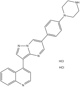
| SMILES | N1=C2C(C=CC=C2)=C(C2=C3N(N=C2)C=C(C2=CC=C(N4CCNCC4)C=C2)C=N3)C=C1.[H]Cl.[H]Cl |
|
| InChi Key | CMQXLLAILGGLRV-UHFFFAOYSA-N | |
| InChi Code | InChI=1S/C25H22N6.2ClH/c1-2-4-24-22(3-1)21(9-10-27-24)23-16-29-31-17-19(15-28-25(23)31)18-5-7-20(8-6-18)30-13-11-26-12-14-30;;/h1-10,15-17,26H,11-14H2;2*1H | |
| Chemical Name | 4-[6-(4-piperazin-1-ylphenyl)pyrazolo[1,5-a]pyrimidin-3-yl]quinoline;dihydrochloride | |
| Synonyms |
|
|
| HS Tariff Code | 2934.99.9001 | |
| Storage |
Powder-20°C 3 years 4°C 2 years In solvent -80°C 6 months -20°C 1 month Note: Please store this product in a sealed and protected environment, avoid exposure to moisture. |
|
| Shipping Condition | Room temperature (This product is stable at ambient temperature for a few days during ordinary shipping and time spent in Customs) |
Biological Activity
| Targets |
ACVR1 (IC50 = 5 nM); BMPR1A (IC50 = 30 nM); ALK2 (IC50 = 5 nM), ALK3 (IC50 = 30 nM)
LDN-193189 2HCl targets bone morphogenetic protein (BMP) type I receptors (ALK1/2/3/6), with an IC50 of ~5 nM for inhibiting BMP4-induced phosphorylation of Smad1/5/8 in PASMCs; it also inhibits TGF-β signaling with an IC50 ≥ 1 μM and targets ActRIIA (type II activin receptor) [1] LDN-193189 2HCl inhibits myostatin/GDF8 signaling by targeting BMP type I receptors (ALK1/2/3/6) and ActRIIA; it represses GDF8-induced Smad2/3 signaling with an IC50 of 0.05 μM for inhibiting GDF8-induced (CAGA)₁₂-luciferase activity and 0.05 μM for inhibiting BMP2-induced BRE-luciferase activity [2] LDN-193189 2HCl targets BMP type I receptors in breast cancer cells, interfering with the BMP pathway regulated by the ZNF217 oncogene [3] |
| ln Vitro |
With IC50 values of 5 nM and 30 nM, respectively, LDN-193189 effectively suppresses the transcriptional activity of the BMP type I receptors ALK2 and ALK3[1]. With IC50 values of less than 500 nM, LDN-193189 exhibits negligible effects on activin and TGF-β type I receptors ALK4, ALK5, and ALK7[1]. ActRIIA is bound by LDN-193189 at a Kd value of 14 nM[2]. LDN-193189 (0.5 μM; 30 min) targeting the suppression of myogenic transcription factors and Smad2/3 signaling triggered by GDF8[2]. Effectively inhibiting GDF8-induced Smad3/4 reporter gene activity is LDN-193189 (0.05, 0.5, and 5 μM)[2]. GDF8-treated myoblasts see their myogenesis restored by LDN-193189 (0–5 μM)[2]. LDN-193189 2HCl potently inhibited BMP4-induced phosphorylation of Smad1/5/8 in PASMCs (IC50 ~5 nM) and showed weak inhibition of TGF-β-induced signaling (IC50 ≥ 1 μM) [1] LDN-193189 2HCl reduced the transcriptional activities of constitutively active ALK2 mutants (ALK2^R206H and ALK2^Q207D) in COS cells, inhibiting Id1 promoter activity in a concentration-dependent manner [1] LDN-193189 2HCl (100 nM) blocked BMP4-induced osteoblast differentiation of C2C12 cells without affecting cell viability [1] LDN-193189 2HCl (0.5 μM) inhibited GDF8-induced phosphorylation of Smad2/3 and p38 in undifferentiated and differentiated primary human myoblasts as well as C2C12 premyoblasts [2] LDN-193189 2HCl (0.05 μM) repressed GDF8-induced (CAGA)₁₂-luciferase activity and BMP2-induced BRE-luciferase activity in C2C12 cells; it repressed TGF-β-induced (CAGA)₁₂-luciferase activity at 0.5 μM [2] LDN-193189 2HCl rescued myogenesis in GDF8-treated myoblasts, upregulated myogenic transcription factors (MyoD, myogenin), and increased MHC-positive myotube formation in C2C12 cells and primary human myoblasts [2] LDN-193189 2HCl (0.5 μM) promoted the contractile activity of myotubular networks in C2C12 cells in vitro, as detected by time-lapse DIC microscopy [2] |
| ln Vivo |
LDN-193189 (ip; 3 mg/kg; daily; 35 days) may have an impact on how breast cancer cells interact with their surroundings in the bone[3]. LDN-193189 (ip; 3 mg/kg; single) reduces functional impairment and ectopic ossification [1]. A selective inhibitor of BMP type I receptor kinases, LDN-193189 (ref. 6), inhibits activation of the BMP signaling effectors SMAD1, SMAD5 and SMAD8 in tissues expressing caALK2 induced by adenovirus specifying Cre (Ad.Cre). This treatment resulted in a reduction in ectopic ossification and functional impairment. In contrast to localized induction of caALK2 by Ad.Cre (which entails inflammation), global postnatal expression of caALK2 (induced without the use of Ad.Cre and thus without inflammation) does not lead to ectopic ossification. However, if in this context an inflammatory stimulus was provided with a control adenovirus, ectopic bone formation was induced. Like LDN-193189, corticosteroid inhibits ossification in Ad.Cre-injected mutant mice, suggesting caALK2 expression and an inflammatory milieu are both required for the development of ectopic ossification in this model. These results support the role of dysregulated ALK2 kinase activity in the pathogenesis of FOP and suggest that small molecule inhibition of BMP type I receptor activity may be useful in treating FOP and heterotopic ossification syndromes associated with excessive BMP signaling.[1] In the present study, researchers aimed at investigating the impact of the LDN-193189 compound, a potent inhibitor of the BMP type I receptor, on metastasis development in vivo. ZNF217-revLuc cells were injected into the left ventricle of nude mice (n = 16) while control mice (n = 13) were inoculated with control pcDNA6-revLuc cells. Mice from each group were treated or not with LDN-193189 for 35 days. We found that systemic LDN-193189 treatment of mice significantly enhanced metastasis development, by increasing both the number and the size of metastases. In pcDNA6-revLuc-injected mice, LDN-193189 also affected the kinetics of metastasis emergence. Altogether, these data suggest that in vivo, LDN-193189 might affect the interaction between breast cancer cells and the bone environment, favoring the emergence and development of multiple metastases. Hence, our report highlights the importance of the choice of drugs and therapeutic strategies used in the management of bone metastases.[3] LDN-193189 2HCl treatment reduced ectopic ossification and joint fusion in conditional caALK2-expressing mice injected with Ad.Cre, preserved joint spaces, and attenuated passive range of motion impairment of the ankle joint (P < 0.001) [1] LDN-193189 2HCl diminished nuclear p-Smad1/5/8 and Runx2 expression in the hindlimb muscles of Ad.Cre-injected caALK2-mutant mice and reduced alkaline phosphatase staining (a marker of osteoblast activity and endochondral bone formation) [1] LDN-193189 2HCl treatment of nude mice injected intracardially with ZNF217-revLuc or pcDNA6-revLuc breast cancer cells significantly enhanced metastasis development, increasing the number and size of metastases and altering the kinetics of metastasis emergence [3] LDN-193189 2HCl increased the total metastases load (measured by bioluminescence) in ZNF217-revLuc-injected mice and pcDNA6-revLuc-injected mice (P < 0.05) [3] |
| Enzyme Assay |
Id1 and plasminogen activator inhibitor-1 promoter luciferase reporter assays[1]
We transiently transfected mouse PASMCs grown to 50% confluence in six-well plates with 0.3 μg Id1 promoter luciferase reporter construct (BRE-Luc30, kindly provided by P. ten Dijke) in combination with 0.6 μg of plasmid expressing constitutively active forms of BMP type I receptors (caALK2, caALK3 or caALK631, kindly provided by K. Miyazono), using Fugene6. To assess activin and TGF-β type I receptor function, we transiently transfected PASMCs with 0.3 μg PAI1 (plasminogen activator inhibitor-1) promoter luciferase reporter construct (CAGA-Luc32, provided by P. ten Dijke) in combination with 0.6 μg of plasmid expressing constitutively active forms of type I receptors (caALK4, caALK5 and caALK733,). For both reporter plasmids, we used 0.2 μg of pRL-TK Renilla luciferase to control for transfection efficiency. We incubated PASMCs with LDN-193189 (2 nM–32 μM) or vehicle starting 1 h after transfection. We harvested cell extracts and quantified relative promoter activity by the ratio of firefly to Renilla luciferase activity with the dual luciferase assay kit. Isothermal titration calorimetry (ITC) was used to measure the binding affinity of LDN-193189 2HCl to ActRIIA; the assay involved titrating the inhibitor into a solution of purified ActRIIA protein, and heat changes during binding were recorded to calculate the dissociation constant (Kd) (dorsomorphin bound ActRIIA with a Kd of 58 nM) [2] Kinase activity assays were performed in PASMCs to evaluate the inhibitory effect of LDN-193189 2HCl on BMP4-induced Smad1/5/8 phosphorylation; cells were treated with BMP4 (10 ng ml⁻¹) and different concentrations of LDN-193189 2HCl, then cell lysates were analyzed by quantitative immunoblotting to determine the IC50 for Smad1/5/8 phosphorylation inhibition [1] |
| Cell Assay |
Immunoblot analysis of Smad1, Smad5 and Smad8 phosphorylation[1]
We mechanically homogenized cell extracts in SDS-lysis buffer (62.5 mM Tris-HCl (pH 6.8), 2% SDS, 10% glycerol, 50 mM dithiothreitol and 0.01% bromophenol blue), separated the proteins by SDS-PAGE, immunoblotted with polyclonal antibodies specific for phosphorylated Smad1, Smad5 and Smad8, phosphorylated Smad2 or rabbit monoclonal antibodies specific for Smad1 or Smad2 , and visualized the immunoreactive proteins with ECL Plus.[1] Primary human myoblasts and C2C12 premyoblasts were serum-starved and pretreated with LDN-193189 2HCl (0.5 μM) for 30 min before stimulation with GDF8 (8 nM for 45 min); cell lysates were subjected to immunoblotting to detect phosphorylated Smad2/3, p38, and total Smad/p38 proteins, with densitometric quantification of band intensities [2] C2C12 cells were transfected with Smad3/4-responsive (CAGA)₁₂-luciferase or Smad1/5-responsive BRE-luciferase reporter constructs and Renilla luciferase; after serum starvation, cells were treated with LDN-193189 2HCl (0.05–0.5 μM) and stimulated with GDF8 (20 nM), BMP2 (10 nM), or TGF-β (100 pM) for 6 h, then luciferase activities were measured and normalized to Renilla activity [2] C2C12 cells and primary human myoblasts were cultured to confluence and switched to differentiation medium containing LDN-193189 2HCl (0.5 μM) with or without GDF8 (8 nM); after 3 days of differentiation, cells were immunostained for skeletal MHC (myosin heavy chain) and DAPI, and myogenic differentiation was quantified by digital image analysis of MHC-positive areas and nuclei [2] PASMCs expressing the conditional caALK2^Q207D transgene were infected with Ad.Cre or Ad.GFP and pretreated with LDN-193189 2HCl (100 nM); cell lysates were analyzed by immunoblotting for phosphorylated Smad1/5/8 and total Smad1 to assess BMP signaling inhibition [1] |
| Animal Protocol |
Dissolved in DMSO and then diluted in water; 3 mg/kg; i.p. injection Ad.Cre on P7 is injected into conditional caALK2–transgenic and wild-type mice Conditionally-expressed, constitutively-active ALK2–transgenic mice[1] The construction of mice expressing a single conditionally expressed allele of the gene encoding constitutively-active ALK2Q207D (CAG-Z-EGFP-caALK2) on a C57BL/6 background was previously described. We obtained CAGGS-CreER mice, which express a tamoxifen-inducible Cre recombinase ubiquitously under the control of the cytomegalovirus immediate-early enhancer and the chicken β-actin promoter/enhancer20, from the Jackson Laboratory. As previously described (Bellanger et al., 2017), pcDNA6-revLuc or ZNF217-revLuc cells (2.5 × 10⁵) were injected into the cardiac left ventricle of n = 18 or n = 20 6-week-old athymic NMRI nude female mice, respectively. Cell implantation was immediately controlled by in vivo bioluminescence imaging. Only mice, the bioluminescent signal of which was diffused throughout the whole body, were considered to be correctly implanted (13/18 and 16/20, respectively, Supplementary Figure 1A ) and were included in the following experimental groups: pcDNA6-revLuc (n = 5), pcDNA6-revLuc + LDN-193189 (n = 8), ZNF217-revLuc (n = 8), and ZNF217-revLuc + LDN-193189 (n = 8). Subsequently, from day 0 to day 35, pcDNA6-revLuc mice or ZNF217-revLuc mice received daily intra-peritoneal (IP) injections of LDN-193189 (3 mg/kg body weight in distilled water) or vehicle (distilled water). The LDN-193189 experimental setup was based on previous in vivo studies (Yu et al., 2008; Lee et al., 2011; Balboni et al., 2013). LDN-193189-treated mice did not exhibit any loss in their body weight, demonstrating that the inhibitor had no severe toxic side effects. Bioluminescence imaging, was performed weekly as previously described (Bellanger et al., 2017). A p value of <0.05 was considered statistically significant.[3] Conditional caALK2-expressing mice (P7) were injected intramuscularly with Ad.Cre in the left gastrocnemius and soleus muscles; LDN-193189 2HCl was administered starting from the time of Ad.Cre injection, and mice were evaluated for ectopic ossification by radiography, μCT, and histology at P13, P15, P30, and P60 [1] Nude mice were injected intracardially with ZNF217-revLuc (n=16) or pcDNA6-revLuc (n=13) breast cancer cells; LDN-193189 2HCl was administered systemically for 35 days, and metastasis development was monitored by bioluminescence imaging and microCT at 35–42 days post-injection [3] Passive range of motion of the ankle joint was assessed in Ad.Cre-injected caALK2-mutant mice treated with LDN-193189 2HCl or vehicle, by measuring the minimum angle formed by the ankle and tibia with passive dorsoflexion at P15 and P30 [1] |
| Toxicity/Toxicokinetics |
LDN-193189 2HCl treatment did not induce fractures, osteopenia, or skeletal abnormalities in conditional caALK2-expressing mice [1] LDN-193189 2HCl (100 nM) did not affect the viability of C2C12 cells in vitro [1] |
| References |
[1]. BMP type I receptor inhibition reduces heterotopic [corrected] ossification. Nat Med, 2008, 14(12), 1363-1369. [2]. Small molecules dorsomorphin and LDN-193189 inhibit myostatin/GDF8 signaling and promote functional myoblast differentiation. J Biol Chem. 2015 Feb 6;290(6):3390-404. [3]. The Bone Morphogenetic Protein Signaling Inhibitor LDN-193189 Enhances Metastasis Development in Mice. Front Pharmacol. 2019 Jun 19;10:667. |
| Additional Infomation |
Fibrodysplasia ossificans progressiva (FOP) is a congenital disorder of progressive and widespread postnatal ossification of soft tissues and is without known effective treatments. Affected individuals harbor conserved mutations in the ACVR1 gene that are thought to cause constitutive activation of the bone morphogenetic protein (BMP) type I receptor, activin receptor-like kinase-2 (ALK2). Here we show that intramuscular expression in the mouse of an inducible transgene encoding constitutively active ALK2 (caALK2), resulting from a glutamine to aspartic acid change at amino acid position 207, leads to ectopic endochondral bone formation, joint fusion and functional impairment, thus phenocopying key aspects of human FOP. A selective inhibitor of BMP type I receptor kinases, LDN-193189 (ref. 6), inhibits activation of the BMP signaling effectors SMAD1, SMAD5 and SMAD8 in tissues expressing caALK2 induced by adenovirus specifying Cre (Ad.Cre). This treatment resulted in a reduction in ectopic ossification and functional impairment. In contrast to localized induction of caALK2 by Ad.Cre (which entails inflammation), global postnatal expression of caALK2 (induced without the use of Ad.Cre and thus without inflammation) does not lead to ectopic ossification. However, if in this context an inflammatory stimulus was provided with a control adenovirus, ectopic bone formation was induced. Like LDN-193189, corticosteroid inhibits ossification in Ad.Cre-injected mutant mice, suggesting caALK2 expression and an inflammatory milieu are both required for the development of ectopic ossification in this model. These results support the role of dysregulated ALK2 kinase activity in the pathogenesis of FOP and suggest that small molecule inhibition of BMP type I receptor activity may be useful in treating FOP and heterotopic ossification syndromes associated with excessive BMP signaling.[1] GDF8, or myostatin, is a member of the TGF-β superfamily of secreted polypeptide growth factors. GDF8 is a potent negative regulator of myogenesis both in vivo and in vitro. We found that GDF8 signaling was inhibited by the small molecule ATP competitive inhibitors dorsomorphin and LDN-193189. These compounds were previously shown to be potent inhibitors of BMP signaling by binding to the BMP type I receptors ALK1/2/3/6. We present the crystal structure of the type II receptor ActRIIA with dorsomorphin and demonstrate that dorsomorphin or LDN-193189 target GDF8 induced Smad2/3 signaling and repression of myogenic transcription factors. As a result, both inhibitors rescued myogenesis in myoblasts treated with GDF8. As revealed by quantitative live cell microscopy, treatment with dorsomorphin or LDN-193189 promoted the contractile activity of myotubular networks in vitro. We therefore suggest these inhibitors as suitable tools to promote functional myogenesis.[2] Breast cancer with bone metastasis is essentially incurable with current anticancer therapies. The bone morphogenetic protein (BMP) pathway is an attractive therapeutic candidate, as it is involved in the bone turnover and in cancer cell formation and their colonization of distant organs such as the bone. We previously reported that in breast cancer cells, the ZNF217 oncogene drives BMP pathway activation, increases the metastatic growth rate in the bone, and accelerates the development of severe osteolytic lesions in mice. [3] LDN-193189 2HCl is a selective ATP-competitive inhibitor of BMP type I receptor kinases and a derivative of dorsomorphin [1] LDN-193189 2HCl inhibits ectopic ossification in a mouse model of fibrodysplasia ossificans progressiva (FOP) by blocking dysregulated ALK2 kinase activity, and its efficacy is comparable to corticosteroid (dexamethasone) treatment in this model [1] LDN-193189 2HCl promotes functional myoblast differentiation by inhibiting myostatin/GDF8 signaling, making it a potential tool for enhancing myogenesis in vitro [2] LDN-193189 2HCl enhances breast cancer metastasis in mice by altering the interaction between cancer cells and the bone microenvironment, highlighting the dual role of BMP pathway inhibition in different pathological contexts [3] |
Solubility Data
| Solubility (In Vitro) |
|
|||
| Solubility (In Vivo) |
Note: Listed below are some common formulations that may be used to formulate products with low water solubility (e.g. < 1 mg/mL), you may test these formulations using a minute amount of products to avoid loss of samples. Injection Formulations (e.g. IP/IV/IM/SC) Injection Formulation 1: DMSO : Tween 80: Saline = 10 : 5 : 85 (i.e. 100 μL DMSO stock solution → 50 μL Tween 80 → 850 μL Saline) *Preparation of saline: Dissolve 0.9 g of sodium chloride in 100 mL ddH ₂ O to obtain a clear solution. Injection Formulation 2: DMSO : PEG300 :Tween 80 : Saline = 10 : 40 : 5 : 45 (i.e. 100 μL DMSO → 400 μLPEG300 → 50 μL Tween 80 → 450 μL Saline) Injection Formulation 3: DMSO : Corn oil = 10 : 90 (i.e. 100 μL DMSO → 900 μL Corn oil) Example: Take the Injection Formulation 3 (DMSO : Corn oil = 10 : 90) as an example, if 1 mL of 2.5 mg/mL working solution is to be prepared, you can take 100 μL 25 mg/mL DMSO stock solution and add to 900 μL corn oil, mix well to obtain a clear or suspension solution (2.5 mg/mL, ready for use in animals). Injection Formulation 4: DMSO : 20% SBE-β-CD in saline = 10 : 90 [i.e. 100 μL DMSO → 900 μL (20% SBE-β-CD in saline)] *Preparation of 20% SBE-β-CD in Saline (4°C,1 week): Dissolve 2 g SBE-β-CD in 10 mL saline to obtain a clear solution. Injection Formulation 5: 2-Hydroxypropyl-β-cyclodextrin : Saline = 50 : 50 (i.e. 500 μL 2-Hydroxypropyl-β-cyclodextrin → 500 μL Saline) Injection Formulation 6: DMSO : PEG300 : castor oil : Saline = 5 : 10 : 20 : 65 (i.e. 50 μL DMSO → 100 μLPEG300 → 200 μL castor oil → 650 μL Saline) Injection Formulation 7: Ethanol : Cremophor : Saline = 10: 10 : 80 (i.e. 100 μL Ethanol → 100 μL Cremophor → 800 μL Saline) Injection Formulation 8: Dissolve in Cremophor/Ethanol (50 : 50), then diluted by Saline Injection Formulation 9: EtOH : Corn oil = 10 : 90 (i.e. 100 μL EtOH → 900 μL Corn oil) Injection Formulation 10: EtOH : PEG300:Tween 80 : Saline = 10 : 40 : 5 : 45 (i.e. 100 μL EtOH → 400 μLPEG300 → 50 μL Tween 80 → 450 μL Saline) Oral Formulations Oral Formulation 1: Suspend in 0.5% CMC Na (carboxymethylcellulose sodium) Oral Formulation 2: Suspend in 0.5% Carboxymethyl cellulose Example: Take the Oral Formulation 1 (Suspend in 0.5% CMC Na) as an example, if 100 mL of 2.5 mg/mL working solution is to be prepared, you can first prepare 0.5% CMC Na solution by measuring 0.5 g CMC Na and dissolve it in 100 mL ddH2O to obtain a clear solution; then add 250 mg of the product to 100 mL 0.5% CMC Na solution, to make the suspension solution (2.5 mg/mL, ready for use in animals). Oral Formulation 3: Dissolved in PEG400 Oral Formulation 4: Suspend in 0.2% Carboxymethyl cellulose Oral Formulation 5: Dissolve in 0.25% Tween 80 and 0.5% Carboxymethyl cellulose Oral Formulation 6: Mixing with food powders Note: Please be aware that the above formulations are for reference only. InvivoChem strongly recommends customers to read literature methods/protocols carefully before determining which formulation you should use for in vivo studies, as different compounds have different solubility properties and have to be formulated differently. (Please use freshly prepared in vivo formulations for optimal results.) |
| Preparing Stock Solutions | 1 mg | 5 mg | 10 mg | |
| 1 mM | 2.0859 mL | 10.4297 mL | 20.8594 mL | |
| 5 mM | 0.4172 mL | 2.0859 mL | 4.1719 mL | |
| 10 mM | 0.2086 mL | 1.0430 mL | 2.0859 mL |