LDH-IN-1 is a novel and potent inhibitor of human lactate dehydrogenase (LDH) with IC50s of 32 and 27 nM for LDHA and LDHB, respectively.
Physicochemical Properties
| Molecular Formula | C30H26N4O4S2 |
| Molecular Weight | 570.681844234467 |
| Exact Mass | 570.139 |
| CAS # | 1964515-43-2 |
| PubChem CID | 131955127 |
| Appearance | White to off-white solid powder |
| LogP | 6.3 |
| Hydrogen Bond Donor Count | 2 |
| Hydrogen Bond Acceptor Count | 8 |
| Rotatable Bond Count | 9 |
| Heavy Atom Count | 40 |
| Complexity | 975 |
| Defined Atom Stereocenter Count | 0 |
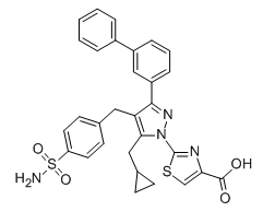
| SMILES | S1C=C(C(O)=O)N=C1N1C(CC2CC2)=C(CC2=CC=C(S(N)(=O)=O)C=C2)C(C2C=CC=C(C3=CC=CC=C3)C=2)=N1 |
| InChi Key | ALJORCZKMBZYCR-UHFFFAOYSA-N |
| InChi Code | InChI=1S/C30H26N4O4S2/c31-40(37,38)24-13-11-19(12-14-24)15-25-27(16-20-9-10-20)34(30-32-26(18-39-30)29(35)36)33-28(25)23-8-4-7-22(17-23)21-5-2-1-3-6-21/h1-8,11-14,17-18,20H,9-10,15-16H2,(H,35,36)(H2,31,37,38) |
| Chemical Name | 2-[5-(cyclopropylmethyl)-3-(3-phenylphenyl)-4-[(4-sulfamoylphenyl)methyl]pyrazol-1-yl]-1,3-thiazole-4-carboxylic acid |
| Synonyms | LDH-IN 1LDH IN-1LDH-IN-1 |
| HS Tariff Code | 2934.99.9001 |
| Storage |
Powder-20°C 3 years 4°C 2 years In solvent -80°C 6 months -20°C 1 month |
| Shipping Condition | Room temperature (This product is stable at ambient temperature for a few days during ordinary shipping and time spent in Customs) |
Biological Activity
| Targets |
1. Lactate dehydrogenase A (LDH-A, also known as LDHA) (IC50 = 11 nM, Ki = 5 nM); the compound also exhibits inhibitory activity against lactate dehydrogenase B (LDH-B) with IC50 = 250 nM, showing selective affinity for LDH-A over LDH-B[1] |
| ln Vitro |
LDH-IN-1 exhibits low nM inhibition of LDHA and LDHB (IC50=32, 27 nM), submicromolar inhibition of deltagenesis, and inhibition of lactate cells in MiaPaCa2 pancreatic cancer and A673 sarcoma (IC50=0.517, 0.854 μM) LDH-IN-1 inhibits the growth of MiaPaCa2 pancreatic cancer cells and A673 sarcoma cells with IC50 of 2.23 and 1.21 μM, respectively). Sample responses to treatment of MiaPaCa-2 cells with LDH-IN-1 exhibited an effect on cell proliferation at doses as low as 250 nM, with practically total reduction of cell growth at 20 μM [1]. 1. Enzyme inhibitory activity: LDH-IN-1 potently inhibited recombinant human LDH-A enzyme activity in a dose-dependent manner with IC50 of 11 nM and Ki of 5 nM; its inhibition against LDH-B was significantly weaker (IC50 = 250 nM), representing a 22.7-fold selectivity for LDH-A over LDH-B. The compound showed no significant inhibitory effect on other related dehydrogenases (such as malate dehydrogenase) at concentrations up to 1 μM, confirming target specificity[1] 2. Cellular lactate production inhibition: In hypoxic A549 lung adenocarcinoma cells, LDH-IN-1 (0.1-10 μM) suppressed lactate secretion in a concentration-dependent manner; at 1 μM, lactate levels in the culture supernatant were reduced by 62% compared with the vehicle control, and at 10 μM, the reduction reached 85%. This effect was reversed by exogenous addition of sodium lactate (20 mM), verifying that the inhibition was LDH-dependent[1] 3. Anti-proliferative activity: In hypoxic cancer cell lines (A549, MDA-MB-231, HCT116), LDH-IN-1 inhibited cell viability with IC50 values of 2.8 μM (A549), 3.2 μM (MDA-MB-231), and 4.1 μM (HCT116) after 72 h of treatment. Under normoxic conditions, the anti-proliferative effect was attenuated (IC50 > 10 μM for all three cell lines), indicating that the compound exerts stronger activity in the hypoxic tumor microenvironment[1] 4. Clonogenic inhibition: In A549 cells cultured under hypoxia, LDH-IN-1 (0.5-5 μM) dose-dependently reduced colony formation; the colony formation rate decreased from 92% (control) to 68% (0.5 μM), 35% (1 μM), and 12% (5 μM). No obvious colony inhibition was observed at 0.5 μM under normoxia[1] |
| ln Vivo | LDH-IN-1 has clearance values of 227 mL/min/kg in vivo, which clearly surpass the mouse species' hepatic blood flow (HBF) of 90 mL/min/kg [1]. |
| Enzyme Assay |
1. Recombinant LDH-A/B activity assay: The assay was performed in a 96-well plate with a reaction buffer containing appropriate concentrations of pyruvate, NADH, and recombinant LDH-A or LDH-B enzyme. LDH-IN-1 was dissolved in DMSO and serially diluted into the reaction system (final DMSO concentration < 0.5%). The reaction was initiated by adding the enzyme, and the decrease in absorbance at 340 nm (corresponding to NADH oxidation) was monitored continuously for 10 min at room temperature. The enzyme activity was calculated based on the absorbance change rate, and the IC50 and Ki values were determined by fitting the dose-response curves with appropriate statistical models. For Ki determination, the assay was repeated with different fixed concentrations of pyruvate, and the data were analyzed using competitive inhibition models[1] 2. Dehydrogenase selectivity assay: The activity of other dehydrogenases (malate dehydrogenase, glyceraldehyde-3-phosphate dehydrogenase, isocitrate dehydrogenase) was detected using corresponding substrate and coenzyme systems. LDH-IN-1 was added at concentrations up to 1 μM, and the enzyme activity was measured using the same absorbance or fluorescence detection method as the LDH assay. The percentage of residual enzyme activity was calculated relative to the vehicle control to evaluate selectivity[1] |
| Cell Assay |
1. Cellular lactate measurement assay: Cancer cells (A549, MDA-MB-231) were seeded in 24-well plates and incubated under hypoxic conditions (1% O₂) for 24 h to adapt. The cells were then treated with different concentrations of LDH-IN-1 (0.1-10 μM) for another 48 h. The culture supernatant was collected, and the lactate concentration was detected using a lactate assay kit based on enzymatic colorimetry. The absorbance at 570 nm was measured, and the lactate concentration was calculated using a standard curve established with known lactate standards. For the rescue experiment, sodium lactate (20 mM) was added to the culture system simultaneously with LDH-IN-1[1] 2. Cell viability assay: Cancer cells were seeded in 96-well plates at a density of 3×10³ cells per well and allowed to attach for 24 h. The cells were then cultured under normoxic or hypoxic conditions and treated with serial dilutions of LDH-IN-1 (0.1-20 μM) for 72 h. A cell viability detection reagent was added to each well and incubated for 2 h at 37℃. The absorbance at the corresponding wavelength was measured, and the cell viability rate was calculated relative to the vehicle control. The IC50 values were obtained by fitting the dose-response curves[1] 3. Colony formation assay: A549 cells were seeded in 6-well plates at a density of 500 cells per well and attached for 24 h. The cells were then treated with LDH-IN-1 (0.5-5 μM) under normoxic or hypoxic conditions and cultured for 14 days. The formed colonies were fixed with fixative solution, stained with a cell staining reagent, and manually counted. The colony formation rate was calculated as the ratio of the number of colonies in the treatment group to that in the control group[1] |
| ADME/Pharmacokinetics |
1. Plasma stability: LDH-IN-1 was incubated with human, rat, and mouse plasma at 37℃ for 0-4 h. After protein precipitation, the remaining compound concentration was detected by LC-MS/MS. The compound showed good plasma stability with a half-life (t1/2) of > 4 h in all three species, and the residual concentration was > 80% after 4 h of incubation[1] 2. Metabolic stability: In human liver microsome incubation experiments, LDH-IN-1 exhibited moderate metabolic stability with a clearance rate of 18 mL/min/kg and an intrinsic half-life of 35 min. The major metabolic pathway was confirmed to be hydroxylation of the pyrazole ring via LC-MS/MS analysis of incubation products[1] 3. Solubility and permeability: The aqueous solubility of LDH-IN-1 was determined to be 12 μM at pH 7.4; in the Caco-2 cell permeability assay, the apparent permeability coefficient (Papp) for apical-to-basolateral transport was 2.1 × 10^-6 cm/s, indicating moderate cell membrane permeability[1] |
| References |
[1]. Discovery and Optimization of Potent, Cell-Active Pyrazole-Based Inhibitors of Lactate Dehydrogenase (LDH). J Med Chem. 2017 Nov 22;60(22):9184-9204. |
| Additional Infomation |
1. LDH-IN-1 is a pyrazole-based small molecule inhibitor designed and optimized for targeting LDH-A, a key enzyme in the glycolytic pathway that is overexpressed in various hypoxic tumors to maintain energy metabolism and acidify the tumor microenvironment[1] 2. The action mechanism of LDH-IN-1 is competitive inhibition of LDH-A by binding to the enzyme's active site, blocking the conversion of pyruvate to lactate, which leads to the accumulation of intracellular pyruvate, reduction of NAD+ regeneration, and subsequent disruption of glycolytic energy supply in hypoxic cancer cells, ultimately inhibiting cell proliferation and colony formation[1] 3. LDH-IN-1 is a lead compound for anti-tumor drug development targeting glycolysis, and its structural optimization was based on initial pyrazole hit compounds to improve enzyme inhibitory activity, selectivity, and ADME properties[1] |
Solubility Data
| Solubility (In Vitro) | DMSO : ~52 mg/mL (~91.12 mM) |
| Solubility (In Vivo) |
Solubility in Formulation 1: ≥ 2.17 mg/mL (3.80 mM) (saturation unknown) in 10% DMSO + 40% PEG300 + 5% Tween80 + 45% Saline (add these co-solvents sequentially from left to right, and one by one), clear solution. For example, if 1 mL of working solution is to be prepared, you can add 100 μL of 21.7 mg/mL clear DMSO stock solution to 400 μL PEG300 and mix evenly; then add 50 μL Tween-80 to the above solution and mix evenly; then add 450 μL normal saline to adjust the volume to 1 mL. Preparation of saline: Dissolve 0.9 g of sodium chloride in 100 mL ddH₂ O to obtain a clear solution. Solubility in Formulation 2: ≥ 2.17 mg/mL (3.80 mM) (saturation unknown) in 10% DMSO + 90% Corn Oil (add these co-solvents sequentially from left to right, and one by one), clear solution. For example, if 1 mL of working solution is to be prepared, you can add 100 μL of 21.7 mg/mL clear DMSO stock solution to 900 μL of corn oil and mix evenly. (Please use freshly prepared in vivo formulations for optimal results.) |
| Preparing Stock Solutions | 1 mg | 5 mg | 10 mg | |
| 1 mM | 1.7523 mL | 8.7615 mL | 17.5230 mL | |
| 5 mM | 0.3505 mL | 1.7523 mL | 3.5046 mL | |
| 10 mM | 0.1752 mL | 0.8761 mL | 1.7523 mL |