Physicochemical Properties
| Molecular Formula | C43H44CL2N10O8 |
| Exact Mass | 898.272 |
| CAS # | 2828438-38-4 |
| PubChem CID | 168432120 |
| Appearance | Light yellow to yellow solid powder |
| LogP | 4.2 |
| Hydrogen Bond Donor Count | 3 |
| Hydrogen Bond Acceptor Count | 13 |
| Rotatable Bond Count | 10 |
| Heavy Atom Count | 63 |
| Complexity | 1680 |
| Defined Atom Stereocenter Count | 0 |
| InChi Key | BUEXWHUMUYQITQ-UHFFFAOYSA-N |
| InChi Code | InChI=1S/C43H44Cl2N10O8/c1-51(43(61)50-38-36(44)31(62-2)21-32(63-3)37(38)45)34-22-33(46-23-47-34)48-25-4-6-26(7-5-25)53-16-18-54(19-17-53)40(58)24-12-14-52(15-13-24)27-8-9-28-29(20-27)42(60)55(41(28)59)30-10-11-35(56)49-39(30)57/h4-9,20-24,30H,10-19H2,1-3H3,(H,50,61)(H,46,47,48)(H,49,56,57) |
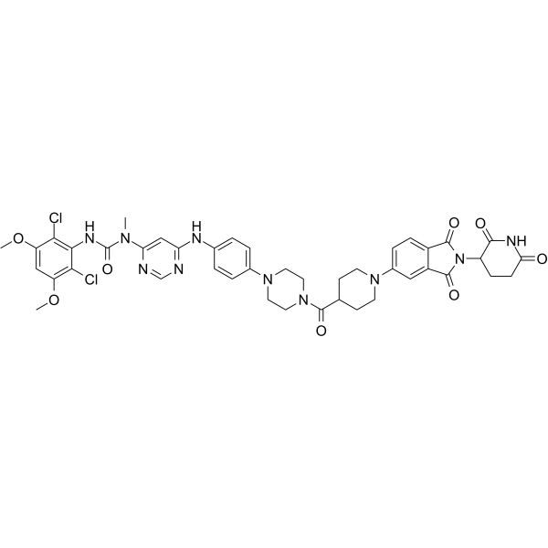
| Chemical Name | 3-(2,6-dichloro-3,5-dimethoxyphenyl)-1-[6-[4-[4-[1-[2-(2,6-dioxopiperidin-3-yl)-1,3-dioxoisoindol-5-yl]piperidine-4-carbonyl]piperazin-1-yl]anilino]pyrimidin-4-yl]-1-methylurea |
| HS Tariff Code | 2934.99.9001 |
| Storage |
Powder-20°C 3 years 4°C 2 years In solvent -80°C 6 months -20°C 1 month |
| Shipping Condition | Room temperature (This product is stable at ambient temperature for a few days during ordinary shipping and time spent in Customs) |
Biological Activity
| Targets | FGFR2 11.8 nM (DC50) |
| ln Vitro | In KATO III, FGFR2 is degraded in a time-dependent manner by LC-MB12 (0.5-10,000 nM, 3-12 hours), with a DC 50 of 11.8 nM [1]. FGFR2 is degraded by 77% in KATO III and 43% in NCI-H1581 by LC-MB12 (100 nM, 6 hours) [1]. With IC50 values of 29.1 nM, 3.7 nM, and 3.2 nM, respectively, LC-MB12 (1-10000 nM, 72 hours) significantly suppresses the growth of KATO III, SNU-16, and NCI-H716 and causes KATO III G0/G1 phase arrest. [1]. |
| ln Vivo | In the SNU-16 nude mouse xenograft model, LC-MB12 (20 mg/kg/day, orally, for 15 days) inhibited tumor growth by 63.1%[1]. LC-MB12 (20 mg/kg, oral) is rapidly absorbed in mice (Cmax: 2.6 h) and has oral bioavailability (F: 13%)[1]. LC-MB12 (20 mg/kg, orally, for 30 days) was well tolerated and showed no significant hepatotoxicity or nephrotoxicity in mice[1]. |
| Cell Assay |
Cell Viability Assay[1] Cell Types: KATO III, SNU-16, NCI-H716 Tested Concentrations: 0.5 nM, 1.5 nM, 4.3 nM, 13 nM, 41 nM, 123 nM, 370 nM, 1111 nM, 3333 nM, 10000 nM Incubation Duration: 72 h Experimental Results: Inhibited cell growth with IC50s value of 29.1 nM (KATO III); 3.7 nM (SNU-16); 3.2 nM (NCI-H716). Cell Cycle Analysis[1] Cell Types: KATO III Tested Concentrations: 29.1 nM Incubation Duration: 72 h Experimental Results: Induced G0/G1 cycle arrest. Immunofluorescence[1] Cell Types: KATO III Tested Concentrations: 100 nM Incubation Duration: 3 h, 6 h Experimental Results: Promoted FGFR2 was relocated from the cell membrane to intracellular vesicles after treated for 3 or 6 h. Induced receptor internalization and re-localization to the perinuclear section after 6 h treatment. Western Blot Analysis[1] Cell Types: KATO III, NCI-H1581 Tested Concentrations: 0.5 nM, 1.5 nM, 4.3 nM, 13 nM, 41 nM, 123 nM, 370 nM, 1111 nM, 3333 nM, 10000 nM Incubation Duration: 6 h Experimental Results: Degraded FGFR2 with a DC50 of 11.8 nM and D max of ∼80% after 6 h of treatment. demonstrated time -dependent e |
| Animal Protocol |
Animal/Disease Models: SNU-16 xenografted in BALB/c-nu (nude) mice [1]. Doses: 20 mg/kg/day Route of Administration: oral administration (po) 15 days Experimental Results: Achieved 63.1% tumor growth inhibition innocuously. Inhibited FGFR phosphorylation and total FGFR2 protein and diminished phosphorylation levels of downstream pPLCγ and ERK1/2. |
| References |
[1]. Discovery of a Selective and Orally Bioavailable FGFR2 Degrader for Treating Gastric Cancer. J Med Chem. 2023 Jun 8;66(11):7438-7453. |
Solubility Data
| Solubility (In Vitro) | May dissolve in DMSO (in most cases), if not, try other solvents such as H2O, Ethanol, or DMF with a minute amount of products to avoid loss of samples |
| Solubility (In Vivo) |
Note: Listed below are some common formulations that may be used to formulate products with low water solubility (e.g. < 1 mg/mL), you may test these formulations using a minute amount of products to avoid loss of samples. Injection Formulations (e.g. IP/IV/IM/SC) Injection Formulation 1: DMSO : Tween 80: Saline = 10 : 5 : 85 (i.e. 100 μL DMSO stock solution → 50 μL Tween 80 → 850 μL Saline) *Preparation of saline: Dissolve 0.9 g of sodium chloride in 100 mL ddH ₂ O to obtain a clear solution. Injection Formulation 2: DMSO : PEG300 :Tween 80 : Saline = 10 : 40 : 5 : 45 (i.e. 100 μL DMSO → 400 μLPEG300 → 50 μL Tween 80 → 450 μL Saline) Injection Formulation 3: DMSO : Corn oil = 10 : 90 (i.e. 100 μL DMSO → 900 μL Corn oil) Example: Take the Injection Formulation 3 (DMSO : Corn oil = 10 : 90) as an example, if 1 mL of 2.5 mg/mL working solution is to be prepared, you can take 100 μL 25 mg/mL DMSO stock solution and add to 900 μL corn oil, mix well to obtain a clear or suspension solution (2.5 mg/mL, ready for use in animals). Injection Formulation 4: DMSO : 20% SBE-β-CD in saline = 10 : 90 [i.e. 100 μL DMSO → 900 μL (20% SBE-β-CD in saline)] *Preparation of 20% SBE-β-CD in Saline (4°C,1 week): Dissolve 2 g SBE-β-CD in 10 mL saline to obtain a clear solution. Injection Formulation 5: 2-Hydroxypropyl-β-cyclodextrin : Saline = 50 : 50 (i.e. 500 μL 2-Hydroxypropyl-β-cyclodextrin → 500 μL Saline) Injection Formulation 6: DMSO : PEG300 : castor oil : Saline = 5 : 10 : 20 : 65 (i.e. 50 μL DMSO → 100 μLPEG300 → 200 μL castor oil → 650 μL Saline) Injection Formulation 7: Ethanol : Cremophor : Saline = 10: 10 : 80 (i.e. 100 μL Ethanol → 100 μL Cremophor → 800 μL Saline) Injection Formulation 8: Dissolve in Cremophor/Ethanol (50 : 50), then diluted by Saline Injection Formulation 9: EtOH : Corn oil = 10 : 90 (i.e. 100 μL EtOH → 900 μL Corn oil) Injection Formulation 10: EtOH : PEG300:Tween 80 : Saline = 10 : 40 : 5 : 45 (i.e. 100 μL EtOH → 400 μLPEG300 → 50 μL Tween 80 → 450 μL Saline) Oral Formulations Oral Formulation 1: Suspend in 0.5% CMC Na (carboxymethylcellulose sodium) Oral Formulation 2: Suspend in 0.5% Carboxymethyl cellulose Example: Take the Oral Formulation 1 (Suspend in 0.5% CMC Na) as an example, if 100 mL of 2.5 mg/mL working solution is to be prepared, you can first prepare 0.5% CMC Na solution by measuring 0.5 g CMC Na and dissolve it in 100 mL ddH2O to obtain a clear solution; then add 250 mg of the product to 100 mL 0.5% CMC Na solution, to make the suspension solution (2.5 mg/mL, ready for use in animals). Oral Formulation 3: Dissolved in PEG400 Oral Formulation 4: Suspend in 0.2% Carboxymethyl cellulose Oral Formulation 5: Dissolve in 0.25% Tween 80 and 0.5% Carboxymethyl cellulose Oral Formulation 6: Mixing with food powders Note: Please be aware that the above formulations are for reference only. InvivoChem strongly recommends customers to read literature methods/protocols carefully before determining which formulation you should use for in vivo studies, as different compounds have different solubility properties and have to be formulated differently. (Please use freshly prepared in vivo formulations for optimal results.) |