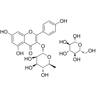
Kaempferol-3-O-glucorhamnoside (astragalin) is a naturally occuring and bioactive flavonoid found in plant Thesium chinense Turcz, that inhibits inflammatory responses via MAPK and NF-κB pathways in vitro and in vivo.
Physicochemical Properties
| Molecular Formula | C27H30O15 |
| Molecular Weight | 594.5181 |
| Exact Mass | 594.158 |
| CAS # | 40437-72-7 |
| Related CAS # | 40437-72-7 |
| PubChem CID | 5318761 |
| Appearance | Light yellow to yellow solid |
| Density | 1.8±0.1 g/cm3 |
| Boiling Point | 945.5±65.0 °C at 760 mmHg |
| Melting Point | 182-185 ºC |
| Flash Point | 314.1±27.8 °C |
| Vapour Pressure | 0.0±0.3 mmHg at 25°C |
| Index of Refraction | 1.744 |
| LogP | 2.66 |
| Hydrogen Bond Donor Count | 9 |
| Hydrogen Bond Acceptor Count | 15 |
| Rotatable Bond Count | 6 |
| Heavy Atom Count | 42 |
| Complexity | 985 |
| Defined Atom Stereocenter Count | 10 |
| SMILES | O(C1([H])C([H])(C([H])(C([H])(C([H])(C([H])([H])[H])O1)O[H])O[H])O[H])C1([H])C([H])(OC2C(C3=C(C([H])=C(C([H])=C3OC=2C2C([H])=C([H])C(=C([H])C=2[H])O[H])O[H])O[H])=O)OC([H])(C([H])([H])O[H])C([H])(C1([H])O[H])O[H] |
| InChi Key | OHOBPOYHROOXEI-OVNVQJKQSA-N |
| InChi Code | InChI=1S/C27H30O15/c1-9-17(32)20(35)22(37)26(38-9)42-25-21(36)18(33)15(8-28)40-27(25)41-24-19(34)16-13(31)6-12(30)7-14(16)39-23(24)10-2-4-11(29)5-3-10/h2-7,9,15,17-18,20-22,25-33,35-37H,8H2,1H3/t9-,15+,17-,18+,20+,21-,22-,25-,26+,27+/m1/s1 |
| Chemical Name | 3-[(2S,3R,4R,5R,6S)-4,5-dihydroxy-6-(hydroxymethyl)-3-[(2S,3R,4S,5S,6R)-3,4,5-trihydroxy-6-methyloxan-2-yl]oxyoxan-2-yl]oxy-5,7-dihydroxy-2-(4-hydroxyphenyl)chromen-4-one |
| Synonyms | Kaempferol-3-O-glucorhamnoside |
| HS Tariff Code | 2934.99.9001 |
| Storage |
Powder-20°C 3 years 4°C 2 years In solvent -80°C 6 months -20°C 1 month Note: This product requires protection from light (avoid light exposure) during transportation and storage. |
| Shipping Condition | Room temperature (This product is stable at ambient temperature for a few days during ordinary shipping and time spent in Customs) |
Biological Activity
| Targets |
The action targets of Kaempferol-3-O-glucorhamnoside are the MAPK signaling pathway (including p38 MAPK, ERK1/2, and JNK) and the NF-κB signaling pathway. [1] |
| ln Vitro |
1. In LPS-stimulated RAW264.7 murine macrophages: Kaempferol-3-O-glucorhamnoside (20 μM, 40 μM) was preincubated with cells for 1 hour before LPS (1 μg/mL) treatment, and after 24 hours of co-incubation, it significantly reduced nitric oxide (NO) production (detected by Griess reagent) and decreased the secretion levels of pro-inflammatory cytokines TNF-α, IL-1β, and IL-6 (measured by ELISA) in a concentration-dependent manner [1] 2. In LPS-stimulated HaCaT human keratinocytes: Kaempferol-3-O-glucorhamnoside (20 μM, 40 μM) was preadministered for 1 hour, followed by LPS (1 μg/mL) stimulation for 6 hours; real-time quantitative PCR (qPCR) results showed that it downregulated the mRNA expression levels of TNF-α, IL-1β, and IL-6 compared with the LPS-only group [1] 3. Mechanism detection in vitro: Western blot analysis revealed that Kaempferol-3-O-glucorhamnoside (20 μM, 40 μM) inhibited the phosphorylation of p38 MAPK, ERK1/2, and JNK in the MAPK pathway, and suppressed the phosphorylation and nuclear translocation of NF-κB p65 subunit as well as the degradation of IκBα in the NF-κB pathway in both LPS-stimulated RAW264.7 cells and HaCaT cells [1] |
| ln Vivo |
1. TPA-induced mouse ear edema model: Male ICR mice (6–8 weeks old) were used; Kaempferol-3-O-glucorhamnoside (10 mg/kg, 20 mg/kg) was administered via intraperitoneal injection 30 minutes before topical application of TPA (2 μg/ear) to the right ear. After 24 hours, the drug significantly reduced ear thickness (measured by a digital caliper) and ear disc weight (punched into 8-mm diameter discs), and decreased myeloperoxidase (MPO) activity (an indicator of neutrophil infiltration) in ear tissue homogenates [1] 2. LPS-induced mouse peritonitis model: Male ICR mice (6–8 weeks old) received intraperitoneal injection of Kaempferol-3-O-glucorhamnoside (20 mg/kg) 30 minutes prior to intraperitoneal injection of LPS (10 mg/kg). After 6 hours, the drug reduced the total number of inflammatory cells in peritoneal exudates (counted by a hemocytometer) and lowered the concentrations of TNF-α, IL-1β, and IL-6 in peritoneal fluid (detected by ELISA) [1] 3. Mechanism detection in vivo: Western blot analysis of liver tissue from LPS-induced mice showed that Kaempferol-3-O-glucorhamnoside (20 mg/kg) inhibited the phosphorylation of p38 MAPK, ERK1/2, JNK, and NF-κB p65 in hepatic tissue compared with the LPS control group [1] |
| Cell Assay |
1. RAW264.7 cell culture and treatment: Cells were maintained in DMEM medium supplemented with 10% fetal bovine serum and 1% penicillin-streptomycin at 37°C in a 5% CO₂ incubator. For experiments, cells were seeded in 96-well plates (for NO and cytokine detection) or 6-well plates (for Western blot) at appropriate densities. After 24 hours of adherence, cells were pretreated with Kaempferol-3-O-glucorhamnoside (20 μM, 40 μM) for 1 hour, then stimulated with LPS (1 μg/mL) for 24 hours (for supernatant collection) or 1 hour (for protein extraction). After treatment, supernatants were collected for NO (Griess reagent) and cytokine (ELISA) detection; cells were lysed to extract total proteins (or nuclear proteins for NF-κB p65) for Western blot [1] 2. HaCaT cell culture and qPCR detection: Cells were cultured in DMEM/F12 medium containing 10% fetal bovine serum and 1% penicillin-streptomycin under 37°C and 5% CO₂. Cells were seeded in 6-well plates and pretreated with Kaempferol-3-O-glucorhamnoside (20 μM, 40 μM) for 1 hour, followed by LPS (1 μg/mL) stimulation for 6 hours. Total RNA was extracted from cells using an RNA extraction kit, reverse-transcribed into cDNA with a reverse transcription kit, and qPCR was performed using specific primers for TNF-α, IL-1β, IL-6, and GAPDH (internal reference) to detect mRNA expression levels [1] 3. Cell viability assay (MTT): RAW264.7 and HaCaT cells were seeded in 96-well plates, treated with Kaempferol-3-O-glucorhamnoside (20 μM, 40 μM) for 25 hours (1 hour pretreatment + 24 hours LPS stimulation), then MTT solution was added and incubated for 4 hours. The supernatant was removed, DMSO was added to dissolve formazan crystals, and absorbance at 570 nm was measured to evaluate cell viability; no significant cytotoxicity was observed in the drug-treated groups [1] |
| Animal Protocol |
1. TPA-induced ear edema experiment: Male ICR mice (n=6 per group) were acclimated for 1 week before the experiment. Kaempferol-3-O-glucorhamnoside was dissolved in dimethyl sulfoxide (DMSO) and then diluted with normal saline to a final DMSO concentration ≤ 0.1% (to avoid solvent toxicity). The drug was administered via intraperitoneal injection at doses of 10 mg/kg and 20 mg/kg; the control group received equal volume of solvent. Thirty minutes after drug administration, 2 μg of TPA (dissolved in acetone) was topically applied to the right ear of each mouse, and the left ear was treated with acetone as a normal control. After 24 hours, mice were sacrificed by cervical dislocation; ear thickness was measured three times at the same site with a digital caliper, and the average value was calculated. Ear discs (8 mm in diameter) were punched from both ears and weighed; the right ear disc weight minus the left ear disc weight was used to evaluate edema degree. Ear tissue was homogenized in physiological saline, centrifuged to collect supernatant, and MPO activity was detected using an MPO assay kit [1] 2. LPS-induced peritonitis experiment: Male ICR mice (n=6 per group) were acclimated for 1 week. Kaempferol-3-O-glucorhamnoside (20 mg/kg) was prepared as the same solvent system as above and administered via intraperitoneal injection; the control group received solvent. Thirty minutes later, mice were injected intraperitoneally with 10 mg/kg LPS (dissolved in normal saline). After 6 hours, mice were sacrificed by cervical dislocation; 5 mL of cold normal saline was injected into the abdominal cavity, and the abdomen was gently massaged for 1 minute. Peritoneal exudate was collected, and the total number of inflammatory cells was counted using a hemocytometer. The remaining exudate was centrifuged, and the supernatant was used to detect TNF-α, IL-1β, and IL-6 concentrations via ELISA. Livers were excised, rinsed with cold normal saline, and stored at -80°C for subsequent Western blot analysis [1] |
| Toxicity/Toxicokinetics |
1. In vitro toxicity: MTT assay showed that Kaempferol-3-O-glucorhamnoside at concentrations of 20 μM and 40 μM had no significant effect on the viability of RAW264.7 macrophages and HaCaT keratinocytes compared with the blank control group (absorbance at 570 nm showed no statistical difference) [1] 2. In vivo toxicity: During the in vivo experiments (TPA ear edema and LPS peritonitis models), mice treated with Kaempferol-3-O-glucorhamnoside (10 mg/kg, 20 mg/kg) showed no abnormal behaviors (such as reduced activity, loss of appetite) or obvious organ damage (visual observation of liver, spleen, kidney after sacrifice). [1] |
| References |
[1]. Kaempferol-3-O-glucorhamnoside inhibits inflammatory responses via MAPK and NF-κB pathways in vitro and in vivo. Toxicol Appl Pharmacol. 2019 Feb 1;364:22-28. |
| Additional Infomation |
Kaempferol 3-neohesperidoside is a member of flavonoids and a glycoside. Kaempferol 3-neohesperidoside has been reported in Diospyros cathayensis, Glycine max, and other organisms with data available. Kaempferol-3-O-glucorhamnoside is a natural flavonoid glycoside derived from plants. The study confirmed that its anti-inflammatory effect is mediated by inhibiting the overactivation of MAPK and NF-κB signaling pathways—two key pathways that regulate the transcription and secretion of pro-inflammatory cytokines. This research provides preclinical experimental evidence for the potential application of Kaempferol-3-O-glucorhamnoside in the treatment of inflammatory diseases (such as dermatitis, peritonitis) [1] |
Solubility Data
| Solubility (In Vitro) | H2O: ~100 mg/mL (~168.2 mM) |
| Solubility (In Vivo) |
Note: Listed below are some common formulations that may be used to formulate products with low water solubility (e.g. < 1 mg/mL), you may test these formulations using a minute amount of products to avoid loss of samples. Injection Formulations (e.g. IP/IV/IM/SC) Injection Formulation 1: DMSO : Tween 80: Saline = 10 : 5 : 85 (i.e. 100 μL DMSO stock solution → 50 μL Tween 80 → 850 μL Saline) *Preparation of saline: Dissolve 0.9 g of sodium chloride in 100 mL ddH ₂ O to obtain a clear solution. Injection Formulation 2: DMSO : PEG300 :Tween 80 : Saline = 10 : 40 : 5 : 45 (i.e. 100 μL DMSO → 400 μLPEG300 → 50 μL Tween 80 → 450 μL Saline) Injection Formulation 3: DMSO : Corn oil = 10 : 90 (i.e. 100 μL DMSO → 900 μL Corn oil) Example: Take the Injection Formulation 3 (DMSO : Corn oil = 10 : 90) as an example, if 1 mL of 2.5 mg/mL working solution is to be prepared, you can take 100 μL 25 mg/mL DMSO stock solution and add to 900 μL corn oil, mix well to obtain a clear or suspension solution (2.5 mg/mL, ready for use in animals). Injection Formulation 4: DMSO : 20% SBE-β-CD in saline = 10 : 90 [i.e. 100 μL DMSO → 900 μL (20% SBE-β-CD in saline)] *Preparation of 20% SBE-β-CD in Saline (4°C,1 week): Dissolve 2 g SBE-β-CD in 10 mL saline to obtain a clear solution. Injection Formulation 5: 2-Hydroxypropyl-β-cyclodextrin : Saline = 50 : 50 (i.e. 500 μL 2-Hydroxypropyl-β-cyclodextrin → 500 μL Saline) Injection Formulation 6: DMSO : PEG300 : castor oil : Saline = 5 : 10 : 20 : 65 (i.e. 50 μL DMSO → 100 μLPEG300 → 200 μL castor oil → 650 μL Saline) Injection Formulation 7: Ethanol : Cremophor : Saline = 10: 10 : 80 (i.e. 100 μL Ethanol → 100 μL Cremophor → 800 μL Saline) Injection Formulation 8: Dissolve in Cremophor/Ethanol (50 : 50), then diluted by Saline Injection Formulation 9: EtOH : Corn oil = 10 : 90 (i.e. 100 μL EtOH → 900 μL Corn oil) Injection Formulation 10: EtOH : PEG300:Tween 80 : Saline = 10 : 40 : 5 : 45 (i.e. 100 μL EtOH → 400 μLPEG300 → 50 μL Tween 80 → 450 μL Saline) Oral Formulations Oral Formulation 1: Suspend in 0.5% CMC Na (carboxymethylcellulose sodium) Oral Formulation 2: Suspend in 0.5% Carboxymethyl cellulose Example: Take the Oral Formulation 1 (Suspend in 0.5% CMC Na) as an example, if 100 mL of 2.5 mg/mL working solution is to be prepared, you can first prepare 0.5% CMC Na solution by measuring 0.5 g CMC Na and dissolve it in 100 mL ddH2O to obtain a clear solution; then add 250 mg of the product to 100 mL 0.5% CMC Na solution, to make the suspension solution (2.5 mg/mL, ready for use in animals). Oral Formulation 3: Dissolved in PEG400 Oral Formulation 4: Suspend in 0.2% Carboxymethyl cellulose Oral Formulation 5: Dissolve in 0.25% Tween 80 and 0.5% Carboxymethyl cellulose Oral Formulation 6: Mixing with food powders Note: Please be aware that the above formulations are for reference only. InvivoChem strongly recommends customers to read literature methods/protocols carefully before determining which formulation you should use for in vivo studies, as different compounds have different solubility properties and have to be formulated differently. (Please use freshly prepared in vivo formulations for optimal results.) |
| Preparing Stock Solutions | 1 mg | 5 mg | 10 mg | |
| 1 mM | 1.6820 mL | 8.4101 mL | 16.8203 mL | |
| 5 mM | 0.3364 mL | 1.6820 mL | 3.3641 mL | |
| 10 mM | 0.1682 mL | 0.8410 mL | 1.6820 mL |