JSH-23 HCl is designed as an inhibitor of NF-κB transcriptional activity with IC50 of 7.1 μM in RAW 264.7 cell line. In LPS-stimulated macrophages RAW 264.7, JSH-23 inhibits NF-κB transcriptional activity in a dose-dependent fashion. Its cytotoxicity is not the cause of this outcome. JSH-23 is discovered to significantly reduce the LPS-induced DNA binding activity of NF-κB while reducing nuclear NF-κB p65 amounts in the same condition. JSH-23 performs these functions without affecting IκB degradation. Additionally, JSH-23 exhibits inhibition effects on the expression of pro-inflammatory transcripts and enzymes, such as IL-6, IL-1β, COX-2, and TNF-α. JSH-23 also prevents the chromatin condensation brought on by apoptosis that is induced by LPS.
Physicochemical Properties
| Molecular Formula | C16H21N2 | |
| Molecular Weight | 276.81 | |
| Exact Mass | 240.162 | |
| CAS # | 749886-87-1 | |
| Related CAS # |
|
|
| PubChem CID | 16760588 | |
| Appearance | Pale purple to purple solid powder | |
| Density | 1.1±0.1 g/cm3 | |
| Boiling Point | 418.7±40.0 °C at 760 mmHg | |
| Melting Point | 104.4-105.0℃ | |
| Flash Point | 245.0±30.9 °C | |
| Vapour Pressure | 0.0±1.0 mmHg at 25°C | |
| Index of Refraction | 1.630 | |
| LogP | 3.66 | |
| Hydrogen Bond Donor Count | 2 | |
| Hydrogen Bond Acceptor Count | 2 | |
| Rotatable Bond Count | 5 | |
| Heavy Atom Count | 18 | |
| Complexity | 223 | |
| Defined Atom Stereocenter Count | 0 | |
| Synonyms | JSH-23 HCL | |
| HS Tariff Code | 2934.99.9001 | |
| Storage |
Powder-20°C 3 years 4°C 2 years In solvent -80°C 6 months -20°C 1 month |
|
| Shipping Condition | Room temperature (This product is stable at ambient temperature for a few days during ordinary shipping and time spent in Customs) |
Biological Activity
| Targets |
NF-κB (IC50 = 7.1 μM) JSH-2 (1-300 μM; 24 hours) at less than <100 μM has no discernible cytotoxic effects on RAW 264.7 cells[1]. After being exposed to LPS for 1 h, NF-B p65 nuclear amount is significantly increased. JSH-23 (30 μM; 1 hour) treatment reduces nuclear NF-κB p65 content in RAW 264.7 cells stimulated by LPS in a dose-dependent manner[1]. |
| ln Vitro |
JSH-2 (1-300 μM; 24 hours) at less than <100 μM has no discernible cytotoxic effects on RAW 264.7 cells[1]. After being exposed to LPS for 1 h, NF-B p65 nuclear amount is significantly increased. JSH-23 (30 μM; 1 hour) treatment reduces nuclear NF-κB p65 content in RAW 264.7 cells stimulated by LPS in a dose-dependent manner[1]. JSH-23 inhibited LPS-induced NF-κB transcriptional activity in a dose-dependent manner in RAW 264.7 macrophages stably transfected with a pNF-κB-SEAP reporter construct, showing 23%, 68%, and 103% inhibition at 3, 10, and 30 μM, respectively.[1] JSH-23 decreased LPS-induced DNA binding activity of NF-κB in a dose-dependent manner, as demonstrated by electrophoretic mobility shift assay (EMSA).[1] Western immunoblot analysis showed that JSH-23 dose-dependently inhibited LPS-induced nuclear translocation of the NF-κB p65 subunit (49%, 75%, and 95% inhibition at 3, 10, and 30 μM, respectively), without affecting LPS-induced IκBα degradation or its cytoplasmic recovery.[1] Semi-quantitative RT-PCR analysis revealed that JSH-23 differentially inhibited LPS-induced mRNA expression of pro-inflammatory mediators: it inhibited IL-6 and inducible nitric oxide synthase (iNOS) at concentrations ≥3 μM, IL-1β and cyclooxygenase-2 (COX-2) at ≥10 μM, and tumor necrosis factor-alpha (TNF-α) at ≥30 μM.[1] JSH-23 inhibited LPS-induced apoptosis (chromatin condensation) in RAW 264.7 cells in a dose-dependent manner, showing 44%, 63%, and 93% inhibition at 3, 10, and 30 μM, respectively.[1] |
| ln Vivo | JSH-23 (orally given at doses of 1 mg/kg or 3 mg/kg twice daily for two weeks) significantly improves nerve conduction and blood flow deficits in diabetic rats[2]. |
| Cell Assay |
To measure NF-κB transcriptional activity, RAW 264.7 macrophages stably harboring a pNF-κB-SEAP-NPT reporter plasmid were treated with LPS (1 μg/mL) and/or JSH-23 for 16 hours. Secreted alkaline phosphatase (SEAP) activity in the cell-free culture medium was then measured as a readout for NF-κB-driven gene expression.[1] For electrophoretic mobility shift assay (EMSA), RAW 264.7 cells were treated with LPS (1 μg/mL) plus JSH-23 for 1 hour. Nuclear extracts were prepared and incubated with a ³²P-labeled oligonucleotide containing a κB binding site. The DNA-protein complexes were resolved on a non-denaturing 6% polyacrylamide gel, followed by autoradiography.[1] For Western immunoblot analysis of NF-κB p65 nuclear translocation, cells were treated with LPS (1 μg/mL) and/or JSH-23 for 1 hour. Nuclear extracts were prepared and subjected to immunoblotting using an antibody against NF-κB p65.[1] For Western immunoblot analysis of IκBα degradation, cells were treated with LPS (1 μg/mL) and/or JSH-23 (30 μM) for various times (10 minutes to 8 hours). Cytoplasmic extracts were prepared and subjected to immunoblotting using an antibody against IκBα.[1] For semi-quantitative RT-PCR analysis of cytokine and enzyme expression, cells were treated with LPS (1 μg/mL) and/or JSH-23 for 6 hours. Total RNA was extracted, reverse transcribed, and amplified by PCR using specific primers for TNF-α, IL-1β, IL-6, iNOS, COX-2, and β-actin (as an internal control). PCR products were separated by agarose gel electrophoresis and visualized with ethidium bromide staining.[1] To assess apoptosis, cells were treated with LPS (1 μg/mL) and/or JSH-23 for 24 hours, then stained with 4',6-diamidino-2-phenylindole (DAPI). Cells were examined under fluorescence microscopy, and nuclei with condensed or fragmented chromatin were counted as apoptotic.[1] To assess cytotoxicity, cells were treated with various concentrations of JSH-23 for 24 hours. Cell viability/proliferation was measured using a WST-1 assay, and absorbance was read at 450 nm.[1] |
| Animal Protocol |
Male Sprague Dawley diabetic rats (250-270 g) 1 mg/kg, 3 mg/kg Orally administered; daily; for 2 weeks |
| Toxicity/Toxicokinetics |
JSH-23 at concentrations up to 100 μM did not show significant cytotoxic effects on RAW 264.7 macrophages over a 24-hour period, as assessed by the WST-1 assay. Cytotoxicity was observed at 300 μM.[1] JSH-23 alone (30 μM) did not induce apoptotic cell death in RAW 264.7 macrophages.[1] |
| References |
[1]. Inhibitory action of novel aromatic diamine compound on lipopolysaccharide-induced nuclear translocation of NF-kappaB without affecting IkappaB degradation. FEBS Lett. 2004 Jul 30;571(1-3):50-4. [2]. JSH-23 targets nuclear factor-kappa B and reverses various deficits in experimental diabetic neuropathy: effect on neuroinflammation and antioxidant defence. Diabetes Obes Metab. 2011 Aug;13(8):750-8. |
| Additional Infomation |
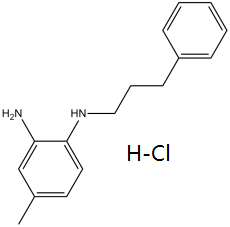
JSH-23 is a diamine that is 1,2-phenylenediamine carrying a methyl substituent at position 4 and a 3-phenylpropyl substituent at position N1. It has a role as a NF-kappaB inhibitor. It is a diamine and a substituted aniline. It is functionally related to a 1,2-phenylenediamine. JSH-23 (4-Methyl-N¹-(3-phenyl-propyl)-benzene-1,2-diamine) is a novel synthetic aromatic diamine compound with a purity of ≥98%.[1] Its mechanism of action is considered rare, as it inhibits NF-κB nuclear translocation without blocking IκBα degradation, potentially by interfering with the nuclear import machinery of NF-κB, similar to the synthetic peptide SN50 which targets the nuclear localization signal (NLS).[1] The compound's inhibitory effects on pro-inflammatory cytokine and enzyme expression suggest potential utility in inflammation-related disorders such as arthritis, cancer, and septic shock.[1] The study was conducted entirely in vitro using the murine macrophage cell line RAW 264.7.[1] |
Solubility Data
| Solubility (In Vitro) |
|
|||
| Solubility (In Vivo) |
Solubility in Formulation 1: ≥ 2.5 mg/mL (10.40 mM) (saturation unknown) in 10% DMSO + 40% PEG300 + 5% Tween80 + 45% Saline (add these co-solvents sequentially from left to right, and one by one), clear solution. For example, if 1 mL of working solution is to be prepared, you can add 100 μL of 25.0 mg/mL clear DMSO stock solution to 400 μL PEG300 and mix evenly; then add 50 μL Tween-80 to the above solution and mix evenly; then add 450 μL normal saline to adjust the volume to 1 mL. Preparation of saline: Dissolve 0.9 g of sodium chloride in 100 mL ddH₂ O to obtain a clear solution. Solubility in Formulation 2: ≥ 2.5 mg/mL (10.40 mM) (saturation unknown) in 10% DMSO + 90% Corn Oil (add these co-solvents sequentially from left to right, and one by one), clear solution. For example, if 1 mL of working solution is to be prepared, you can add 100 μL of 25.0 mg/mL clear DMSO stock solution to 900 μL of corn oil and mix evenly. Solubility in Formulation 3: 0.5% hydroxyethyl cellulose: 30 mg/mL (Please use freshly prepared in vivo formulations for optimal results.) |
| Preparing Stock Solutions | 1 mg | 5 mg | 10 mg | |
| 1 mM | 3.6126 mL | 18.0629 mL | 36.1259 mL | |
| 5 mM | 0.7225 mL | 3.6126 mL | 7.2252 mL | |
| 10 mM | 0.3613 mL | 1.8063 mL | 3.6126 mL |