Physicochemical Properties
| Molecular Formula | C25H27CL4IN4 |
| Molecular Weight | 652.2235 |
| Exact Mass | 650.003 |
| CAS # | 3520-43-2 |
| Related CAS # | 3520-43-2; |
| PubChem CID | 5492929 |
| Appearance | Purple to purplish red solid powder |
| LogP | 4.157 |
| Hydrogen Bond Donor Count | 0 |
| Hydrogen Bond Acceptor Count | 3 |
| Rotatable Bond Count | 6 |
| Heavy Atom Count | 34 |
| Complexity | 625 |
| Defined Atom Stereocenter Count | 0 |
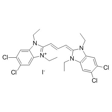
| SMILES | CCN1C2=CC(=C(C=C2[N+](=C1/C=C/C=C3N(C4=CC(=C(C=C4N3CC)Cl)Cl)CC)CC)Cl)Cl.[I-] |
| InChi Key | FYNNIUVBDKICAX-UHFFFAOYSA-M |
| InChi Code | InChI=1S/C25H27Cl4N4.HI/c1-5-30-20-12-16(26)17(27)13-21(20)31(6-2)24(30)10-9-11-25-32(7-3)22-14-18(28)19(29)15-23(22)33(25)8-4/h9-15H,5-8H2,1-4H31H/q+1/p-1 |
| Chemical Name | 1H-Benzimidazolium, 5,6-dichloro-2-(3-(5,6-dichloro-1,3-diethyl-1,3-dihydro-2H-benzimidazol-2-ylidene)-1-propenyl)-1,3-diethyl-, iodide |
| Synonyms | JC 1 JC-1 JC1 |
| HS Tariff Code | 2934.99.9001 |
| Storage |
Powder-20°C 3 years 4°C 2 years In solvent -80°C 6 months -20°C 1 month Note: Please store this product in a sealed and protected environment (e.g. under nitrogen), avoid exposure to moisture and light. |
| Shipping Condition | Room temperature (This product is stable at ambient temperature for a few days during ordinary shipping and time spent in Customs) |
Biological Activity
| ln Vitro | JC-1 staining solution composition 1.1 Make the stock solution with DMSO to achieve a concentration of 5 mg/mL of JC-1. For instance, dissolve 5 mg of JC-1 in 1 mL of DMSO. It is advised to aliquot the JC-1 storage solution and store it in the dark at -20°C or -80°C. 1.2 Working solution replacement: Make a JC-1 working solution at a concentration of 1–20 μg/mL by mixing PBS or previously prepared serum-free cell bone marrow with the storage solution. Note: 1) Please utilize the existing configuration and modify the JC-1's working liquid concentration in accordance with the real circumstances. 2) You can add 20% diluent Pluronic F127 solution to the working solution, with a final concentration of 0.02-0.05%, if the effect of JC-1 entering the cells is not good. Pluronic F127 can assist JC-1 in entering the cell by preventing it from aggregating in the buffer. JC-1 staining 1) Plate cells at a density of 5×10⁵ cells/mL using a 6-well plate as an example. In an incubator with 5% CO2 and 37°C, cultivate overnight. Note: During induction, it is advised that the cell density not surpass 1×10⁶/mL. Additionally, you can change the density to a density that suits your own culture type. 2) Once sterile, take 0.5 mL of the cell suspension. 3) Centrifuge for three to five minutes at 400 g; remove supernatant. 4) After resuspending the cells in 1 milliliter of JC-1 working solution, incubate for 15 to 30 minutes on the side wall of an incubator set at 37°C with 5% CO2. 5) After that, 6) resuspend the cells in 2 milliliters of buffer or cell culture media, and centrifuge for an additional 5 minutes at 400 g. Discard the supernatant and repeat twice. 7) Re-suspend the cells in 1 mL of new culture medium or buffer, and then go straight to the observation of fluorescence microscopy or flow cytometry analysis. 8) Data analysis (flow cytometry): Red JC-1 aggregates from healthy cell mitochondria are detected using the FL2 channel; green ones containing green Note: If the cells are to be used for microplate reader detection, resuspend them with 300 μL buffer and proceed as directed. Transfer the stained cells into a 100 μL volume of a light-tight 96-well plate, and then carry out fluorescence microplate analysis. |
| References |
[1]. JC-1: alternative excitation wavelengths facilitate mitochondrial membrane potential cytometry. Cell Death Dis. 2012 Nov 22;3:e430. [2]. Ratiometric high-resolution imaging of JC-1 fluorescence reveals the subcellular heterogeneity of astrocytic mitochondria. Pflügers Archiv - European Journal of Physiology. 2011,462(5): 693-708. [3]. Real-time analysis of amyloid fibril formation of α-synuclein using a fibrillation-state-specific fluorescent probe of JC-1. Biochem. J. 2009, 418:311-323. [4]. JC-1, but not DiOC6(3) or rhodamine 123, is a reliable fluorescent probe to assess delta psi changes in intact cells: implications for studies on mitochondrial functionality during apoptosis. FEBS Lett. 1997 Jul 7;411(1):77-82. |
| Additional Infomation | 1,1',3,3'-tetraethyl-5,5',6,6'-tetrachloroimidacarbocyanine iodide is a cyanine dye and an organic iodide salt. It has a role as a fluorochrome. It contains a 1,1',3,3'-tetraethyl-5,5',6,6'-tetrachloroimidacarbocyanine. |
Solubility Data
| Solubility (In Vitro) |
DMSO : ~5 mg/mL (~7.67 mM) H2O : < 0.1 mg/mL |
| Solubility (In Vivo) |
Solubility in Formulation 1: 1.25 mg/mL (1.92 mM) in 10% DMSO + 40% PEG300 + 5% Tween80 + 45% Saline (add these co-solvents sequentially from left to right, and one by one), suspension solution; with sonication. For example, if 1 mL of working solution is to be prepared, you can add 100 μL of 12.5 mg/mL clear DMSO stock solution to 400 μL PEG300 and mix evenly; then add 50 μL Tween-80 to the above solution and mix evenly; then add 450 μL normal saline to adjust the volume to 1 mL. Preparation of saline: Dissolve 0.9 g of sodium chloride in 100 mL ddH₂ O to obtain a clear solution. Solubility in Formulation 2: 1.25 mg/mL (1.92 mM) in 10% DMSO + 90% (20% SBE-β-CD in Saline) (add these co-solvents sequentially from left to right, and one by one), suspension solution; with ultrasonication. For example, if 1 mL of working solution is to be prepared, you can add 100 μL of 12.5 mg/mL clear DMSO stock solution to 900 μL of 20% SBE-β-CD physiological saline solution and mix evenly. Preparation of 20% SBE-β-CD in Saline (4°C,1 week): Dissolve 2 g SBE-β-CD in 10 mL saline to obtain a clear solution. (Please use freshly prepared in vivo formulations for optimal results.) |
| Preparing Stock Solutions | 1 mg | 5 mg | 10 mg | |
| 1 mM | 1.5332 mL | 7.6661 mL | 15.3322 mL | |
| 5 mM | 0.3066 mL | 1.5332 mL | 3.0664 mL | |
| 10 mM | 0.1533 mL | 0.7666 mL | 1.5332 mL |