Physicochemical Properties
| Molecular Formula | C48H44CLFN8O11 |
| Molecular Weight | 963.36 |
| Exact Mass | 962.28 |
| Elemental Analysis | C, 59.84; H, 4.60; Cl, 3.68; F, 1.97; N, 11.63; O, 18.27 |
| CAS # | 2705844-82-0 |
| PubChem CID | 153835264 |
| Appearance | Light yellow to yellow solid powder |
| Density | 1.49±0.1 g/cm3(Predicted) |
| LogP | 3.7 |
| Hydrogen Bond Donor Count | 4 |
| Hydrogen Bond Acceptor Count | 16 |
| Rotatable Bond Count | 19 |
| Heavy Atom Count | 69 |
| Complexity | 1870 |
| Defined Atom Stereocenter Count | 0 |
| InChi Key | GYKNPXCQINZRLL-UHFFFAOYSA-N |
| InChi Code | InChI=1S/C48H44ClFN8O11/c1-65-35-7-4-6-33(50)41(35)43-32-21-27(49)9-11-29(32)42-26(23-53-43)24-54-48(57-42)55-28-10-12-30(37(22-28)66-2)44(61)52-16-18-68-20-19-67-17-15-51-39(60)25-69-36-8-3-5-31-40(36)47(64)58(46(31)63)34-13-14-38(59)56-45(34)62/h3-12,21-22,24,34H,13-20,23,25H2,1-2H3,(H,51,60)(H,52,61)(H,54,55,57)(H,56,59,62) |
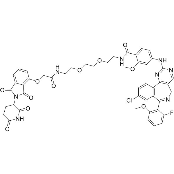
| Chemical Name | 4-[[9-chloro-7-(2-fluoro-6-methoxyphenyl)-5H-pyrimido[5,4-d][2]benzazepin-2-yl]amino]-N-[2-[2-[2-[[2-[2-(2,6-dioxopiperidin-3-yl)-1,3-dioxoisoindol-4-yl]oxyacetyl]amino]ethoxy]ethoxy]ethyl]-2-methoxybenzamide |
| Synonyms | 2705844-82-0; CHEMBL5278315; 4-((9-chloro-7-(2-fluoro-6-methoxyphenyl)-5H-benzo[c]pyrimido[4,5-e]azepin-2-yl)amino)-N-(2-(2-(2-(2-((2-(2,6-dioxopiperidin-3-yl)-1,3-dioxoisoindolin-4-yl)oxy)acetamido)ethoxy)ethoxy)ethyl)-2-methoxybenzamide; SCHEMBL25163855; EX-A7164; BDBM50609429; 4-[[9-chloro-7-(2-fluoro-6-methoxyphenyl)-5H-pyrimido[5,4-d][2]benzazepin-2-yl]amino]-N-[2-[2-[2-[[2-[2-(2,6-dioxopiperidin-3-yl)-1,3-dioxoisoindol-4-yl]oxyacetyl]amino]ethoxy]ethoxy]ethyl]-2-methoxybenzamide; |
| HS Tariff Code | 2934.99.9001 |
| Storage |
Powder-20°C 3 years 4°C 2 years In solvent -80°C 6 months -20°C 1 month |
| Shipping Condition | Room temperature (This product is stable at ambient temperature for a few days during ordinary shipping and time spent in Customs) |
Biological Activity
| Targets | Aurora A 28 nM (DC50) Aurora A 99 nM (Kd) Aurora A 193 nM (EC50) Cereblon |
| ln Vitro | JB170 (1 μM; 24-72 hours; MV4-11 cells) decreases the survival of cancer cells via mediating the depletion of Aurora-A [1]. AURORA-A levels are lowered by JB170 (0.01-10 μM; 6 hours; MV4-11 cells) [1]. JB170 (0.5 μM; 12 hours; MV4-11 cells) inhibits or slows the advancement of the S phase [1]. JB170 (0.5 μM; 0-72 hours; MV4-11 cells) exclusively targets AURORA-A to trigger apoptosis[1]. JB170 (0.1 μM; 0-9 hours; IMR5 cells) exhibits a quick AURORA-A depletion. In comparison to AURORA-A, JB170 (0~1 μM; 6 hours; MV4-11 cells) was significantly diminished in the mutants. In MV4-11 cells, JB170 (0.1 μM; 18 hours) does not cause AURORA-A activation. JB170 (0~1 μM; 24 hours; IMR5 cells) significantly eliminates the depletion of AURORA-AT217D. JB170 (1 μM; 4 days; IMR5 cells) mediates the reduction of Aurora-A, which prevents the survival of cancer cells. By lowering AURORA-A mRNA levels, JB170 (IMR5 cells) lowers AURORA-A levels [1]. |
| Enzyme Assay |
Isothermal titration calorimetry[1] All titrations were performed on a Nano ITC calorimeter at 25 °C. The titrations of the binary complexes (AURORA-A into JB170 and CEREBLON-TBD into JB170) were performed as reverse titrations. Protein concentrations were determined spectroscopically at 280 nm using calculated extinction coefficients and a Thermo Scientific NanoDrop spectrophotometer and a buffer of 25 mM HEPES pH 7.5, 200 mM NaCl, 0.5 mM TCEP, 5% glycerol was used. For AURORA-A, concentrations in the injector (between 57 and 110 µM) had to be optimized due to protein stability issues matching JB170 concentrations between 1.0 and 10.0 µM. Values were calculated from four titrations. Best conditions were achieved at 110 µM AURORA-A and 10 µM JB170. For the CEREBLON (TBD) titration concentrations between 88 and 100 µM were used for the protein and 2.0 and 3.5 µM for JB170. Dissociation constants were calculated from three independent titrations. Titrations for the ternary complexes were determined as previously described41. Briefly, CEREBLON(TBD) at 0.1 µM was titrated as described above. The binary complex remained in the calorimeter and the excess of solution after the titration was removed using a syringe. AURORA-A (110 µM) was titrated into the binary complex which had a JB170 concentration of 3.2 µM and 2.8 µM in two independent titration experiments. All data were fitted using a single binding site model in NanoAnalyse software to obtain Kd values and thermodynamic binding parameters. |
| Cell Assay |
Cell Viability Assay[1] Cell Types: MV4-11 cells Tested Concentrations: 1 µM Incubation Duration: 24-72 hrs (hours) Experimental Results: After 72 hrs (hours), the number of viable cells was 32% of control levels. Western Blot Analysis[1] Cell Types: MV4-11 cells Tested Concentrations: 0.01~10 μM Incubation Duration: 6 hrs (hours) Experimental Results: Substantial degradation was observed at 100 nM and 1 µM. Apoptosis Analysis[1] Cell Types: MV4-11 cells Tested Concentrations: 0.5 µM Incubation Duration: 0~72 hrs (hours) Experimental Results: Apoptosis was exclusively caused by targeting AURORA-A. Cell Cycle Analysis[1] Cell Types: MV4-11 cells Tested Concentrations: 0.5 µM Incubation Duration: 12 hrs (hours) Experimental Results: Delayed or arrested S-phase progression. |
| References |
[1]. PROTAC-mediated degradation reveals a non-catalytic function of AURORA-A kinase. Nat Chem Biol. 2020;16(11):1179-1188. |
| Additional Infomation | The mitotic kinase AURORA-A is essential for cell cycle progression and is considered a priority cancer target. Although the catalytic activity of AURORA-A is essential for its mitotic function, recent reports indicate an additional non-catalytic function, which is difficult to target by conventional small molecules. We therefore developed a series of chemical degraders (PROTACs) by connecting a clinical kinase inhibitor of AURORA-A to E3 ligase-binding molecules (for example, thalidomide). One degrader induced rapid, durable and highly specific degradation of AURORA-A. In addition, we found that the degrader complex was stabilized by cooperative binding between AURORA-A and CEREBLON. Degrader-mediated AURORA-A depletion caused an S-phase defect, which is not the cell cycle effect observed upon kinase inhibition, supporting an important non-catalytic function of AURORA-A during DNA replication. AURORA-A degradation induced rampant apoptosis in cancer cell lines and thus represents a versatile starting point for developing new therapeutics to counter AURORA-A function in cancer.[1] |
Solubility Data
| Solubility (In Vitro) | DMSO : 100 mg/mL (103.80 mM) |
| Solubility (In Vivo) |
Solubility in Formulation 1: ≥ 2.5 mg/mL (2.60 mM) (saturation unknown) in 10% DMSO + 90% Corn Oil (add these co-solvents sequentially from left to right, and one by one), clear solution. For example, if 1 mL of working solution is to be prepared, you can add 100 μL of 25.0 mg/mL clear DMSO stock solution to 900 μL of corn oil and mix evenly. (Please use freshly prepared in vivo formulations for optimal results.) |
| Preparing Stock Solutions | 1 mg | 5 mg | 10 mg | |
| 1 mM | 1.0380 mL | 5.1902 mL | 10.3803 mL | |
| 5 mM | 0.2076 mL | 1.0380 mL | 2.0761 mL | |
| 10 mM | 0.1038 mL | 0.5190 mL | 1.0380 mL |