Physicochemical Properties
| Molecular Formula | C14H10N3NA4O12PS2 |
| Molecular Weight | 599.30 |
| Exact Mass | 552.96 |
| CAS # | 207572-67-6 |
| Related CAS # | PPADS tetrasodium;192575-19-2 |
| PubChem CID | 135458049 |
| Appearance | Typically exists as solid at room temperature |
| Hydrogen Bond Donor Count | 1 |
| Hydrogen Bond Acceptor Count | 15 |
| Rotatable Bond Count | 5 |
| Heavy Atom Count | 36 |
| Complexity | 918 |
| Defined Atom Stereocenter Count | 0 |
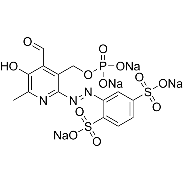
| SMILES | [Na+].[Na+].[Na+].[Na+].O=P(OCC1=C(C=O)C(=O)C(C)=N/C/1=N/NC1=CC(S([O-])(=O)=O)=CC=C1S([O-])(=O)=O)([O-])[O-] |
| InChi Key | SDCXIPVOUDBGDK-UHFFFAOYSA-J |
| InChi Code | InChI=1S/C14H14N3O12PS2.4Na/c1-7-13(19)9(5-18)10(6-29-30(20,21)22)14(15-7)17-16-11-4-8(31(23,24)25)2-3-12(11)32(26,27)28;;;;/h2-5,19H,6H2,1H3,(H2,20,21,22)(H,23,24,25)(H,26,27,28);;;;/q;4*+1/p-4 |
| Chemical Name | tetrasodium;2-[[4-formyl-5-hydroxy-6-methyl-3-(phosphonatooxymethyl)pyridin-2-yl]diazenyl]benzene-1,4-disulfonate |
| Synonyms | iso-PPADS tetrasodium salt; 207572-67-6; Pyridoxalphosphate-6-azophenyl-2',5'-disulfonic acid tetrasodium salt; tetrasodium;2-[[4-formyl-5-hydroxy-6-methyl-3-(phosphonatooxymethyl)pyridin-2-yl]diazenyl]benzene-1,4-disulfonate; HB1951; DA-54441; J-013597; Sodium 2-((4-formyl-5-hydroxy-6-methyl-3-((phosphonatooxy)methyl)pyridin-2-yl)diazenyl)benzene-1,4-disulfonate |
| HS Tariff Code | 2934.99.9001 |
| Storage |
Powder-20°C 3 years 4°C 2 years In solvent -80°C 6 months -20°C 1 month |
| Shipping Condition | Room temperature (This product is stable at ambient temperature for a few days during ordinary shipping and time spent in Customs) |
Biological Activity
| Targets | IC50 = 43 nM and 84 nM for P2X1 and P2X3 receptor, respectively |
| ln Vitro | Seven PPADS (Pyridoxal-5'-Phosphate 6-Azophenyl 2',4'-DiSulfonate) analogs were investigated at Group 1 P2X receptors expressed in Xenopus oocytes. All seven analogs potently inhibited P2X1 (IC50 range, 5-32 nM) and P2X3 (IC50 range, 22-345 nM), the two Group I P2X receptor subtypes. Analogs showed greater inhibitory activity where the pyridoxal moiety of PPADS contained a 5'-phosphonate group, rather than a 5'-phosphate group. Analogs also showed greater potency where disulfonate groups were removed from, or substituted at, the azophenyl moiety. The most active analog was MRS 2257 (pyridoxal-5'-phosphonate 6-azophenyl 3',5'-bismethylenephosphonate) at P2X1 (IC50, 5 nM) and P2X3 (IC50, 22 nM) receptors, being 14-fold and 10-fold more potent than PPADS itself. MRS 2257 produced a nonsurmountable inhibition when tested against a range of ATP concentrations, although blockade was reversed by about 85% after 20 minutes of washout. TNP-ATP and Ip5I were equipotent with MRS 2257 at P2X1 receptors, whereas TNP-ATP was 64-fold more potent than MRS 2257 at P2X3 receptors. In conclusion, the PPADS template can be altered at the pyridoxal and phenyl moieties to produce P2X1 and P2X3 receptor antagonists showing higher potency and greater degree of reversibility than the parent compound at these Group I P2X receptors[2]. |
| ln Vivo | Hippocampus functional MRI analysis revealed considerable changes in response to the increase in tidal volume during mechanical ventilation. Intratracheal lidocaine, iso-PPADS , and TRPV4 genetic deficiency protected mice against ventilationinduced hippocampal pro-apoptotic signaling. Mechanical stretch in both, BEAS-2B cells and ventilated wild-type mice, resulted in TRPV4 activation and reduced Trpv4 and P2x expression. Intratracheal replenishment of adenosine triphosphate in Trpv4 mice abrogated the protective effect of TRPV4 deficiency. Autopsy lung tissue from ventilated patients showed decreased lung TRPV4 levels compared with nonventilated CONCLUSIONS:: TRPV4 mechanosensors and purinergic receptors are involved in the mechanisms of ventilator-induced brain injury. Inhibition of this neural signaling, either using nonspecific or specific inhibitors targeting the TRPV4/adenosine triphosphate/P2X signaling axis, may represent a novel strategy to prevent or treat ventilator-induced brain injury [1]. |
| Enzyme Assay | Electrophysiology [2] Nucleotide-evoked membrane currents were recorded from cRNA-injected oocytes studied under voltage-clamp conditions using a twin-electrode amplifier. Intracellular microelectrodes had a resistance of 1–2 MΩ when filled with KCl (3 M). Oocytes were perfused constantly (at 5 ml min−1) with an extracellular solution containing (mM): NaCl 110, KCl 2.5, HEPES 5, BaCl2 1.8, pH 7.4–7.5. All recordings were made at room temperature (18°C) at a holding potential between −60 and −90 mV. Electrophysiological data were filtered initially at 3 kHz, captured at a rate of 20 Hz on a computer connected to an MP100WSW interface and displayed using commercial software. |
| Cell Assay | Oocyte Preparation and P2X Receptor Expression [2] Xenopus laevis were anaesthetised with Tricaine (0.2%, wt/vol) and killed by decapitation (in accordance with Institution regulations). The dissection and removal of ovaries, as well as the preparation of defolliculated Xenopus oocytes, have been described in detail elsewhere [King et al., 1997]. Defolliculated oocytes do not possess native P1 or P2 receptors that could otherwise complicate the analysis of agonist activity [King et al., 1996a,b]. Also, defolliculated oocytes are largely devoid of ecto-ATPase activity, so avoiding the complicating issue of ectoenzyme inhibition by P2 receptor antagonists [Ziganshin et al., 1995]. Mature oocytes (stages V and VI) were injected (40 nl) cytosolically with capped ribonucleic acid (cRNA, 1 mg/ml) encoding either rat P2X1 or rat P2X3 receptor subunits. Injected oocytes were incubated at 18°C in a bathing solution (pH 7.5) containing (mM): NaCl 110, KCl 1, NaHCO3 2.4, Tris-HCl 7.5, Ca(NO3)2 0.33, CaCl2 0.41, MgSO4 0.82, supplemented with gentamycin sulphate 50 μg/l for 48 h to allow full receptor expression, then stored at 4°C for up to 12 days. |
| Animal Protocol |
Subjects: Wild-type, TRPV4-deficient C57BL/6J mice, 8-10 weeks old. Human postmortem lung tissue and human lung epithelial cell line BEAS-2B. Intervention: Mice subjected to mechanical ventilation were studied using functional MRI to assess hippocampal activity. The effects of lidocaine (a nonselective ion-channel inhibitor), P2X-purinoceptor antagonist (iso-PPADS), or genetic TRPV4 deficiency on hippocampal dopamine-dependent pro-apoptotic signaling were studied in mechanically ventilated mice. Human lung epithelial cells (BEAS-2B) were used to study the effects of mechanical stretch on TRPV4 and P2X expression and activation. TRPV4 levels were measured in postmortem lung tissue from ventilated and nonventilated patients [1]. |
| References |
[1]. Lung Purinoceptor Activation Triggers Ventilator-Induced Brain Injury. Crit Care Med. 2019 Nov;47(11):e911-e918. [2]. Actions of a Series of PPADS Analogs at P2X1 and P2X3 Receptors. Drug Dev Res. 2001 Aug;53(4):281-291. |
| Additional Infomation | Objectives: Mechanical ventilation can cause ventilator-induced brain injury via afferent vagal signaling and hippocampal neurotransmitter imbalances. The triggering mechanisms for vagal signaling during mechanical ventilation are unknown. The objective of this study was to assess whether pulmonary transient receptor potential vanilloid type-4 (TRPV4) mechanoreceptors and vagal afferent purinergic receptors (P2X) act as triggers of ventilator-induced brain injury. Design: Controlled, human in vitro and ex vivo studies, as well as murine in vivo laboratory studies. Setting: Research laboratory.[1] |
Solubility Data
| Solubility (In Vitro) | May dissolve in DMSO (in most cases), if not, try other solvents such as H2O, Ethanol, or DMF with a minute amount of products to avoid loss of samples |
| Solubility (In Vivo) |
Note: Listed below are some common formulations that may be used to formulate products with low water solubility (e.g. < 1 mg/mL), you may test these formulations using a minute amount of products to avoid loss of samples. Injection Formulations (e.g. IP/IV/IM/SC) Injection Formulation 1: DMSO : Tween 80: Saline = 10 : 5 : 85 (i.e. 100 μL DMSO stock solution → 50 μL Tween 80 → 850 μL Saline) *Preparation of saline: Dissolve 0.9 g of sodium chloride in 100 mL ddH ₂ O to obtain a clear solution. Injection Formulation 2: DMSO : PEG300 :Tween 80 : Saline = 10 : 40 : 5 : 45 (i.e. 100 μL DMSO → 400 μLPEG300 → 50 μL Tween 80 → 450 μL Saline) Injection Formulation 3: DMSO : Corn oil = 10 : 90 (i.e. 100 μL DMSO → 900 μL Corn oil) Example: Take the Injection Formulation 3 (DMSO : Corn oil = 10 : 90) as an example, if 1 mL of 2.5 mg/mL working solution is to be prepared, you can take 100 μL 25 mg/mL DMSO stock solution and add to 900 μL corn oil, mix well to obtain a clear or suspension solution (2.5 mg/mL, ready for use in animals). Injection Formulation 4: DMSO : 20% SBE-β-CD in saline = 10 : 90 [i.e. 100 μL DMSO → 900 μL (20% SBE-β-CD in saline)] *Preparation of 20% SBE-β-CD in Saline (4°C,1 week): Dissolve 2 g SBE-β-CD in 10 mL saline to obtain a clear solution. Injection Formulation 5: 2-Hydroxypropyl-β-cyclodextrin : Saline = 50 : 50 (i.e. 500 μL 2-Hydroxypropyl-β-cyclodextrin → 500 μL Saline) Injection Formulation 6: DMSO : PEG300 : castor oil : Saline = 5 : 10 : 20 : 65 (i.e. 50 μL DMSO → 100 μLPEG300 → 200 μL castor oil → 650 μL Saline) Injection Formulation 7: Ethanol : Cremophor : Saline = 10: 10 : 80 (i.e. 100 μL Ethanol → 100 μL Cremophor → 800 μL Saline) Injection Formulation 8: Dissolve in Cremophor/Ethanol (50 : 50), then diluted by Saline Injection Formulation 9: EtOH : Corn oil = 10 : 90 (i.e. 100 μL EtOH → 900 μL Corn oil) Injection Formulation 10: EtOH : PEG300:Tween 80 : Saline = 10 : 40 : 5 : 45 (i.e. 100 μL EtOH → 400 μLPEG300 → 50 μL Tween 80 → 450 μL Saline) Oral Formulations Oral Formulation 1: Suspend in 0.5% CMC Na (carboxymethylcellulose sodium) Oral Formulation 2: Suspend in 0.5% Carboxymethyl cellulose Example: Take the Oral Formulation 1 (Suspend in 0.5% CMC Na) as an example, if 100 mL of 2.5 mg/mL working solution is to be prepared, you can first prepare 0.5% CMC Na solution by measuring 0.5 g CMC Na and dissolve it in 100 mL ddH2O to obtain a clear solution; then add 250 mg of the product to 100 mL 0.5% CMC Na solution, to make the suspension solution (2.5 mg/mL, ready for use in animals). Oral Formulation 3: Dissolved in PEG400 Oral Formulation 4: Suspend in 0.2% Carboxymethyl cellulose Oral Formulation 5: Dissolve in 0.25% Tween 80 and 0.5% Carboxymethyl cellulose Oral Formulation 6: Mixing with food powders Note: Please be aware that the above formulations are for reference only. InvivoChem strongly recommends customers to read literature methods/protocols carefully before determining which formulation you should use for in vivo studies, as different compounds have different solubility properties and have to be formulated differently. (Please use freshly prepared in vivo formulations for optimal results.) |
| Preparing Stock Solutions | 1 mg | 5 mg | 10 mg | |
| 1 mM | 1.6686 mL | 8.3431 mL | 16.6861 mL | |
| 5 mM | 0.3337 mL | 1.6686 mL | 3.3372 mL | |
| 10 mM | 0.1669 mL | 0.8343 mL | 1.6686 mL |