Physicochemical Properties
| Molecular Formula | C19H15BRCLF3N2O3S |
| Molecular Weight | 523.751212358475 |
| Exact Mass | 521.96 |
| Elemental Analysis | C, 43.57; H, 2.89; Br, 15.26; Cl, 6.77; F, 10.88; N, 5.35; O, 9.16; S, 6.12 |
| CAS # | 1207660-00-1 |
| PubChem CID | 46504926 |
| Appearance | White to off-white solid powder |
| LogP | 4.5 |
| Hydrogen Bond Donor Count | 1 |
| Hydrogen Bond Acceptor Count | 7 |
| Rotatable Bond Count | 4 |
| Heavy Atom Count | 30 |
| Complexity | 774 |
| Defined Atom Stereocenter Count | 0 |
| InChi Key | OGXJZCDFFBDSJJ-UHFFFAOYSA-N |
| InChi Code | InChI=1S/C19H15BrClF3N2O3S/c20-14-7-11-5-6-26(18(27)10-1-2-10)16(11)9-17(14)30(28,29)25-12-3-4-15(21)13(8-12)19(22,23)24/h3-4,7-10,25H,1-2,5-6H2 |
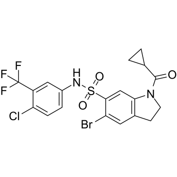
| Chemical Name | 5-bromo-N-[4-chloro-3-(trifluoromethyl)phenyl]-1-(cyclopropanecarbonyl)-2,3-dihydroindole-6-sulfonamide |
| Synonyms | Indophagolin, |
| HS Tariff Code | 2934.99.9001 |
| Storage |
Powder-20°C 3 years 4°C 2 years In solvent -80°C 6 months -20°C 1 month Note: This product requires protection from light (avoid light exposure) during transportation and storage. |
| Shipping Condition | Room temperature (This product is stable at ambient temperature for a few days during ordinary shipping and time spent in Customs) |
Biological Activity
| Targets |
Indophagolin targets membrane-bound purinergic receptor P2X4 [1] |
| ln Vitro |
In MCF7 cells, autophagosome production is inhibited by 10 μM of indophagolin [1]. Indophagolin potently inhibited autophagy in multiple cancer cell lines (HeLa, U2OS, A549, MCF-7) and non-cancerous cells (HEK293T), as evidenced by accumulated LC3-II (western blot) and p62/SQSTM1 (immunofluorescence and western blot) [1] Indophagolin directly bound to P2X4 receptor, confirmed by thermal proteome profiling (TPP), surface plasmon resonance (SPR), and co-immunoprecipitation (Co-IP) assays [1] Knockdown of P2X4 (siRNA) or P2X4 antagonist (5-BDBD) abolished Indophagolin-mediated autophagy inhibition, while overexpression of P2X4 enhanced its effect [1] Indophagolin (1-10 μM) dose-dependently inhibited autophagic flux: blocked LC3-II turnover (in presence of bafilomycin A1) and reduced autophagosome-lysosome fusion (mCherry-GFP-LC3 reporter assay) [1] Indophagolin did not affect apoptosis (Annexin V/PI staining) or necrosis at concentrations up to 10 μM, showing selectivity for autophagy inhibition [1] Indophagolin (5 μM) suppressed proliferation of cancer cells (A549, MCF-7) by ~40% compared to control, which was reversed by P2X4 knockdown [1] |
| ln Vivo |
In nude mice bearing A549 lung cancer xenografts, intraperitoneal administration of Indophagolin (10 mg/kg, once daily for 14 days) significantly inhibited tumor growth: tumor volume reduced by ~55% and tumor weight by ~50% compared to vehicle control [1] Indophagolin accumulated LC3-II and p62 in xenograft tumor tissues (IHC staining), confirming autophagy inhibition in vivo [1] Indophagolin (10 mg/kg, ip, daily for 14 days) did not alter P2X4 expression in normal tissues (liver, kidney, spleen) of mice, as detected by western blot [1] |
| Enzyme Assay |
Thermal Proteome Profiling (TPP): Cancer cells (HeLa) were lysed, and cell lysates were incubated with Indophagolin (10 μM) or vehicle for 30 minutes at 4°C. Lysates were subjected to a temperature gradient (42-67°C) for thermal denaturation, followed by centrifugation to remove aggregated proteins. Soluble proteins were analyzed by liquid chromatography-tandem mass spectrometry (LC-MS/MS), and P2X4 was identified as a target with increased thermal stability in presence of Indophagolin [1] Surface Plasmon Resonance (SPR): Recombinant human P2X4 protein was immobilized on a sensor chip. Serial dilutions of Indophagolin (0.1-20 μM) were injected over the chip surface at a constant flow rate. Binding responses were recorded as resonance units (RU), and binding affinity was determined by fitting sensorgrams to a 1:1 binding model [1] Co-Immunoprecipitation (Co-IP): HeLa cells were treated with Indophagolin (5 μM) for 4 hours, then lysed in immunoprecipitation buffer. Cell lysates were incubated with anti-P2X4 antibody overnight at 4°C, followed by addition of protein A/G beads. After washing, bound proteins were eluted, separated by SDS-PAGE, and detected by western blot with anti-Indophagolin antibody (custom-generated) [1] |
| Cell Assay |
Autophagy Inhibition Assay: Cells were seeded in 6-well plates (5×10⁵ cells/well) and cultured overnight. Indophagolin (0.5-10 μM) was added, and cells were incubated for 24 hours. For autophagic flux analysis, cells were co-treated with Indophagolin (5 μM) and bafilomycin A1 (100 nM) for 16 hours. LC3-II and p62 levels were detected by western blot; p62 puncta were visualized by immunofluorescence microscopy [1] mCherry-GFP-LC3 Reporter Assay: Cells transfected with mCherry-GFP-LC3 plasmid were treated with Indophagolin (5 μM) for 24 hours. Autophagosomes (yellow puncta, mCherry+GFP+) and autolysosomes (red puncta, mCherry+GFP-) were counted by confocal microscopy to assess fusion efficiency [1] P2X4 Knockdown/Overexpression Assay: Cells were transfected with P2X4 siRNA (30 nM) or P2X4 overexpression plasmid for 48 hours, then treated with Indophagolin (5 μM) for 24 hours. Autophagy markers (LC3-II, p62) and cell proliferation (CCK-8 assay) were measured [1] Apoptosis/Necrosis Assay: Cells were treated with Indophagolin (1-10 μM) for 48 hours, stained with Annexin V-FITC and PI, and analyzed by flow cytometry to quantify apoptotic/necrotic cells [1] |
| Animal Protocol |
A549 Xenograft Model: 6-8 week-old male nude mice were subcutaneously injected with 2×10⁶ A549 cells into the right flank. When tumors reached 100-150 mm³, mice were randomized into 2 groups (n=6/group): Vehicle control (DMSO:PEG400:PBS = 5:45:50, v/v/v, ip, daily) and Indophagolin treatment group (10 mg/kg, dissolved in DMSO:PEG400:PBS = 5:45:50, ip, once daily for 14 days) [1] Tumor Measurements: Tumor volume was calculated every 2 days using calipers (volume = length × width² / 2). At the end of treatment, mice were euthanized, tumors were excised and weighed. Tumor tissues were fixed in formalin for IHC staining or frozen for western blot analysis [1] Tissue Collection: Normal tissues (liver, kidney, spleen) were collected from mice to detect P2X4 expression and assess potential toxicity [1] |
| Toxicity/Toxicokinetics |
In xenograft studies, Indophagolin (10 mg/kg, ip, daily for 14 days) caused no significant toxicity: mice showed no body weight loss (>5%), abnormal behavior, or histopathological damage in liver, kidney, or spleen [1] |
| References |
[1]. Thermal proteome profiling identifies the membrane-bound purinergic receptor P2X4 as a target of the autophagy inhibitor indophagolin [published online ahead of print, 2021 Mar 9]. Cell Chem Biol. 2021;S2451-9456(21)00102-1. |
| Additional Infomation |
Indophagolin is a small-molecule autophagy inhibitor that targets P2X4 receptor, representing a novel mechanism for autophagy modulation [1] Indophagolin’s autophagy inhibitory effect is mediated by P2X4 binding, which blocks autophagosome-lysosome fusion without affecting early autophagosome formation [1] Indophagolin shows potential as an anti-cancer agent by inhibiting autophagy and suppressing tumor growth in vivo [1] Indophagolin exhibits high selectivity for P2X4, with no cross-reactivity to other P2X family members (P2X1, P2X2, P2X3, P2X7) confirmed by TPP and SPR [1] |
Solubility Data
| Solubility (In Vitro) | DMSO : ~250 mg/mL (~477.33 mM) |
| Solubility (In Vivo) |
Solubility in Formulation 1: 2.08 mg/mL (3.97 mM) in 10% DMSO + 40% PEG300 + 5% Tween80 + 45% Saline (add these co-solvents sequentially from left to right, and one by one), suspension solution; with sonication. For example, if 1 mL of working solution is to be prepared, you can add 100 μL of 20.8 mg/mL clear DMSO stock solution to 400 μL PEG300 and mix evenly; then add 50 μL Tween-80 to the above solution and mix evenly; then add 450 μL normal saline to adjust the volume to 1 mL. Preparation of saline: Dissolve 0.9 g of sodium chloride in 100 mL ddH₂ O to obtain a clear solution. Solubility in Formulation 2: ≥ 2.08 mg/mL (3.97 mM) (saturation unknown) in 10% DMSO + 90% Corn Oil (add these co-solvents sequentially from left to right, and one by one), clear solution. For example, if 1 mL of working solution is to be prepared, you can add 100 μL of 20.8 mg/mL clear DMSO stock solution to 900 μL of corn oil and mix evenly. (Please use freshly prepared in vivo formulations for optimal results.) |
| Preparing Stock Solutions | 1 mg | 5 mg | 10 mg | |
| 1 mM | 1.9093 mL | 9.5465 mL | 19.0931 mL | |
| 5 mM | 0.3819 mL | 1.9093 mL | 3.8186 mL | |
| 10 mM | 0.1909 mL | 0.9547 mL | 1.9093 mL |