Indirubin-3'-monoxime is a novel, potent and selective inhibitor of GSK-3β (glycogen synthase kinase 3β) which also weakly inhibits 5-Lipoxygenase with IC50s of 22 nM and 7.8-10 µM, respectively; Indirubin-3'-monoxime also shows inhibitory activities against CDK5/p25 and CDK1/cyclin B, with IC50s of 100 and 180 nM. As GSK3β phosphorylates tau protein, indirubin-
Physicochemical Properties
| Molecular Formula | C16H11N3O2 |
| Molecular Weight | 277.2774 |
| Exact Mass | 277.085 |
| Elemental Analysis | C, 69.31; H, 4.00; N, 15.15; O, 11.54 |
| CAS # | 160807-49-8 |
| Related CAS # | Indirubin;479-41-4 |
| PubChem CID | 3707 |
| Appearance | Brown to red solid powder |
| Density | 1.5±0.1 g/cm3 |
| Boiling Point | 532.2±50.0 °C at 760 mmHg |
| Melting Point | 241 °C |
| Flash Point | 275.7±30.1 °C |
| Vapour Pressure | 0.0±1.5 mmHg at 25°C |
| Index of Refraction | 1.772 |
| LogP | 1.08 |
| Hydrogen Bond Donor Count | 3 |
| Hydrogen Bond Acceptor Count | 3 |
| Rotatable Bond Count | 1 |
| Heavy Atom Count | 21 |
| Complexity | 405 |
| Defined Atom Stereocenter Count | 0 |
| SMILES | O([H])C1=C(C2=C([H])C([H])=C([H])C([H])=C2N1[H])C1=C(C2=C([H])C([H])=C([H])C([H])=C2N1[H])N=O |
| InChi Key | HBDSHCUSXQATPO-BRNLPKLHSA-N |
| InChi Code | InChI=1S/C16H11N3O2/c20-16-13(9-5-1-3-7-11(9)18-16)15-14(19-21)10-6-2-4-8-12(10)17-15/h1-8,17,21H,(H,18,20)/b15-13-,19-14- |
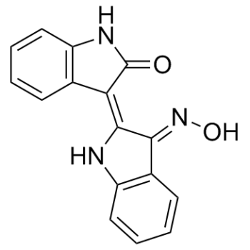
| Chemical Name | 3-[1,3-dihydro-3-(hydroxyimino)-2H-indol-2-ylidene]-1,3-dihydro-2H-indol-2-one |
| Synonyms | Indirubin-3’-oxime |
| HS Tariff Code | 2934.99.9001 |
| Storage |
Powder-20°C 3 years 4°C 2 years In solvent -80°C 6 months -20°C 1 month |
| Shipping Condition | Room temperature (This product is stable at ambient temperature for a few days during ordinary shipping and time spent in Customs) |
Biological Activity
| Targets |
GSK-3β (IC50 = 22 nM); CDK5/p25 (IC50 = 100 nM); CDK1/cyclin B (IC50 = 180 nM); 5-LOX (IC50 = 7.8-10 μM)
Glycogen Synthase Kinase-3β (GSK-3β) (IC₅₀ = 0.05 μM) [1] Cyclin-Dependent Kinase 5/p25 (CDK5/p25) (IC₅₀ = 0.07 μM) [1] Cyclin-Dependent Kinase 1/Cyclin B (CDK1/Cyclin B) (IC₅₀ = 0.12 μM) [1] Cyclin-Dependent Kinase 2/Cyclin A (CDK2/Cyclin A) (IC₅₀ = 0.18 μM) [1] Cyclin-Dependent Kinase 2/Cyclin E (CDK2/Cyclin E) (IC₅₀ = 0.21 μM) [1] Histone Deacetylase (HDAC) 1 (IC₅₀ = 2.3 μM) [4] Histone Deacetylase (HDAC) 3 (IC₅₀ = 3.1 μM) [4] Histone Deacetylase (HDAC) 6 (IC₅₀ = 1.8 μM) [4] Leukotriene B4 Receptor 1 (BLT1) (no definite IC₅₀ data provided) [3] |
| ln Vitro |
Indirubin-3'-monoxime has a Ki of 0.85 μM and a Km of 110 μM. It inhibits GSK-3β by complementation with ATP. Even with an IC50 value of about 100 nM, indirubin-3'-monoxime still prevents tau phosphorylation by GSK-3{. At the AT100 epitope, phosphorylation is totally inhibited by indiviubunin-3'-monoxime [1]. An IC50 of approximately 2 μM is observed for indinabinin-3'-monoxime's inhibition of vascular smooth muscle cell (VSMC) proliferation. VSMC migration induced by PDGF was inhibited by indorubin-3'-monoxime]. Both the generation of LT in monocytes that promotes migration and the migration response in VSMC are disrupted by indorubin-3'-monoxime. Furthermore, with comparable efficacy (IC50 values of 5.0±1.1 and 3.7±1.2μM, respectively), indirubin-3'-monoxime impedes the production of 5-lipoxygenase (5-LO) products in monocytes and neutrophils. With an IC50 of 7.8-10 μM in cell-free experiments, indorubin-3'-monoxime is a 5-LO reagent [3]. 1. Dual inhibition of kinases and HDACs: Indirubin-3'-monoxime potently inhibited GSK-3β and CDK5/p25 (IC₅₀ = 0.05 μM and 0.07 μM, respectively) and moderately inhibited other CDKs (CDK1/Cyclin B, CDK2/Cyclin A/E) [1]. It also exhibited inhibitory activity against HDAC1, HDAC3, and HDAC6 (IC₅₀ = 2.3 μM, 3.1 μM, and 1.8 μM, respectively), acting as a dual kinase-HDAC inhibitor [4] 2. Inhibition of abnormal tau phosphorylation: In SH-SY5Y neuroblastoma cells treated with okadaic acid (OA) to induce tau hyperphosphorylation, Indirubin-3'-monoxime (0.1-1 μM) dose-dependently reduced phosphorylation of tau at Ser396 and Thr231 (Western blot), with 1 μM reducing p-tau/Total tau ratios by 68% and 72%, respectively. This effect was mediated by GSK-3β and CDK5 inhibition [1, 2] 3. Antiproliferative and pro-apoptotic activity in cancer cells: Indirubin-3'-monoxime (0.5-10 μM) inhibited proliferation of multiple cancer cell lines (HeLa, MCF-7, A549, HepG2) with IC₅₀ values ranging from 1.2 to 3.5 μM (MTT assay). It induced G2/M cell cycle arrest (flow cytometry: G2/M phase cells increased from 18% to 42% at 5 μM) and apoptosis (Annexin V-FITC/PI staining: apoptotic rate increased from 4% to 35% at 10 μM) in HeLa cells. Western blot detected activation of caspase-3/7 and downregulation of anti-apoptotic protein Bcl-2, and upregulation of pro-apoptotic protein Bax [4] 4. Inhibition of vascular smooth muscle cell (VSMC) migration: In PDGF-BB-stimulated rat aortic VSMCs, Indirubin-3'-monoxime (0.1-10 μM) dose-dependently inhibited cell migration (Transwell assay: migration rate reduced by 70% at 10 μM) and wound healing (scratch assay: healing rate reduced by 65% at 10 μM). It blocked leukotriene B4 (LTB4)-mediated BLT1 signaling, reducing downstream ERK1/2 phosphorylation and MMP-9 expression [3] 5. Neuroprotective effects against oxidative stress: In H₂O₂-treated SH-SY5Y cells, Indirubin-3'-monoxime (0.5-2 μM) dose-dependently increased cell viability (MTT assay: viability increased from 45% to 82% at 2 μM) and reduced reactive oxygen species (ROS) production (DCFH-DA staining: ROS levels reduced by 60% at 2 μM). It upregulated antioxidant enzymes (SOD1, CAT) and reduced lipid peroxidation (MDA levels reduced by 55% at 2 μM) [2] |
| ln Vivo |
In breast milk fed on a high-fat diet, indirubin-3'-monoxime (0.1, 0.2, and 0.4 mg/kg, ip) preventatively cures cognitive impairment and counteracts heightened oxidative midday indicators. In addition, dose-dependent reductions in insulin, TG, TC, and plasma glucose were observed when lactations given a high-fat diet (HFD) had better β-cell function. In addition, HOMA-IR levels were considerably lower in the Indirubin-3'-monoxime group as compared to the HFD group. The elevated EL in the HFD group was considerably reduced by -3'-Monoxime (0.4 mg/kg) [2]. 1. Improvement of cognitive impairment in high-fat diet (HFD)-induced mice: C57BL/6 mice fed HFD for 8 weeks to induce cognitive dysfunction were treated with Indirubin-3'-monoxime (5 mg/kg or 10 mg/kg, intraperitoneal injection, once daily) for 8 weeks. Morris water maze tests showed: (1) Escape latency reduced by 42% (5 mg/kg) and 58% (10 mg/kg) compared to HFD control; (2) Time spent in target quadrant increased by 35% (5 mg/kg) and 48% (10 mg/kg); (3) Number of platform crossings increased by 2.3-fold (10 mg/kg). Brain tissue analysis: (1) Hippocampal p-tau (Ser396/Thr231) levels reduced by 45% (10 mg/kg); (2) GSK-3β phosphorylation (Ser9, inactive form) increased by 60% (10 mg/kg); (3) ROS and MDA levels reduced by 50% and 48% (10 mg/kg); (4) SOD1 and CAT activities increased by 42% and 38% (10 mg/kg) [2] |
| Enzyme Assay |
GSK-3β is expressed in and purified from insect Sf9 cells. It is measured with a final volume of 30 μL of 15 μM[γ-32P]ATP (3000 Ci/mmol; 1 mCi/L) after a 1/100 dilution in 1 mg/mL BSA, 10 mM DTT, and 5 μL of 40 μM GS-1 peptide as a substrate. Following a 30-minute incubation at 30°C, 25-μL aliquots of the supernatant are spotted onto pieces of Whatman P81 phosphocellulose paper that are 2.5 x 3 cm. Twenty seconds later, the filters are washed five times (for at least five minutes each time) in a solution of 10 mL of phosphoric acid/liter of water. A 1 mL sample of ACS scintillation fluid is used to count the wet filters[1]. 1. GSK-3β kinase activity assay: Prepare recombinant human GSK-3β and glycogen synthase-derived substrate peptide. Set up reaction mixtures containing 50 nM GSK-3β, 10 μM ATP, 50 μM substrate peptide, 10 mM MgCl₂, and varying concentrations of Indirubin-3'-monoxime (0.01-1 μM) in kinase buffer (25 mM Tris-HCl, pH 7.5, 0.1 mM EGTA, 1 mM DTT). Incubate at 30°C for 30 minutes, terminate with 20 mM EDTA. Measure substrate phosphorylation by [γ-³²P]ATP incorporation (P81 paper filtration and scintillation counting). Calculate IC₅₀ from inhibition percentage vs. concentration curves [1] 2. CDK5/p25 kinase activity assay: Recombinant CDK5/p25 complex (50 nM) was incubated with 10 μM ATP, 50 μM histone H1 substrate, 10 mM MgCl₂, and Indirubin-3'-monoxime (0.01-1 μM) in kinase buffer. Incubate at 30°C for 30 minutes, terminate with EDTA. Detect phosphorylation via autoradiography after SDS-PAGE. Quantify band intensity to calculate IC₅₀ [1] 3. HDAC activity assay: Use a fluorogenic HDAC assay kit with acetylated histone peptide substrate. Incubate recombinant HDAC1/3/6 (20 nM) with varying concentrations of Indirubin-3'-monoxime (0.1-10 μM) and substrate in assay buffer (50 mM Tris-HCl, pH 8.0, 137 mM NaCl, 2.7 mM KCl, 1 mM MgCl₂) at 37°C for 1 hour. Add developer to release fluorescence, measure intensity at 460 nm (excitation 360 nm). Calculate IC₅₀ by comparing to inhibitor-free controls [4] |
| Cell Assay |
Cytotoxicity of Indirubin-3'-monoxime in monocytes is analysed by MTT assay in a 96-well format using a multi-well scanning spectrophotometer. Following a 30-minute incubation with Indirubin-3'-monoxime, neutrophils (5 106 cells/mL) or monocytes (2 106 cells/mL) are tested for cell viability using the MTT assay. There is no detectable acute cytotoxicity when compared to vehicle (0.3% DMSO) in neutrophils (103.94.4%; 129.45.4%; n=3, each)[3]. 1. Neuroblastoma cell tau phosphorylation assay: Seed SH-SY5Y cells (5×10⁴ cells/well) in 24-well plates, incubate overnight. Pre-treat with Indirubin-3'-monoxime (0.1-1 μM) for 1 hour, then add okadaic acid (100 nM) for 24 hours. Lyse cells, perform Western blot with anti-p-tau (Ser396/Thr231), anti-total tau, and anti-tubulin antibodies. Quantify p-tau/total tau ratios using ImageJ [1, 2] 2. Cancer cell proliferation and apoptosis assay: For proliferation: Seed cancer cells (HeLa, MCF-7, 1×10⁴ cells/well) in 96-well plates, treat with Indirubin-3'-monoxime (0.5-10 μM) for 72 hours, measure viability via MTT assay. For apoptosis: Seed HeLa cells (5×10⁵ cells/well) in 6-well plates, treat with 5-10 μM drug for 48 hours, stain with Annexin V-FITC/PI, analyze by flow cytometry. For Western blot: Detect caspase-3/7, Bcl-2, Bax, and tubulin [4] 3. VSMC migration assay: For Transwell: Seed PDGF-BB-stimulated VSMCs (1×10⁴ cells/well) in upper chambers, add Indirubin-3'-monoxime (0.1-10 μM) to both chambers, incubate for 24 hours. Fix lower chamber cells with methanol, stain with crystal violet, count migrated cells. For scratch assay: Seed VSMCs to confluence, create scratch with pipette tip, treat with drug and PDGF-BB, image at 0 and 24 hours, calculate healing rate [3] 4. Oxidative stress assay: Seed SH-SY5Y cells (5×10⁴ cells/well) in 24-well plates, pre-treat with Indirubin-3'-monoxime (0.5-2 μM) for 1 hour, then add H₂O₂ (200 μM) for 24 hours. Measure viability via MTT assay. For ROS: Load cells with DCFH-DA (10 μM) for 30 minutes, measure fluorescence intensity. For antioxidant enzymes: Lyse cells, assay SOD1 and CAT activities; measure MDA levels via thiobarbituric acid reaction [2] |
| Animal Protocol |
Male mice (5–6 weeks old) are divided into five groups (n=10) at random. Groups 1 and 2 receive a normal pellet diet (NPD); Groups 3 through 5 receive a HFD; and Groups 6 through 9 receive an Indirubin-3'-monoxime treatment (0.1, 0.2, and 0.4 mg/kg i.p., respectively) once daily for one week after receiving the HFD for 8 weeks. In (2.5% v/v) DMSO in saline, indorubin-3'-monoxime is dissolved. Equal volumes of vehicle (2.5% v/v DMSO in saline) are administered to the mice in the NPD and HFD groups. Indirubin-3'-monoxime dosages are decided upon. Mice are kept for eight weeks in standard husbandry conditions (22°C and 60% humidity) with free access to food and water on a 12/12-hour light/dark cycle. Throughout the course of the experiment, weekly weight checks are made[2]. 1. HFD-induced cognitive impairment mouse model: Male C57BL/6 mice (6 weeks old) were divided into 4 groups (n=10/group): Normal diet (ND) + vehicle, HFD + vehicle, HFD + Indirubin-3'-monoxime 5 mg/kg, HFD + 10 mg/kg. HFD groups were fed HFD for 16 weeks (8 weeks induction + 8 weeks treatment). Drug was dissolved in DMSO (5% final volume) + 0.9% saline, administered via intraperitoneal injection once daily for 8 weeks. Vehicle groups received DMSO/saline. Morris water maze tests were performed during weeks 14-16. After sacrifice, hippocampus and cortex were dissected for Western blot (p-tau, GSK-3β), ROS/MDA detection, and antioxidant enzyme assays [2] |
| Toxicity/Toxicokinetics |
1. In vitro cytotoxicity: Indirubin-3'-monoxime showed low cytotoxicity to normal cells (human umbilical vein endothelial cells, HUVECs) with IC₅₀ > 20 μM, while exhibiting potent antiproliferative activity against cancer cells (IC₅₀ = 1.2-3.5 μM), indicating a favorable therapeutic index [4] 2. In vivo safety: In the HFD mouse model, Indirubin-3'-monoxime (5-10 mg/kg, i.p., 8 weeks) did not cause significant changes in body weight, liver function (ALT, AST), or kidney function (BUN, creatinine) compared to vehicle groups. Histopathological examination of liver, kidney, and brain showed no drug-related lesions [2] |
| References |
[1]. Indirubins inhibit glycogen synthase kinase-3 beta and CDK5/p25, two protein kinases involved in abnormal tau phosphorylation in Alzheimer's disease. A property common to most cyclin-dependent kinase inhibitors? J Biol Chem. 2001 Jan 5;276(1):251-60. [2]. Neuroprotective role of Indirubin-3'-monoxime, a GSKβ inhibitor in high fat diet induced cognitive impairment in mice. Biochem Biophys Res Commun. 2014 Oct 3;452(4):1009-15. [3]. Indirubin-3'-monoxime exerts a dual mode of inhibition towards leukotriene-mediated vascular smooth muscle cell migration. Cardiovasc Res. 2014 Mar 1;101(3):522-32. [4]. Indirubin Derivatives as Dual Inhibitors Targeting Cyclin-Dependent Kinase and Histone Deacetylase for Treating Cancer [published online ahead of print, 2021 Oct 8]. J Med Chem. 2021;10.1021/acs.jmedchem.1c01311. |
| Additional Infomation |
Indirubin-3'-monoxime is a member of the class of biindoles that is indirubin in which the keto group at position 3' has undergone condensation with hydroxylamine to form the corresponding oxime. It has a role as an EC 2.7.11.22 (cyclin-dependent kinase) inhibitor, an EC 2.7.11.1 (non-specific serine/threonine protein kinase) inhibitor, an osteogenesis regulator, a neuroprotective agent and an anti-obesity agent. It is a member of oxindoles, a bisindole, a ring assembly, a ketoxime and an alkaloid. 1. Chemical background: Indirubin-3'-monoxime is a synthetic derivative of indirubin, a natural product isolated from Indigofera tinctoria. It is a yellow crystalline powder, soluble in DMSO and ethanol, slightly soluble in water [1, 4] 2. Mechanism of action: It acts as a dual inhibitor of cyclin-dependent kinases (CDKs) and histone deacetylases (HDACs), blocking cell cycle progression and inducing apoptosis in cancer cells. It also inhibits GSK-3β and CDK5/p25, reducing abnormal tau phosphorylation in neurocells, and modulates BLT1-ERK1/2-MMP-9 signaling to inhibit VSMC migration [1, 3, 4] 3. Therapeutic potential: Potential applications include: (1) Neurodegenerative diseases (Alzheimer's disease) via reducing tau hyperphosphorylation and oxidative stress [1, 2]; (2) Cancer (cervical, breast, lung, liver cancer) as a dual CDK-HDAC inhibitor [4]; (3) Cardiovascular diseases (atherosclerosis) via inhibiting VSMC migration [3] 4. Selectivity profile: It exhibits higher selectivity for GSK-3β, CDK5, and HDAC1/3/6 over other kinases (e.g., ERK1/2, JNK) and HDAC isoforms (e.g., HDAC2, HDAC4) [1, 4] |
Solubility Data
| Solubility (In Vitro) | DMSO: ~125 mg/mL (450.8 mM) |
| Solubility (In Vivo) |
Solubility in Formulation 1: ≥ 2.5 mg/mL (9.02 mM) (saturation unknown) in 10% DMSO + 40% PEG300 +5% Tween-80 + 45% Saline (add these co-solvents sequentially from left to right, and one by one), clear solution. For example, if 1 mL of working solution is to be prepared, you can add 100 μL of 25.0 mg/mL clear DMSO stock solution to 400 μL PEG300 and mix evenly; then add 50 μL Tween-80 + to the above solution and mix evenly; then add 450 μL normal saline to adjust the volume to 1 mL. Preparation of saline: Dissolve 0.9 g of sodium chloride in 100 mL ddH₂ O to obtain a clear solution. (Please use freshly prepared in vivo formulations for optimal results.) |
| Preparing Stock Solutions | 1 mg | 5 mg | 10 mg | |
| 1 mM | 3.6065 mL | 18.0323 mL | 36.0646 mL | |
| 5 mM | 0.7213 mL | 3.6065 mL | 7.2129 mL | |
| 10 mM | 0.3606 mL | 1.8032 mL | 3.6065 mL |