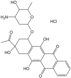
Physicochemical Properties
| Molecular Formula | C26H27NO9.HCL |
| Molecular Weight | 533.95 |
| Exact Mass | 533.145 |
| Elemental Analysis | C, 58.49; H, 5.29; Cl, 6.64; N, 2.62; O, 26.97 |
| CAS # | 57852-57-0 |
| Related CAS # | 58957-92-9 |
| PubChem CID | 636362 |
| Appearance | Orange to red solid powder |
| Boiling Point | 725.4ºC at 760 mmHg |
| Melting Point | 183-185ºC |
| Flash Point | 392.5ºC |
| LogP | 2.522 |
| Hydrogen Bond Donor Count | 6 |
| Hydrogen Bond Acceptor Count | 10 |
| Rotatable Bond Count | 3 |
| Heavy Atom Count | 37 |
| Complexity | 912 |
| Defined Atom Stereocenter Count | 6 |
| SMILES | OC(C(C(C1=CC=CC=C21)=O)=C3C2=O)=C4[C@H](C[C@@](C(C)=O)(O)CC4=C3O)O[C@@](O[C@@H](C)[C@H]5O)([H])C[C@@H]5N.Cl |
| InChi Key | JVHPTYWUBOQMBP-RVFAQHLVSA-N |
| InChi Code | InChI=1S/C26H27NO9.ClH/c1-10-21(29)15(27)7-17(35-10)36-16-9-26(34,11(2)28)8-14-18(16)25(33)20-19(24(14)32)22(30)12-5-3-4-6-13(12)23(20)31;/h3-6,10,15-17,21,29,32-34H,7-9,27H2,1-2H3;1H/t10-,15-,16-,17-,21+,26-;/m0./s1 |
| Chemical Name | (7S,9S)-9-acetyl-7-[(2R,4S,5S,6S)-4-amino-5-hydroxy-6-methyloxan-2-yl]oxy-6,9,11-trihydroxy-8,10-dihydro-7H-tetracene-5,12-dione;hydrochloride |
| Synonyms | 4-demethoxydaunorubicin (NSC256439, 4-DMDR) HCl; IMI30; NSC256439; IMI-30; NSC-256439; IMI 30; NSC 256439; 4-DMDR; IDA; 4-Demethoxydaunomycin; brand name: Idamycin; IDAMYCIN PFS |
| HS Tariff Code | 2934.99.9001 |
| Storage |
Powder-20°C 3 years 4°C 2 years In solvent -80°C 6 months -20°C 1 month Note: Please store this product in a sealed and protected environment (e.g. under nitrogen), avoid exposure to moisture and light. |
| Shipping Condition | Room temperature (This product is stable at ambient temperature for a few days during ordinary shipping and time spent in Customs) |
Biological Activity
| Targets | Topo II (MCF-7 cells) ( IC50 = 3.3 ng/mL ); Multicellular spheroids ( IC50 = 7.9 ng/mL ) |
| ln Vitro | Idarubicin possesses potent anticancer action against multicellular spheroids that is similar to the effects on monolayer cells' ability to inhibit proliferation.[1] CYP450 2D6 is inhibited by idarubicin.[2] Compared to doxorubicin and epirubicin, it is approximately 57.5-fold and 25-fold more active, respectively. Multidrug resistance mediated by P-glycoprotein can be overcome by idarubicin.[3] Idarubicin prevents the production of PMN superoxide radicals.[4] Idarubicin could be combined with monoclonal antibodies that are active against Thy-1, anti-Ly-2.1, or anti-L3T4 while maintaining the protein's solubility.[5] At an IC50 of 12 nM, idarubicin suppresses the growth of NALM-6 cells.[6] |
| ln Vivo | Idarubicin reduction is reliant on ketone reductases and proceeds more stereoselectively than that of most ketones, almost exclusively producing the (13S)-epimer. Because Idarubicin has asymmetric centers close to its carbonyl group, chiral induction may be the cause of the reduction's high stereospecificity.[7] |
| Enzyme Assay | Completed with isolated human CYP450 proteins for each isoform, the metabolism of idarubicin by the CYP450 isoenzymes 3A4, 2D6, 2C8, 2C9, and 1A2 is assessed. This evaluation is conducted using the high throughput P450 inhibition testing method. Each drug's following characteristics are intended to be examined by the metabolism experiments: (1) if Idarubicin is a substrate of the CYP450 3A4, 2C8, 2C9, 1A2 or 2D6 isoenzymes; (2) if the metabolism of each isoenzyme is impacted by known inhibitors; (3) if Idarubicin is an inhibitor of CYP450 isoenzymes; and (4) if the CYP450 metabolism of Idarubicin is inhibited by caspofungin or itraconazole. The known substrates used as controls to verify the corresponding isoenzyme activity and assess the impact of idarubicin on the isoenzyme activity are dibenzylfluorescein (DBF) (CYP3A4, CYP2C8, CYP2C9), 3-cyano-7-ethoxycoumarin (Cyp1A2), and 7-methoxy-4-(aminomethyl)-coumarin (MAMC) (CYP2D6). In addition, the control CYP450 inhibitors for the 3A4, 2C8, 2C9, 1A2, or 2D6 isoenzymes are quercetin, quinidine, furafylline, ketoconazole, or suflaphenazole, in that order. For the duration of the manufacturer's recommended incubation period of 20 to 60 minutes at 37°C, the substrate, inhibitor, and idarubicin are added to each protein sample. When needed, samples are examined using a fluorescence plate reader after reactions are halted with an organic solvent solution. Control samples are prepared for qualitative comparisons for each experiment, containing a known quantity of substrate and synthesized metabolite, without the isoenzyme. Every experiment is run in triplicate. |
| Cell Assay | The inhibition of [3H]thymidine uptake is used to compare the anti-proliferative activity of the conjugate Idarubicin to that of the free drug. In summary, 100 μL/well of a flat-bottomed microtitre plate containing 1.5 × 10⁶/mL of NALM-6 cells is added, and the plate is then incubated for one hour at 37ºC. Filtered free idarubicin and idarubicin-mAb conjugates are diluted in sterile PBS and added to the wells in duplicate at different concentrations (100 μL/well); the plates are then incubated for 24 hours at 37ºC with 7% CO2. After 4 hours of incubation, each well is filled with 50 μL medium containing 1 μCi [3H]thymidine, and the plates are left to incubate for an additional 4 hours. Cells are collected using glass fiber filter paper, dried, and counted using a scintillation counter. Using the same methodology, studies of specificity are carried out in which the cytotoxicity of insignificant Idarubicin-JGT conjugates is contrasted with the killing capacity of Idarubicin-anti-CD19 conjugates against CD19 + cells. For thirty rain on ice, NALM-6 cells (1.5 x 10⁶/mL, 300 μL tube) are incubated with different concentrations of Idarubicin-anti-CD 19 or Idarubicin-JGT conjugates. After three cycles of ice-cold RPMI-1640 medium (4 mL/cycle), the cells are reconstituted in new medium and put into 96-well plates (100 μL/well). There are two wells plated out of each duplicate tube, for a total of four wells per drug concentration. Two days later, [3H]thymidine is pulsed into the cells, and they are harvested. |
| Animal Protocol | Rat, rabbit, mouse, dog |
| References |
[1]. J Chemother . 2005 Dec;17(6):663-7. [2]. Hematology . 2004 Jun;9(3):217-21. [3]. Eur Urol . 1997;31(3):365-70. [4]. J Leukoc Biol . 1990 Mar;47(3):224-33. [5]. Transplantation . 1988 Jul;46(1):126-31. [6]. Cancer Immunol Immunother . 1993 Aug;37(3):195-202. [7]. Xenobiotica . 1991 Apr;21(4):473-80. |
| Additional Infomation |
Idarubicin Hydrochloride can cause developmental toxicity and male reproductive toxicity according to state or federal government labeling requirements. Idarubicin hydrochloride is an anthracycline. Idarubicin Hydrochloride is the hydrochloride salt of the anthracycline antineoplastic antibiotic idarubicin. Idarubicin intercalates into DNA and inhibits topoisomerase II, thereby inhibiting DNA replication and ultimately, interfering with RNA and protein synthesis. Due to its high lipophilicity, idarubicin penetrates cell membranes more efficiently than other anthracycline antibiotic compounds An orally administered anthracycline antineoplastic. The compound has shown activity against BREAST NEOPLASMS; LYMPHOMA; and LEUKEMIA. See also: Idarubicin (has active moiety). |
Solubility Data
| Solubility (In Vitro) |
|
|||
| Solubility (In Vivo) |
Solubility in Formulation 1: ≥ 2.08 mg/mL (3.90 mM) (saturation unknown) in 10% DMSO + 40% PEG300 + 5% Tween80 + 45% Saline (add these co-solvents sequentially from left to right, and one by one), clear solution. For example, if 1 mL of working solution is to be prepared, you can add 100 μL of 20.8 mg/mL clear DMSO stock solution to 400 μL PEG300 and mix evenly; then add 50 μL Tween-80 to the above solution and mix evenly; then add 450 μL normal saline to adjust the volume to 1 mL. Preparation of saline: Dissolve 0.9 g of sodium chloride in 100 mL ddH₂ O to obtain a clear solution. Solubility in Formulation 2: ≥ 2.08 mg/mL (3.90 mM) (saturation unknown) in 10% DMSO + 90% (20% SBE-β-CD in Saline) (add these co-solvents sequentially from left to right, and one by one), clear solution. For example, if 1 mL of working solution is to be prepared, you can add 100 μL of 20.8 mg/mL clear DMSO stock solution to 900 μL of 20% SBE-β-CD physiological saline solution and mix evenly. Preparation of 20% SBE-β-CD in Saline (4°C,1 week): Dissolve 2 g SBE-β-CD in 10 mL saline to obtain a clear solution. (Please use freshly prepared in vivo formulations for optimal results.) |
| Preparing Stock Solutions | 1 mg | 5 mg | 10 mg | |
| 1 mM | 1.8728 mL | 9.3642 mL | 18.7283 mL | |
| 5 mM | 0.3746 mL | 1.8728 mL | 3.7457 mL | |
| 10 mM | 0.1873 mL | 0.9364 mL | 1.8728 mL |