ITE is a novel and potent agonist of aryl hydrocarbon receptor (AhR) with immunosuppressive activity. It activates AHR with a Ki of 3 nM.
Physicochemical Properties
| Molecular Formula | C14H10N2O3S |
| Molecular Weight | 286.3058 |
| Exact Mass | 286.041 |
| CAS # | 448906-42-1 |
| PubChem CID | 4668801 |
| Appearance | Light yellow to yellow solid powder |
| Density | 1.427g/cm3 |
| Boiling Point | 520.4ºC at 760 mmHg |
| Flash Point | 268.5ºC |
| Index of Refraction | 1.688 |
| LogP | 2.642 |
| Hydrogen Bond Donor Count | 1 |
| Hydrogen Bond Acceptor Count | 5 |
| Rotatable Bond Count | 4 |
| Heavy Atom Count | 20 |
| Complexity | 404 |
| Defined Atom Stereocenter Count | 0 |
| InChi Key | KDDXOGDIPZSCTM-UHFFFAOYSA-N |
| InChi Code | InChI=1S/C14H10N2O3S/c1-19-14(18)11-7-20-13(16-11)12(17)9-6-15-10-5-3-2-4-8(9)10/h2-7,15H,1H3 |
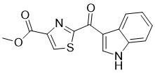
| Chemical Name | methyl 2-(1H-indole-3-carbonyl)-1,3-thiazole-4-carboxylate |
| HS Tariff Code | 2934.99.9001 |
| Storage |
Powder-20°C 3 years 4°C 2 years In solvent -80°C 6 months -20°C 1 month |
| Shipping Condition | Room temperature (This product is stable at ambient temperature for a few days during ordinary shipping and time spent in Customs) |
Biological Activity
| Targets |
- ITE (2-(1'H-indole-3'-carbonyl)-thiazole-4-carboxylic acid methyl ester) targets the aryl hydrocarbon receptor (AHR) [1] - ITE targets the aryl hydrocarbon receptor (AHR) to regulate T-cell-mediated immunity [2] - ITE targets the aryl hydrocarbon receptor (AHR) to inhibit pulmonary artery endothelial cell function [3] |
| ln Vitro |
ITE, an endogenous agonist of AhR, binds to AHR directly at a concentration of 3 nM [1]. There is a T cell proliferation response when ITE (0.03-30 mg/mL) decreases [2]. At 10 and 20 μM, ITE efficiently inhibits the development of human pulmonary artery endothelial cells (HPAEC), whereas at 0.01–5 μM, it has no effect. ITE had no effect on HPAECs' cell process cycle at 10 or 20 μM, and at 20 μM, it did not cause caspase to be induced in HPAEC hubs. Additionally, in HPAECs, ITE (20 μM) decreased AhR protein levels and raised CYP1A1 and CYP1B1 mRNA levels [3]. - For human bronchial epithelial cells (BEAS-2B): ITE (10–100 nM) induced AHR nuclear translocation (immunofluorescence) and activated AHR-mediated transcription. At 100 nM, it increased luciferase activity by ~3.5-fold in AHR-luciferase HEK293 cells [1] - For mouse splenic T cells: ITE (10–100 nM) dose-dependently inhibited ConA-induced proliferation (CFSE staining). At 100 nM, it reduced proliferation rate by ~60% and decreased IFN-γ/IL-17 secretion by ~55%、~65% (ELISA) [2] - For human pulmonary artery endothelial cells (HPAEC): ITE (0.1–5 μM) inhibited proliferation (IC50: ~1 μM, MTT assay). At 2 μM, it reduced migration by ~50% (Transwell) and tube formation by ~45% (Matrigel assay), and downregulated CYP1A1/VCAM-1 (western blot) [3] |
| ln Vivo |
In mice, ITE (200 μg, ip) dramatically prevents the onset of experimental autoimmune uveitis (EAU). In mice, ITE decreased the percentage of cells producing IL-17, IL-10, or IFN-γ. Moreover, ITE reduced the small cell ratio of inflammatory cytokines in mouse LN cells [2]. - For experimental autoimmune uveitis (EAU) mice: C57BL/6 mice were immunized with IRBP to induce EAU. ITE (100 μg/mouse, 5% DMSO + 95% saline) was intraperitoneally injected twice weekly for 3 weeks. It reduced EAU clinical scores by ~70%, decreased retinal T-cell infiltration (immunohistochemistry), and lowered ocular IFN-γ/IL-17 (ELISA) [2] |
| Enzyme Assay |
- AHR luciferase reporter assay: AHR-luciferase HEK293 cells (1×10⁴/well, 96-well plate) were treated with ITE (1–100 nM) for 24 hours. Luciferase substrate was added, and luminescence intensity was measured to calculate activation rate vs. vehicle [1] - AHR nuclear translocation assay: BEAS-2B cells on coverslips were treated with 100 nM ITE for 2 hours, fixed, permeabilized, and incubated with anti-AHR antibody + fluorescent secondary antibody. DAPI stained nuclei, and translocation was observed via fluorescence microscope [1] |
| Cell Assay |
- Mouse splenic T-cell proliferation assay: Purified T cells (CFSE-stained, 5×10⁵/well) were treated with ITE (10, 50, 100 nM) + ConA (5 μg/mL) for 72 hours. CFSE intensity was measured via flow cytometry to calculate proliferation rate [2] - HPAEC proliferation assay: HPAEC (3×10³/well, 96-well plate) were treated with ITE (0.1–5 μM) for 72 hours. MTT reagent was added, DMSO dissolved formazan, and absorbance at 570 nm was measured to calculate viability and IC50 [3] - HPAEC migration/tube formation assay: Migrated cells (Transwell, 2 μM ITE, 6 hours) were stained and counted. Tubes (Matrigel, 2 μM ITE, 6 hours) were observed and quantified [3] |
| Animal Protocol |
- EAU mouse protocol: Female C57BL/6 mice (6–8 weeks) were subcutaneously immunized with 200 μg IRBP. ITE (100 μg/mouse) or vehicle was intraperitoneally injected on day 0,7,14. On day 21, eyes were enucleated for histological analysis and retinal cytokine detection [2] |
| Toxicity/Toxicokinetics |
- In vitro toxicity: ≤100 nM ITE had no cytotoxicity on BEAS-2B cells/mouse T cells (viability >90%) [1,2]; ≤5 μM ITE had no cytotoxicity on MRC-5 cells (viability >85%) [3] - In vivo toxicity: 100 μg/mouse ITE (3 weeks) caused no abnormal weight, liver/kidney function, or organ pathology in EAU mice [2] |
| References |
[1]. A ligand for the aryl hydrocarbon receptor isolated from lung. Proc Natl Acad Sci U S A. 2002 Nov 12;99(23):14694-9. [2]. ITE, a novel endogenous nontoxic aryl hydrocarbon receptor ligand, efficiently suppresses EAU and T-cell-mediated immunity. Invest Ophthalmol Vis Sci. 2013 Nov 13;54(12):7463-9. [3]. ITE inhibits growth of human pulmonary artery endothelial cells. Exp Lung Res. 2017 Oct;43(8):283-292. |
| Additional Infomation |
2-[1H-indol-3-yl(oxo)methyl]-4-thiazolecarboxylic acid methyl ester is an indolyl carboxylic acid. - ITE is an endogenous AHR ligand isolated from human lung tissue, with no toxicity of exogenous AHR ligands (e.g., TCDD) [1] - ITE suppresses autoimmunity (e.g., EAU) by activating AHR to inhibit T-cell function [2] - ITE inhibits HPAEC function via AHR signaling, with potential for pulmonary vascular diseases [3] |
Solubility Data
| Solubility (In Vitro) | DMSO : ≥ 41 mg/mL (~143.20 mM) |
| Solubility (In Vivo) |
Solubility in Formulation 1: ≥ 2.67 mg/mL (9.33 mM) (saturation unknown) in 10% DMSO + 90% Corn Oil (add these co-solvents sequentially from left to right, and one by one), clear solution. For example, if 1 mL of working solution is to be prepared, you can add 100 μL of 26.7 mg/mL clear DMSO stock solution to 900 μL of corn oil and mix evenly. Solubility in Formulation 2: 2.08 mg/mL (7.26 mM) in 10% DMSO + 40% PEG300 + 5% Tween80 + 45% Saline (add these co-solvents sequentially from left to right, and one by one), suspension solution; with ultrasonication. For example, if 1 mL of working solution is to be prepared, you can add 100 μL of 20.8 mg/mL clear DMSO stock solution to 400 μL PEG300 and mix evenly; then add 50 μL Tween-80 to the above solution and mix evenly; then add 450 μL normal saline to adjust the volume to 1 mL. Preparation of saline: Dissolve 0.9 g of sodium chloride in 100 mL ddH₂ O to obtain a clear solution. Solubility in Formulation 3: 10 mg/mL (34.93 mM) in 17% Polyethylene glycol 12-hydroxystearate in Saline (add these co-solvents sequentially from left to right, and one by one), suspension solution; with ultrasonication. Preparation of saline: Dissolve 0.9 g of sodium chloride in 100 mL ddH₂ O to obtain a clear solution. (Please use freshly prepared in vivo formulations for optimal results.) |
| Preparing Stock Solutions | 1 mg | 5 mg | 10 mg | |
| 1 mM | 3.4927 mL | 17.4636 mL | 34.9272 mL | |
| 5 mM | 0.6985 mL | 3.4927 mL | 6.9854 mL | |
| 10 mM | 0.3493 mL | 1.7464 mL | 3.4927 mL |