IPI-3063 is a potent and selective PI3K p110δ inhibitor with IC50 of 2.5 ± 1.2 nM and IC50 values of >1000 nM for other classes of PI3K isoforms (p110α, p110β, p110γ). IPI-3063 inhibits p110α, p110β, and p110γ with respective IC50 values of 1171±533 nM, 1508 nM, and 2187±1529 nM. IPI-3063 significantly increases antibody class switching to IgG1 while significantly decreasing mouse B cell proliferation, survival, and plasmablast differentiation. IPI-3063 has a significant effect at 1 nM and is very effective at lowering p-AKT. Additionally, at 10 nM, IPI-3063 significantly lowers p-ERK1/2. IPI-3063 is extremely potent; at 10 nM, it significantly reduces B cell survival.
Physicochemical Properties
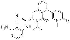
| Molecular Formula | C25H25N7O2 | |
| Molecular Weight | 455.52 | |
| Exact Mass | 455.206 | |
| Elemental Analysis | C, 65.92; H, 5.53; N, 21.52; O, 7.02 | |
| CAS # | 1425043-73-7 | |
| Related CAS # | IPI-3063; IPI 3063; IPI3063.; | |
| PubChem CID | 71276090 | |
| Appearance | White to off-white solid powder | |
| Density | 1.4±0.1 g/cm3 | |
| Boiling Point | 817.8±65.0 °C at 760 mmHg | |
| Flash Point | 448.4±34.3 °C | |
| Vapour Pressure | 0.0±2.9 mmHg at 25°C | |
| Index of Refraction | 1.688 | |
| LogP | 3.1 | |
| Hydrogen Bond Donor Count | 2 | |
| Hydrogen Bond Acceptor Count | 7 | |
| Rotatable Bond Count | 5 | |
| Heavy Atom Count | 34 | |
| Complexity | 962 | |
| Defined Atom Stereocenter Count | 1 | |
| SMILES | C1(=O)C=CC(C2=C3C(=O)N(C(C)C)C([C@@H](NC4=NC=NC(N)=C4C#N)C)=CC3=CC=C2)=CN1C |
|
| InChi Key | OBHAYOJCPNWKBL-HNNXBMFYSA-N | |
| InChi Code | InChI=1S/C25H25N7O2/c1-14(2)32-20(15(3)30-24-19(11-26)23(27)28-13-29-24)10-16-6-5-7-18(22(16)25(32)34)17-8-9-21(33)31(4)12-17/h5-10,12-15H,1-4H3,(H3,27,28,29,30)/t15-/m0/s1 | |
| Chemical Name | 4-amino-6-[[(1S)-1-[8-(1-methyl-6-oxopyridin-3-yl)-1-oxo-2-propan-2-ylisoquinolin-3-yl]ethyl]amino]pyrimidine-5-carbonitrile | |
| Synonyms |
|
|
| HS Tariff Code | 2934.99.9001 | |
| Storage |
Powder-20°C 3 years 4°C 2 years In solvent -80°C 6 months -20°C 1 month |
|
| Shipping Condition | Room temperature (This product is stable at ambient temperature for a few days during ordinary shipping and time spent in Customs) |
Biological Activity
| Targets |
p110δ (IC50 = 2.5 nM); p110α (IC50 = 1170 nM); p110β (IC50 = 1508 nM); p110γ (IC50 = 2187 nM)
The target of IPI-3063 is the phosphoinoside-3-kinase (PI3K) p110δ isoform. In biochemical assays, its IC₅₀ value for p110δ is 2.5 ± 1.2 nM (n = 5), while for other PI3K isoforms, the IC₅₀ values are significantly higher: 1,171 ± 533 nM (n = 6) for p110α, 1,508 ± 624 nM (n = 5) for p110β, and 2,187 ± 1,529 nM (n = 4) for p110γ [1] In cell-based assays, the cellular IC₅₀ of IPI-3063 for p110δ is 0.1 ± 0.01 nM (n = 6), and for other class I PI3K isoforms, the cellular IC₅₀ values are at least 1,000-fold higher: 1,901 ± 1,318 nM (n = 4) for p110α, 102.8 ± 35.7 nM (n = 4) for p110β, and 418.8 ± 117.2 nM (n = 2) for p110γ [1] |
| ln Vitro |
IPI-3063 is a p110δ selective compound with an IC50 = 0.1 nM in p110δ-specific cell-based assays and cellular IC50 values for the other class I PI3K isoforms are at least 1,000-fold higher. IPI-3063 significantly lowers mouse B cell survival, proliferation, and plasmablast differentiation[1]. 1. Effect on mouse B cell signaling: When mouse primary B cells were stimulated with αIgM + IL-4, IPI-3063 potently reduced the phosphorylation of AKT at serine 473 (p-AKT S473) with a significant effect at 1 nM, and also reduced the phosphorylation of ERK1/2 at Thr202/Tyr204 (p-ERK1/2) with a significant effect at 10 nM. Similar results were observed in B cells stimulated with LPS (for p-AKT), but LPS did not induce ERK1/2 phosphorylation [1] 2. Effect on mouse B cell survival: In assays assessing mouse B cell survival, IPI-3063 reduced BAFF-dependent survival in a dose-dependent manner, achieving a significant decrease when present at 10 nM, and the effect approached that of the pan-PI3K inhibitor GDC-0941. A similar trend was observed in cells incubated with IL-4, while the p110γ inhibitor AS-252424 had no significant effect on survival [1] 3. Effect on mouse B cell proliferation: For mouse B cell proliferation, IPI-3063 blocked proliferation in αIgM + IL-4 stimulated B cells at the lowest concentration tested (1 nM). It significantly reduced cell accumulation at all concentrations tested in αIgM + IL-4 stimulated cells, and had a similar dose-dependent inhibitory effect in LPS-stimulated B cells. However, it did not affect B cell proliferation following stimulation with α-CD40 + IL-4. In LPS + IL-4 stimulated B cells, the inhibitor showed a similar trend but with greater variability [1] 4. Effect on mouse B cell differentiation and antibody class switching: IPI-3063 potently decreased plasmablast differentiation in LPS-stimulated mouse B cells starting at 1 nM (measured by the percentage of the CD138⁺ B220ˡᵒ population), and the highest concentrations inhibited plasmablast differentiation to the same degree as GDC-0941. In B cells stimulated with αCD40 + IL-4, it increased the percentage of B220⁺ cells switching to IgG1 starting at 1 nM, approaching the effect of GDC-0941, but had no significant effect on IgG1 switching in LPS + IL-4 activated cells. Additionally, measuring IgM secretion by ELISA showed a similar trend to plasmablast differentiation inhibition, and the p110γ inhibitor AS-252424 had no significant effect in these assays [1] 5. Effect on human B cell proliferation: In human B cell proliferation assays, IPI-3063 blocked proliferation of human B cells stimulated with human CD40L + anti-human IgM/IgG + hIL-2 + hIL-21 at 1 nM, and significantly reduced proliferation starting at 1 nM when measuring the total number of divided cells and the percent divided. The p110γ inhibitor AS-252424 had no effect on human B cell proliferation [1] |
| ln Vivo | IPI-3063 has good pharmacokinetics in mice[1]. |
| Enzyme Assay |
Human recombinant PI3K-α, PI3K-β, PI3K-δ, and PI3K-γ are used. Phosphatidylinositol 4,5 bis phosphate (diC8-PtdIns(4,5)P2) is used. PI3K-α, β, and δ and are heterodimers made up of the p85α regulatory subunit and the full-length p110α, p110β, or p110δ catalytic subunit. The catalytic subunit p110γ has a monomer called PI3K-γ. Samples of kinase (10 nM-α, β, and δ; 20 nM-γ) are incubated with IPI-3063 for 30 min at room temperature in reaction buffer (15 mM HEPES pH 7.4, 20 mM NaCl, 1 mM EGTA, 0.02% Tween 20, 10 mM MgCl2, 0.2 mg/mL bovine-γ-globulins) followed by addition of ATP/diC8-PtdIns(4,5)P2 mixture to give final concentrations of 3 mM ATP and 500 µM diC8-PtdIns(4,5)P2. Reactions are incubated for 2 hours at room temperature, and PI3K activity is measured. Plate readers are used to read plates in luminescence mode. 1. Preparation of reagents and samples: Human recombinant PI3K-α, -β, -δ, and -γ were used. PI3K-α, β, and δ are heterodimers consisting of full-length p110α, p110β, or p110δ catalytic subunit and the p85α regulatory subunit, while PI3K-γ is a monomer of the p110γ catalytic subunit. Phosphatidylinositol 4,5 bis phosphate (diC8-PtdIns(4,5)P2) was used as a substrate [1] 2. Incubation process: Kinase samples (10 nM for α, β, and δ; 20 nM for γ) were incubated with IPI-3063 for 30 minutes at room temperature in reaction buffer (composed of 15 mM HEPES pH 7.4, 20 mM NaCl, 1 mM EGTA, 0.02% Tween 20, 10 mM MgCl₂, 0.2 mg/mL bovine-γ-globulins). After that, an ATP/diC8-PtdIns(4,5)P2 mixture was added to achieve final concentrations of 3 mM ATP and 500 µM diC8-PtdIns(4,5)P2 [1] 3. Reaction and detection: The reactions were incubated at room temperature for 2 hours. PI3K activity was assessed using an ADP-Glo Max assay kit, and plates were read on an Envision plate reader in luminescence mode [1] |
| Cell Assay |
Purified mouse B cells are incubated for 48 hours in either interleukin-4 (IL-4) or B-cell activating factor (BAFF), along with varying concentrations of IPI-3063 and IPI-443. 1. Mouse B cell purification and culture: Mouse splenic B cells were purified by negative selection, with purity >95% verified by FACS analysis using anti-B220 antibody. Purified B cells were seeded at a final concentration of 0.5 or 0.25 × 10⁶ cells/mL in RPMI 1640 medium supplemented with 10% (vol/vol) heat-inactivated FCS, 5 mM Hepes, 2 mM l-glutamine, 100 U/mL penicillin, 100 µg/mL streptomycin, and 50 µM 2-mercaptoethanol [1] 2. Western blot analysis for signaling molecules: Purified mouse B cells were pretreated with IPI-3063 at various concentrations for 30 minutes, then activated with 5 µg/mL αIgM + 10 ng/mL IL-4 or 5 µg/mL LPS for 1 hour before harvest. Cell lysates were prepared, and western blot was performed to detect p-AKT S473 and p-ERK1/2. ImageJ was used to measure mean fluorescence intensities of each band, and phospho-signal was normalized with actin measurements, with fold change calculated using the stimulated/no drug control [1] 3. B cell survival assay: Total splenocytes were pretreated with IPI-3063 at different concentrations, then cultured with 10 ng/mL IL-4 or 60 ng/mL BAFF for 48 hours. The percentage of viable B cells was calculated by measuring the percentage of B220⁺ 7AAD⁻ cells using FACS [1] 4. B cell proliferation assay (CFSE labeling): Mouse splenocytes or purified B cells were pretreated with IPI-3063 for 30 minutes, then stimulated with different stimuli (αIgM + IL-4 for 72 h, LPS for 72 h, αCD40 + IL-4 for 96 h, LPS + IL-4 for 96 h). B cells were labeled with 2.5 µM CFSE before stimulation. FACS was used to analyze CFSE fluorescence, and the total number of divided cells was determined by the number of B220⁺ 7AAD⁻ CFSEˡᵒ cells [1] 5. B cell differentiation and antibody class switching assay: Purified mouse B cells were pretreated with IPI-3063, then stimulated with 5 µg/mL LPS for 72 h (for plasmablast differentiation) or 5 µg/mL anti-CD40 + 5 ng/mL mIL-4 or 5 µg/mL LPS + 5 ng/mL mIL-4 for 96 h (for IgG1 class switching). FACS was used to detect plasmablasts (7AAD⁻ CFSEˡᵒ CD138⁺ B220ˡᵒ population) and IgG1-switched B cells (7AAD⁻ CFSEˡᵒ B220⁺ IgG1⁺ cells). For IgM secretion, supernatants from LPS-stimulated B cells were collected after 3 days, diluted 1:1,000, and detected by ELISA using anti-mouse IgM coating antibody and HRP-conjugated rabbit anti-mouse IgM secondary antibody [1] 6. Human B cell purification and proliferation assay: Peripheral blood mononuclear cells (PBMCs) were purified from human peripheral blood by density gradient centrifugation using Ficoll-Paque. Human B cells were then purified from PBMCs by negative selection, with purity increased from 4% to >70% verified by FACS using anti-CD19 PE conjugated antibody. Purified human B cells were seeded at 0.1 × 10⁶ cells/mL and cultured with 2 µg/mL human CD40L + 5 µg/mL anti-human IgM/IgG + 100 µg/mL hIL-2 + 100 µg/mL hIL-21 in RPMI 1640 medium with the same supplements as mouse B cell culture. B cells were labeled with CFSE, and after 120 hours of stimulation, FACS was used to analyze the total number of divided cells (CD19⁺ 7AAD⁻ CFSEˡᵒ cells) and the percent divided [1] 7. pAKT S473 ELISA assay: SKOV3 and 786.0 cells were seeded into 96-well plates at 2 million per 200 µl culture media per well; Raji and Raw264.7 cells were seeded at the same density in FBS-free media. After overnight incubation at 5% CO₂ and 37°C, cells were treated with IPI-3063 for 30 minutes. Raji cells were stimulated with 10 µg/mL anti-human IgM for 30 minutes, Raw264.7 cells with 25 nM C5a for 3 minutes (both in the presence of the inhibitor), while SKOV3 and 786.0 cells were not stimulated. Medium was aspirated, 50 µL/well of ice-cold lysis buffer was added, and pAKT level was determined using a phospho-Akt1 (S473) sandwich ELISA antibody kit [1] |
| Animal Protocol |
Brown Norway rats 50 mg/kg oral administration |
| ADME/Pharmacokinetics |
It was mentioned that IPI-3063 has good pharmacokinetics in mice, but no specific parameters such as absorption, distribution, metabolism, excretion, half-life, or oral bioavailability were provided [1] |
| References |
[1]. Front Immunol. 2017, 8: 747. [2]. Chem Biol . 2013 Nov 21;20(11):1364-74. |
| Additional Infomation |
1. IPI-3063 is a potent and selective inhibitor of the p110δ isoform of PI3K. It is a useful tool for studying p110δ function in immune cells, especially in B cells, as it can effectively modulate B cell responses (survival, proliferation, differentiation, antibody class switching) at low nanomolar concentrations [1] 2. The class I PI3Ks are important enzymes that relay signals from cell surface receptors to downstream mediators driving cellular functions. Elevated PI3K signaling is found in B cell malignancies and lymphocytes of patients with autoimmune disease. The p110δ catalytic isoform is critical for B lymphocyte development, survival, activation, and differentiation, making it a rational target. IPI-3063 was developed to target this isoform, and its potent and selective inhibition of p110δ provides a means to study the role of p110δ in B cell biology and potential therapeutic applications in B cell-driven diseases [1] 3. Currently, idelalisib is the only selective p110δ inhibitor FDA-approved to treat certain B cell malignancies. IPI-3063, as a novel selective p110δ inhibitor, has similar effects on B cell functions as the pan-PI3K inhibitor GDC-0941 in vitro, indicating its potential as a research tool and a potential lead compound for B cell-driven diseases (such as B cell malignancies and B cell-mediated autoimmune diseases) [1] |
Solubility Data
| Solubility (In Vitro) |
DMSO: ~91 mg/mL (~117.1 mM) Ethanol: ~12 mg/mL (~26.3 mM) |
|
| Solubility (In Vivo) |
|
| Preparing Stock Solutions | 1 mg | 5 mg | 10 mg | |
| 1 mM | 2.1953 mL | 10.9765 mL | 21.9529 mL | |
| 5 mM | 0.4391 mL | 2.1953 mL | 4.3906 mL | |
| 10 mM | 0.2195 mL | 1.0976 mL | 2.1953 mL |