Physicochemical Properties
| Molecular Formula | C10H12O2 |
| Molecular Weight | 164.2011 |
| Exact Mass | 164.083 |
| CAS # | 499-44-5 |
| PubChem CID | 3611 |
| Appearance | Off-white to yellow solid powder |
| Density | 1.1±0.1 g/cm3 |
| Boiling Point | 303.4±35.0 °C at 760 mmHg |
| Melting Point | 50-52 °C(lit.) |
| Flash Point | 128.1±18.5 °C |
| Vapour Pressure | 0.0±1.4 mmHg at 25°C |
| Index of Refraction | 1.554 |
| LogP | 1.89 |
| Hydrogen Bond Donor Count | 1 |
| Hydrogen Bond Acceptor Count | 2 |
| Rotatable Bond Count | 1 |
| Heavy Atom Count | 12 |
| Complexity | 280 |
| Defined Atom Stereocenter Count | 0 |
| InChi Key | FUWUEFKEXZQKKA-UHFFFAOYSA-N |
| InChi Code | InChI=1S/C10H12O2/c1-7(2)8-4-3-5-9(11)10(12)6-8/h3-7H,1-2H3,(H,11,12) |
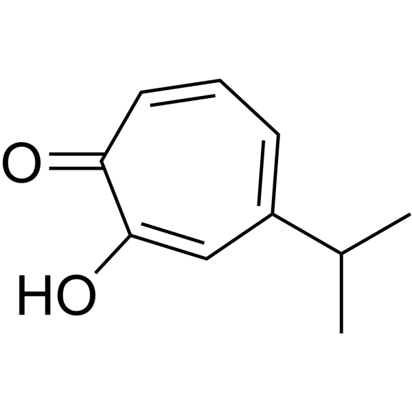
| Chemical Name | 2-hydroxy-6-propan-2-ylcyclohepta-2,4,6-trien-1-one |
| HS Tariff Code | 2934.99.9001 |
| Storage |
Powder-20°C 3 years 4°C 2 years In solvent -80°C 6 months -20°C 1 month |
| Shipping Condition | Room temperature (This product is stable at ambient temperature for a few days during ordinary shipping and time spent in Customs) |
Biological Activity
| Targets |
- Hinokitiol targets the nuclear factor erythroid 2-related factor 2 (Nrf2) pathway in glioma stem cells.[1] - Hinokitiol targets DNA methyltransferase 1 (DNMT1) and ubiquitin-like with PHD and ring finger domains 1 (UHRF1) in colon cancer cells, with an IC50 value of 12.5 μM for DNMT1 inhibition [2] |
| ln Vitro |
Hinokitol demonstrated a dose-dependent reduction in viability in the glioma cell lines T98G and U87MG, with IC50 values of 152.5 ± 25.3 µM and 316.5 ± 35.5 µM, respectively. In vitro carcinogenesis is inhibited by junperol, which also reduces ALDH activity and glioma stem cells' ability to self-renew. Additionally, in glioma stem cells, hikitol dose-dependently lowers Nrf2 expression [1]. Colon cancer cell proliferation is inhibited by hortikitiol (0-100 μM) in a time- and dose-dependent manner. Hinokitiol (5, 10 μM) decreases the expression of UHRF1 and DNMT1 mRNA and protein in HCT-116 cells while raising 5hmC levels to promote TET1 expression. Hinokitiol can also restore the mRNA expression of the MGMT, CHST10, and BTG4 genes and lower their methylation status [2]. - In glioma stem cells (GSCs) isolated from U87MG and U251MG glioma cell lines, treatment with Hinokitiol (1–10 μM) for 72 h dose-dependently reduced sphere formation capacity. At 10 μM, the number of spheres was decreased by ~70% (U87MG GSCs) and ~65% (U251MG GSCs) compared to the control group. Additionally, Hinokitiol (5–10 μM) inhibited GSC invasion (detected via Transwell assay) by ~55%–60% and downregulated the expression of stemness markers (CD133, Nestin) and Nrf2 target genes (HO-1, NQO1) at the protein level (Western blot) [1] - In human colon cancer cell lines (HCT116 and SW480), Hinokitiol (5–20 μM) for 48 h dose-dependently inhibited cell proliferation (MTT assay), with IC50 values of 8.2 μM (HCT116) and 9.5 μM (SW480). It also reduced global DNA methylation levels (detected via 5-methylcytosine ELISA) by ~40%–45% at 15 μM, and downregulated DNMT1 and UHRF1 protein expression (Western blot) and mRNA levels (RT-PCR) in a dose-dependent manner [2] |
| ln Vivo |
- Female BALB/c nude mice (4–6 weeks old) were subcutaneously inoculated with U87MG glioma stem cells (1×10⁶ cells/mouse). When tumors reached ~100 mm³, mice were randomly divided into control and Hinokitiol treatment groups. Hinokitiol was administered intraperitoneally at 20 mg/kg every 2 days for 3 weeks. At the end of treatment, tumor volume in the Hinokitiol group was ~55% smaller than that in the control group, and tumor weight was reduced by ~50%. Immunohistochemical staining of tumor tissues showed decreased expression of Nrf2, CD133, and Ki-67 (proliferation marker) in the Hinokitiol group [1] |
| Enzyme Assay |
- For DNMT activity assay: Recombinant human DNMT1 was incubated with a methyl donor (S-adenosylmethionine, SAM) and a biotinylated DNA substrate in a reaction buffer. Hinokitiol (0.1–50 μM) was added to the reaction system, and the mixture was incubated at 37°C for 1 h. The methylated DNA product was captured by streptavidin-coated plates, and the amount of methylated cytosine was detected using a specific antibody and a horseradish peroxidase (HRP)-conjugated secondary antibody. The absorbance at 450 nm was measured, and the IC50 value of Hinokitiol for DNMT1 inhibition was calculated based on the dose-response curve [2] |
| Cell Assay |
- For GSC sphere formation assay (Reference [1]): Glioma stem cells were seeded in ultra-low attachment 6-well plates at a density of 1×10³ cells/well in serum-free medium supplemented with growth factors. Hinokitiol (1–10 μM) was added, and the cells were cultured for 7 days. The number of spheres with diameter >50 μm was counted under a light microscope, and the sphere formation efficiency was calculated as (number of spheres / number of seeded cells) × 100% [1] - For GSC invasion assay (Reference [1]): Transwell chambers with 8-μm pores were coated with Matrigel. Glioma stem cells (5×10⁴ cells/well) treated with Hinokitiol (5–10 μM) for 24 h were seeded in the upper chamber with serum-free medium, and the lower chamber was filled with medium containing 10% fetal bovine serum. After 24 h of incubation, cells on the upper surface of the membrane were removed, and cells that invaded to the lower surface were fixed with 4% paraformaldehyde, stained with crystal violet, and counted under a microscope [1] - For colon cancer cell proliferation assay (Reference [2]): HCT116 and SW480 cells were seeded in 96-well plates at a density of 5×10³ cells/well. After 24 h of attachment, Hinokitiol (5–20 μM) was added, and the cells were cultured for 48 h. MTT reagent was added to each well, and the mixture was incubated for 4 h. The formazan crystals were dissolved with dimethyl sulfoxide, and the absorbance at 570 nm was measured using a microplate reader. Cell viability was calculated as (absorbance of treatment group / absorbance of control group) × 100% [2] - For DNA methylation detection (Reference [2]): HCT116 cells treated with Hinokitiol (15 μM) for 48 h were harvested, and genomic DNA was extracted. The DNA was denatured, and 5-methylcytosine levels were detected using an ELISA kit. The absorbance at 450 nm was measured, and the relative methylation level was calculated based on a standard curve [2] |
| Animal Protocol |
- For glioma xenograft experiment (Reference [1]): Female BALB/c nude mice (4–6 weeks old) were maintained under specific pathogen-free conditions. U87MG glioma stem cells (1×10⁶ cells in 100 μL of phosphate-buffered saline mixed with Matrigel at a 1:1 ratio) were subcutaneously injected into the right flank of each mouse. When tumors grew to ~100 mm³, mice were divided into two groups (n=6 per group): control group (intraperitoneal injection of vehicle: 10% dimethyl sulfoxide + 90% physiological saline) and Hinokitiol treatment group (intraperitoneal injection of Hinokitiol at 20 mg/kg, dissolved in the same vehicle). Injections were given every 2 days for 3 weeks. Tumor volume was measured every 3 days using a caliper, calculated as (length × width²) / 2. At the end of the experiment, mice were euthanized, tumors were excised and weighed, and tumor tissues were fixed in 4% paraformaldehyde for immunohistochemical analysis [1] |
| References |
[1]. Hinokitiol suppresses cancer stemness and oncogenicity in glioma stem cells by Nrf2 regulation. Cancer Chemother Pharmacol. 2017 Aug;80(2):411-419. [2]. Hinokitiol induces DNA demethylation via DNMT1 and UHRF1 inhibition in colon cancer cells. BMC Cell Biol. 2017 Feb 27;18(1):14. |
| Additional Infomation |
Beta-thujaplicin is a monoterpenoid that is cyclohepta-2,4,6-trien-1-one substituted by a hydroxy group at position 2 and an isopropyl group at position 4. Isolated from Thuja plicata and Chamaecyparis obtusa, it exhibits antimicrobial activities. It has a role as an antifungal agent, an antibacterial agent, an antiplasmodial drug, an antineoplastic agent and a plant metabolite. It is an enol, a cyclic ketone and a monoterpenoid. It derives from a hydride of a cyclohepta-1,3,5-triene. Hinokitiol has been reported in Thujopsis dolabrata, Chamaecyparis formosensis, and other organisms with data available. - Hinokitiol (β-thujaplicin) is a natural monoterpenoid phenol isolated from the heartwood of Cupressaceae family plants, with known antioxidant and antimicrobial activities [1, 2] - In glioma stem cells, Hinokitiol suppresses cancer stemness and oncogenicity by downregulating the Nrf2 pathway, which is hyperactivated in GSCs and promotes stemness maintenance and tumorigenesis [1] - In colon cancer cells, Hinokitiol induces DNA demethylation by inhibiting DNMT1 (a key DNA methyltransferase) and UHRF1 (a regulator of DNMT1 recruitment), which may reactivate tumor suppressor genes silenced by hypermethylation [2] |
Solubility Data
| Solubility (In Vitro) |
DMSO : ~100 mg/mL (~609.01 mM) H2O : ~0.67 mg/mL (~4.08 mM) |
| Solubility (In Vivo) |
Solubility in Formulation 1: ≥ 2.5 mg/mL (15.23 mM) (saturation unknown) in 10% DMSO + 40% PEG300 + 5% Tween80 + 45% Saline (add these co-solvents sequentially from left to right, and one by one), clear solution. For example, if 1 mL of working solution is to be prepared, you can add 100 μL of 25.0 mg/mL clear DMSO stock solution to 400 μL PEG300 and mix evenly; then add 50 μL Tween-80 to the above solution and mix evenly; then add 450 μL normal saline to adjust the volume to 1 mL. Preparation of saline: Dissolve 0.9 g of sodium chloride in 100 mL ddH₂ O to obtain a clear solution. Solubility in Formulation 2: ≥ 2.5 mg/mL (15.23 mM) (saturation unknown) in 10% DMSO + 90% (20% SBE-β-CD in Saline) (add these co-solvents sequentially from left to right, and one by one), clear solution. For example, if 1 mL of working solution is to be prepared, you can add 100 μL of 25.0 mg/mL clear DMSO stock solution to 900 μL of 20% SBE-β-CD physiological saline solution and mix evenly. Preparation of 20% SBE-β-CD in Saline (4°C,1 week): Dissolve 2 g SBE-β-CD in 10 mL saline to obtain a clear solution. Solubility in Formulation 3: ≥ 2.5 mg/mL (15.23 mM) (saturation unknown) in 10% DMSO + 90% Corn Oil (add these co-solvents sequentially from left to right, and one by one), clear solution. For example, if 1 mL of working solution is to be prepared, you can add 100 μL of 25.0 mg/mL clear DMSO stock solution to 900 μL of corn oil and mix evenly. (Please use freshly prepared in vivo formulations for optimal results.) |
| Preparing Stock Solutions | 1 mg | 5 mg | 10 mg | |
| 1 mM | 6.0901 mL | 30.4507 mL | 60.9013 mL | |
| 5 mM | 1.2180 mL | 6.0901 mL | 12.1803 mL | |
| 10 mM | 0.6090 mL | 3.0451 mL | 6.0901 mL |