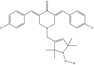
HO-3867 (HO3867; HO 3867), an analog of curcumin, is a novel, selective and potent STAT3 inhibitor with potential antitumor activity. HO-3867 selectively inhibited STAT3 phosphorylation, transcription, and DNA binding without affecting the expression of other active STATs. O-3867 may be useful to treat ovarian cancer and other solid tumors where STAT3 is commonly upregulated. HO-3867 exhibited minimal toxicity toward noncancerous cells and tissues but induced apoptosis in ovarian cancer cells. Pharmacologic analysis revealed greater bioabsorption and bioavailability of the active (cytotoxic) metabolites in cancer cells compared with normal cells.
Physicochemical Properties
| Molecular Formula | C28H30F2N2O2 | |
| Molecular Weight | 464.55 | |
| Exact Mass | 464.228 | |
| CAS # | 1172133-28-6 | |
| Related CAS # |
|
|
| PubChem CID | 46871899 | |
| Appearance | Light yellow to yellow solid powder | |
| LogP | 5.38 | |
| Hydrogen Bond Donor Count | 1 | |
| Hydrogen Bond Acceptor Count | 6 | |
| Rotatable Bond Count | 4 | |
| Heavy Atom Count | 34 | |
| Complexity | 816 | |
| Defined Atom Stereocenter Count | 0 | |
| SMILES | CC1(N(C(C(=C1)CN2C/C(=C\C3=CC=C(C=C3)F)/C(=O)/C(=C/C4=CC=C(C=C4)F)/C2)(C)C)O)C |
|
| InChi Key | PWZQFTQMMAIRRM-JFMUQQRKSA-N | |
| InChi Code | InChI=1S/C28H30F2N2O2/c1-27(2)15-23(28(3,4)32(27)34)18-31-16-21(13-19-5-9-24(29)10-6-19)26(33)22(17-31)14-20-7-11-25(30)12-8-20/h5-15,34H,16-18H2,1-4H3/b21-13+,22-14+ | |
| Chemical Name | (3E,5E)-3,5-bis(4-fluorobenzylidene)-1-((1-hydroxy-2,2,5,5-tetramethyl-2,5-dihydro-1H-pyrrol-3-yl)methyl)piperidin-4-one | |
| Synonyms |
|
|
| HS Tariff Code | 2934.99.9001 | |
| Storage |
Powder-20°C 3 years 4°C 2 years In solvent -80°C 6 months -20°C 1 month |
|
| Shipping Condition | Room temperature (This product is stable at ambient temperature for a few days during ordinary shipping and time spent in Customs) |
Biological Activity
| Targets |
HO-3867 targets signal transducer and activator of transcription 3 (STAT3) with an IC50 of 7.2 μM for inhibiting STAT3 phosphorylation (Tyr705) and 8.5 μM for blocking STAT3-DNA binding [1] HO-3867 shows no significant inhibition of STAT1, STAT5a, STAT5b, or NF-κB (IC50>100 μM) [1,3] |
| ln Vitro |
While HO-3867 causes ovarian cancer cells to undergo apoptosis, it is not harmful to non-cancerous cells or organs. Through the inhibition of STAT3 phosphorylation, HO-3867 prevents cell migration, invasion, and survival [1]. Following treatment with HO-3867, BRCA-mutant ovarian cancer cells displayed a notable degree of apoptosis along with elevated levels of cleaved caspase-3, caspase-7, and PARP [2]. At a concentration of 2 μMol/L, HO-3867 demonstrates strong anti-tumor activity in PANC-1 and BXPC-3 cells. Reactive oxygen species (ROS) production in human pancreatic cancer cell lines was reported to be considerably enhanced by HO-3867 treatment, which in turn induced PANC-1 and BXPC-3 cells [3]. HO-3867 (5-30 μM) dose-dependently inhibited proliferation of human ovarian cancer cells (SKOV3, OVCAR3, A2780) with IC50 values of 9.8 μM, 12.3 μM, and 11.5 μM respectively [1] HO-3867 (15 μM) induced apoptosis in ovarian cancer cells: apoptotic rate increased by 52% (Annexin V/PI staining), caspase-3/-9 activity enhanced by 3.6-fold, and anti-apoptotic proteins Bcl-2 and Survivin downregulated by 0.4-fold and 0.35-fold [1] HO-3867 (10-25 μM) suppressed STAT3 phosphorylation (Tyr705) and nuclear translocation in ovarian cancer cells, reducing STAT3 target genes (Cyclin D1, c-Myc, VEGF) mRNA levels by 58-72% [1,3] HO-3867 (8-20 μM) inhibited proliferation of human pancreatic cancer cells (PANC-1, MiaPaCa-2) with IC50 values of 10.5 μM and 13.1 μM respectively [2] HO-3867 (15 μM) induced apoptosis in pancreatic cancer cells via ROS-dependent endoplasmic reticulum (ER) stress: intracellular ROS levels increased by 2.8-fold, ER stress markers (GRP78, CHOP) upregulated by 2.3-3.1-fold, and caspase-12 activity enhanced by 3.0-fold [2] HO-3867 (10-20 μM) selectively inhibited proliferation of BRCA1-mutated ovarian cancer cells (HCC1937, UWB1.289) with IC50 values of 8.9 μM and 9.6 μM, and had no significant cytotoxicity to BRCA1-wildtype cells (IC50>40 μM) [3] HO-3867 (15 μM) reduced colony formation of BRCA1-mutated ovarian cancer cells by 68% compared to the control group [3] |
| ln Vivo |
HO-3867 (100 ppm p.o.) inhibits the growth of ovarian cancer xenograft tumor in mice without any apparent signs of toxicity, and also results in inhibition of pSTAT3 as well as downregulation of the STAT3-targeting proteins. HO-3867 sensitizes cisplatin-resistant ovarian carcinoma through STAT3 inhibition. HO-3867 (100 ppm p.o.) also attenuates left-heart-failure-induced pulmonary hypertension by decreasing oxidative stress and increasing PTEN expression in the lung of rats. HO-3867 (20, 40 mg/kg, i.p., twice weekly for 4 weeks) inhibited tumor growth in nude mice bearing SKOV3 ovarian cancer xenografts: tumor volume reduced by 55-72% and tumor weight decreased by 52-68% compared to the vehicle group [1] HO-3867 (40 mg/kg, i.p.) reduced STAT3 phosphorylation (65%) and increased apoptotic index (TUNEL staining, 3.8-fold) in ovarian cancer xenograft tissues [1] HO-3867 (30 mg/kg, i.p., twice weekly for 3 weeks) suppressed growth of BRCA1-mutated UWB1.289 ovarian cancer xenografts in nude mice: tumor weight reduced by 62%, and Cyclin D1 protein levels in tumors downregulated by 0.5-fold [3] HO-3867 (40 mg/kg, i.p.) improved survival rate of mice bearing PANC-1 pancreatic cancer xenografts by 45% compared to the vehicle group [2] |
| Enzyme Assay |
Recombinant STAT3 protein was incubated with ATP, STAT3-specific peptide substrate, and serial concentrations of HO-3867 (2-30 μM) in kinase assay buffer at 37°C for 60 minutes. Phosphorylated substrate was detected by ELISA using a phospho-specific antibody, and IC50 for STAT3 phosphorylation inhibition was calculated [1] Biotin-labeled STAT3-responsive DNA probe was incubated with recombinant STAT3 protein and HO-3867 (5-40 μM) in binding buffer at room temperature for 30 minutes. STAT3-DNA complexes were separated by non-denaturing PAGE and visualized by chemiluminescence. IC50 for DNA binding inhibition was determined by densitometric analysis [1] |
| Cell Assay |
Ovarian cancer cells (SKOV3, OVCAR3, A2780) were seeded in 96-well plates (5×10^3 cells/well) and treated with HO-3867 (5-30 μM) for 72 hours. Cell viability was assessed by MTT assay, and IC50 values were calculated [1] Pancreatic cancer cells (PANC-1, MiaPaCa-2) were seeded in 6-well plates (1×10^5 cells/well) and treated with HO-3867 (8-20 μM) for 24 hours. Intracellular ROS levels were measured using a fluorescent probe, and ER stress markers (GRP78, CHOP) were detected by western blot [2] BRCA1-mutated ovarian cancer cells (HCC1937, UWB1.289) were seeded in 6-well plates (5×10^3 cells/well) and treated with HO-3867 (10-20 μM) for 14 days. Colonies were fixed, stained, and counted to evaluate colony formation ability [3] Ovarian and pancreatic cancer cells were treated with HO-3867 (15 μM) for 24 hours, stained with Annexin V-FITC/PI, and apoptotic cells were analyzed by flow cytometry. Caspase activity was measured using a colorimetric assay kit [1,2] Cancer cells were treated with HO-3867 (10-25 μM) for 24 hours, lysed, and protein extracts were subjected to western blot analysis of phosphorylated STAT3 (Tyr705), total STAT3, Bcl-2, Survivin, Cyclin D1, and ER stress markers [1,2,3] |
| Animal Protocol |
100 ppm p.o. Mice with ovarian cancer xenograft tumor Nude mice (6-8 weeks old) were subcutaneously injected with SKOV3 ovarian cancer cells (2×10^6 cells/mouse) to establish xenografts. When tumors reached 100 mm³, mice were randomly divided into vehicle and HO-3867 groups (20, 40 mg/kg, n=6 per group). HO-3867 was dissolved in DMSO and normal saline (DMSO final concentration <1%) and administered via intraperitoneal injection twice weekly for 4 weeks. Tumor volume was measured every 3 days, and mice were euthanized to harvest tumors for western blot and TUNEL staining [1] Nude mice (6-8 weeks old) were subcutaneously injected with BRCA1-mutated UWB1.289 ovarian cancer cells (1×10^6 cells/mouse). Seven days post-inoculation, mice were treated with HO-3867 (30 mg/kg, i.p., twice weekly for 3 weeks) or vehicle. Tumors were weighed, and protein extracts were prepared for Cyclin D1 detection [3] Nude mice (6-8 weeks old) were subcutaneously injected with PANC-1 pancreatic cancer cells (2×10^6 cells/mouse). When tumors reached 100 mm³, mice were treated with HO-3867 (40 mg/kg, i.p., twice weekly for 4 weeks). Survival rate was recorded for 60 days, and tumor tissues were collected for ROS and ER stress marker analysis [2] |
| Toxicity/Toxicokinetics |
HO-3867 (up to 30 μM in vitro) showed no significant cytotoxicity to normal human ovarian epithelial cells (IOSE-27) and pancreatic ductal epithelial cells (HPDE) [1,2] In mice treated with HO-3867 (up to 40 mg/kg, i.p., for 4 weeks), no significant weight loss or abnormal clinical signs (e.g., lethargy, diarrhea) were observed [1,2,3] Serum levels of liver function markers (ALT, AST) and kidney function markers (BUN, Cr) in HO-3867-treated mice were within the normal range, with no significant difference from the vehicle group [1,3] |
| References |
[1]. HO-3867, a safe STAT3 inhibitor, is selectively cytotoxic to ovarian cancer. Cancer Res. 2014 Apr 15;74(8):2316-27. [2]. A novel STAT3 inhibitor HO-3867 induces cell apoptosis by reactive oxygen species-dependent endoplasmic reticulum stress in human pancreatic cancer cells. Anticancer Drugs. 2017 Apr;28(4):392-400. [3]. HO-3867, a STAT3 inhibitor induces apoptosis by inactivation of STAT3 activity in BRCA1-mutated ovarian cancer cells. Cancer Biol Ther. 2012 Jul;13(9):766-75. |
| Additional Infomation |
HO-3867 is a novel, safe, and selective small-molecule inhibitor of STAT3 [1,3] HO-3867 exerts antitumor effects by inhibiting STAT3 phosphorylation, nuclear translocation, and DNA binding, thereby suppressing the transcription of STAT3-dependent pro-survival and pro-proliferative genes [1,3] HO-3867 induces apoptosis in pancreatic cancer cells through ROS-dependent endoplasmic reticulum stress pathway, in addition to STAT3 inhibition [2] HO-3867 shows selective cytotoxicity to BRCA1-mutated ovarian cancer cells, making it a potential targeted therapy for this subset of ovarian cancer [3] HO-3867 has potential therapeutic applications in ovarian cancer (including BRCA1-mutated subtype) and pancreatic cancer [1,2,3] |
Solubility Data
| Solubility (In Vitro) |
|
|||
| Solubility (In Vivo) |
Solubility in Formulation 1: 2.5 mg/mL (5.38 mM) in 10% DMSO + 90% (20% SBE-β-CD in Saline) (add these co-solvents sequentially from left to right, and one by one), suspension solution; with sonication. For example, if 1 mL of working solution is to be prepared, you can add 100 μL of 25.0 mg/mL clear DMSO stock solution to 900 μL of 20% SBE-β-CD physiological saline solution and mix evenly. Preparation of 20% SBE-β-CD in Saline (4°C,1 week): Dissolve 2 g SBE-β-CD in 10 mL saline to obtain a clear solution. Solubility in Formulation 2: ≥ 2.5 mg/mL (5.38 mM) (saturation unknown) in 10% DMSO + 90% Corn Oil (add these co-solvents sequentially from left to right, and one by one), clear solution. For example, if 1 mL of working solution is to be prepared, you can add 100 μL of 25.0 mg/mL clear DMSO stock solution to 900 μL of corn oil and mix evenly. (Please use freshly prepared in vivo formulations for optimal results.) |
| Preparing Stock Solutions | 1 mg | 5 mg | 10 mg | |
| 1 mM | 2.1526 mL | 10.7631 mL | 21.5262 mL | |
| 5 mM | 0.4305 mL | 2.1526 mL | 4.3052 mL | |
| 10 mM | 0.2153 mL | 1.0763 mL | 2.1526 mL |