HLY78 is a novel and potent small molecule activator of the Wnt/β-catenin signaling pathway which targets the DIX domain of Axin and potentiates the Axin-LRP6 association to promote Wnt signaling transduction.
Physicochemical Properties
| Molecular Formula | C17H17NO2 |
| Molecular Weight | 267.322 |
| Exact Mass | 267.126 |
| Elemental Analysis | C, 76.38; H, 6.41; N, 5.24; O, 11.97 |
| CAS # | 854847-61-3 |
| PubChem CID | 44422459 |
| Appearance | Off-white to light yellow solid powder |
| LogP | 3.659 |
| Hydrogen Bond Donor Count | 0 |
| Hydrogen Bond Acceptor Count | 3 |
| Rotatable Bond Count | 1 |
| Heavy Atom Count | 20 |
| Complexity | 361 |
| Defined Atom Stereocenter Count | 0 |
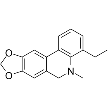
| SMILES | O1COC2C1=CC1=C(C=2)CN(C)C2C1=CC=CC=2CC |
| InChi Key | FAZZYPIBZBGQSH-UHFFFAOYSA-N |
| InChi Code | InChI=1S/C17H17NO2/c1-3-11-5-4-6-13-14-8-16-15(19-10-20-16)7-12(14)9-18(2)17(11)13/h4-8H,3,9-10H2,1-2H3 |
| Chemical Name | 4-ethyl-5-methyl-6H-[1,3]dioxolo[4,5-j]phenanthridine |
| Synonyms | HLY78; HL Y78; HLY-78 |
| HS Tariff Code | 2934.99.9001 |
| Storage |
Powder-20°C 3 years 4°C 2 years In solvent -80°C 6 months -20°C 1 month |
| Shipping Condition | Room temperature (This product is stable at ambient temperature for a few days during ordinary shipping and time spent in Customs) |
Biological Activity
| Targets |
HLY78 acts as an agonist of the Wnt/β-catenin signaling pathway by targeting the DIX domain of Axin, thereby promoting the association between Axin and low-density lipoprotein receptor-related protein 5/6 (LRP5/6), which leads to phosphorylation of LRP6 and activation of downstream signaling. [2] |
| ln Vitro |
By triggering the Wnt/β-catenin pathway, HLY78 prevents tumor and embryonic cell death brought on by carbon ion radiation [2]. In HGC-27 and AGS cells, HLY78 (20 μM, 0-48 h) significantly enhanced the ability to form colonies by 2.78 and 2.88 times, respectively, in comparison to the control group [3]. HGC-27 and AGS cells' ability to migrate can be enhanced by HLY78 (20 μM, 0-48 hours) [3]. Dihydroartemisinin can enhance TNKS expression, which is markedly increased by HLY78 [3]. In human gastric cancer cell lines (HGC-27 and AGS), HLY78 treatment decreased cell viability in a concentration-dependent manner as assessed by the CCK-8 assay. The viability of both cell lines decreased with increasing concentrations of HLY78 (0.5 to 100 µM), and 20 µM was selected as the optimal concentration for subsequent experiments. [3] HLY78 (20 µM) significantly increased the colony formation ability of HGC-27 and AGS cells compared to the control group, as shown in the colony formation assay. [3] HLY78 (20 µM) promoted the migration of HGC-27 and AGS cells, as evidenced by reduced wound distance in the wound healing assay and an increased number of migratory cells in the Transwell assay. [3] Western blot analysis showed that treatment with HLY78 (20 µM) in gastric cancer cells led to increased protein expression levels of TNKS, AXIN2, β-catenin, N-cadherin, Vimentin, TWIST, and MMP2, while it decreased the expression of E-cadherin. These effects indicate that HLY78 activates the Wnt/β-catenin pathway and promotes the epithelial-to-mesenchymal transition (EMT) process. [3] Immunofluorescence analysis confirmed that HLY78 treatment increased the expression levels of TNKS, AXIN2, and β-catenin in the nuclei and cytoplasm of HGC-27 and AGS cells. [3] |
| ln Vivo |
Through the LRP6/GSK3β/β-catenin signaling pathway, HLY78 (0-1.8 mg/kg, intranasal injection, once) improves neurological deficits following subarachnoid hemorrhage in rats. In a rat model of subarachnoid hemorrhage (SAH), intranasal administration of HLY78 (0.6 mg/kg and 1.8 mg/kg) significantly improved short-term neurological function (modified Garcia score and beam-balance score) at 24 hours post-SAH compared to the vehicle-treated SAH group. [2] HLY78 (0.6 mg/kg) treatment attenuated neuronal apoptosis in the ipsilateral cortex at 24 hours post-SAH, as demonstrated by decreased TUNEL-positive cells and Western blot analysis showing increased Bcl-2 levels and decreased Bax and cleaved caspase-3 levels. [2] Long-term administration of HLY78 (0.6 mg/kg) improved neurobehavioral outcomes up to 21 days post-SAH, as shown by enhanced performance in the rotarod test and Morris water maze test (reduced escape latency and swimming distance, increased time spent in the target quadrant). [2] The neuroprotective and anti-apoptotic effects of HLY78 were mediated through the LRP6/GSK3β/β-catenin pathway, as evidenced by increased levels of p-LRP6, p-GSK3β (Ser9), and β-catenin, and decreased levels of p-β-catenin, Bax, and cleaved caspase-3 in the ipsilateral hemisphere. These effects were reversed by LRP6 siRNA knockdown. [2] |
| Cell Assay |
For the Cell Counting Kit-8 (CCK-8) assay, HGC-27 and AGS cells were seeded into 96-well plates at a density of 5x10³ cells per well. After cell adherence, the culture medium was replaced with fresh medium containing different concentrations of HLY78 (0.5, 10, 20, 30, 40, 50, 100 µM). Following 48 hours of incubation, CCK-8 reagent was added to each well and incubated for 1 hour. Absorbance was then measured at 450 nm using a microplate reader to determine cell viability. [3] For the colony formation assay, HGC-27 and AGS cells were seeded into 6-well plates at a density of 5x10³ cells per well and treated with HLY78 (20 µM) for 24 hours. The medium was changed every 3-4 days. After one week, cells were fixed with methanol and stained with 0.1% crystal violet. Colonies were observed and counted under a light microscope. [3] For the wound healing assay, HGC-27 and AGS cells were seeded into 6-well plates and grown to 95% confluence. A sterile pipette tip was used to create a scratch. After washing, cells were treated with HLY78 (20 µM) in serum-free medium. Images of the scratch were taken at 0 and 48 hours, and the migration distance was measured. [3] For the Transwell migration assay, HGC-27 and AGS cells in serum-free medium were plated into the upper chambers of Transwell inserts (5x10³ cells/well). The lower chambers were filled with medium containing 10% fetal bovine serum as a chemoattractant. After 12 hours of incubation, cells that migrated to the lower surface were fixed, stained, and counted under a microscope. [3] For western blot analysis, HGC-27 and AGS cells treated with HLY78 (20 µM) were lysed. Proteins were separated by SDS-PAGE, transferred to PVDF membranes, and probed with specific primary antibodies overnight at 4°C, followed by incubation with HRP-conjugated secondary antibodies. Protein bands were visualized using an ECL reagent. [3] For immunofluorescence analysis, HGC-27 and AGS cells grown on coverslips were treated with HLY78 (20 µM), fixed, permeabilized, and blocked. Cells were then incubated with primary antibodies against TNKS, AXIN2, or β-catenin overnight at 4°C, followed by incubation with fluorophore-conjugated secondary antibodies. Nuclei were stained with DAPI, and images were captured using a fluorescence microscope. [3] |
| Animal Protocol |
Animal/Disease Models: Adult male SD (SD (Sprague-Dawley)) rats (280-310 g, n=9/group, SAH model) [2] Doses: 0, 0.2, 0.6 and 1.8 mg/kg Route of Administration: intranasal injection, 1 time , results after 1 hour SAH (subarachnoid hemorrhage): 0.6 mg/kg Dramatically diminished short-term and long-term neurobehavioral deficits and neuronal apoptosis after SAH. Successful delivery to the brain via intranasal administration at 0.6 mg/kg was sufficient to Dramatically increase LRP6 phosphorylation. Reversal of changes in Bcl-2, Bax and cleaved caspase 3 levels. The SAH model was induced in adult male Sprague-Dawley rats (280–310 g) by endovascular perforation of the left internal carotid artery to puncture the bifurcation of the anterior and middle cerebral arteries. Sham-operated rats underwent the same procedure without perforation. [2] HLY78 was administered intranasally at doses of 0.2, 0.6, and 1.8 mg/kg for dose-response evaluation in short-term neurobehavioral tests. Based on the results, 0.6 mg/kg was selected for subsequent long-term and mechanistic studies. [2] For short-term assessment, neurological function was evaluated at 24 hours post-SAH using the modified Garcia score and beam-balance test. For long-term assessment, the rotarod test was performed at 7, 14, and 21 days post-SAH, and the Morris water maze test was conducted from 21 to 25 days post-SAH. [2] For mechanistic studies, some animals received intracerebroventricular injection of LRP6 siRNA or scrambled siRNA 24 hours before SAH induction to knockdown LRP6 expression. [2] Animals were euthanized at various time points (3, 6, 12, 24, 72 hours or up to 25 days post-SAH) for brain tissue collection for Western blot, immunofluorescence, and TUNEL staining. [2] |
| References |
[1]. Small-molecule modulation of Wnt signaling via modulating the Axin-LRP5/6 interaction. Nat Chem Biol. 2013 Sep;9(9):579-85. [2]. HLY78 Attenuates Neuronal Apoptosis via the LRP6/GSK3β/β-Catenin Signaling Pathway After Subarachnoid Hemorrhage in Rats. Neurosci Bull. 2020 Oct;36(10):1171-1181. [3]. Dihydroartemisinin suppresses proliferation, migration, the Wnt/β-catenin pathway and EMT via TNKS in gastric cancer. Oncol Lett. 2021 Oct;22(4):688. |
| Additional Infomation |
HLY78 (4-ethyl-5-methyl-5,6-dihydro-1,3-dioxolo[4,5-j]phenanthridine) is a small-molecule lycorine derivative with a molecular weight of 267.32. [2] It functions by synergizing with Wnt ligands to promote Axin-LRP5/6 interaction, leading to LRP6 phosphorylation and activation of the canonical Wnt/β-catenin signaling pathway. [2] The study proposes HLY78 as a potential therapeutic agent for attenuating early brain injury after subarachnoid hemorrhage by reducing neuronal apoptosis via the LRP6/GSK3β/β-catenin pathway. [2] Limitations of the study include the lack of pharmacokinetic data, unknown systemic effects and safety profile of HLY78 in other organs, and the need for further research on other potential roles of LRP6 (e.g., in angiogenesis and neurite growth) after SAH. [2] |
Solubility Data
| Solubility (In Vitro) | DMSO : ~26 mg/mL (~97.26 mM) |
| Solubility (In Vivo) |
Note: Listed below are some common formulations that may be used to formulate products with low water solubility (e.g. < 1 mg/mL), you may test these formulations using a minute amount of products to avoid loss of samples. Injection Formulations (e.g. IP/IV/IM/SC) Injection Formulation 1: DMSO : Tween 80: Saline = 10 : 5 : 85 (i.e. 100 μL DMSO stock solution → 50 μL Tween 80 → 850 μL Saline) *Preparation of saline: Dissolve 0.9 g of sodium chloride in 100 mL ddH ₂ O to obtain a clear solution. Injection Formulation 2: DMSO : PEG300 :Tween 80 : Saline = 10 : 40 : 5 : 45 (i.e. 100 μL DMSO → 400 μLPEG300 → 50 μL Tween 80 → 450 μL Saline) Injection Formulation 3: DMSO : Corn oil = 10 : 90 (i.e. 100 μL DMSO → 900 μL Corn oil) Example: Take the Injection Formulation 3 (DMSO : Corn oil = 10 : 90) as an example, if 1 mL of 2.5 mg/mL working solution is to be prepared, you can take 100 μL 25 mg/mL DMSO stock solution and add to 900 μL corn oil, mix well to obtain a clear or suspension solution (2.5 mg/mL, ready for use in animals). Injection Formulation 4: DMSO : 20% SBE-β-CD in saline = 10 : 90 [i.e. 100 μL DMSO → 900 μL (20% SBE-β-CD in saline)] *Preparation of 20% SBE-β-CD in Saline (4°C,1 week): Dissolve 2 g SBE-β-CD in 10 mL saline to obtain a clear solution. Injection Formulation 5: 2-Hydroxypropyl-β-cyclodextrin : Saline = 50 : 50 (i.e. 500 μL 2-Hydroxypropyl-β-cyclodextrin → 500 μL Saline) Injection Formulation 6: DMSO : PEG300 : castor oil : Saline = 5 : 10 : 20 : 65 (i.e. 50 μL DMSO → 100 μLPEG300 → 200 μL castor oil → 650 μL Saline) Injection Formulation 7: Ethanol : Cremophor : Saline = 10: 10 : 80 (i.e. 100 μL Ethanol → 100 μL Cremophor → 800 μL Saline) Injection Formulation 8: Dissolve in Cremophor/Ethanol (50 : 50), then diluted by Saline Injection Formulation 9: EtOH : Corn oil = 10 : 90 (i.e. 100 μL EtOH → 900 μL Corn oil) Injection Formulation 10: EtOH : PEG300:Tween 80 : Saline = 10 : 40 : 5 : 45 (i.e. 100 μL EtOH → 400 μLPEG300 → 50 μL Tween 80 → 450 μL Saline) Oral Formulations Oral Formulation 1: Suspend in 0.5% CMC Na (carboxymethylcellulose sodium) Oral Formulation 2: Suspend in 0.5% Carboxymethyl cellulose Example: Take the Oral Formulation 1 (Suspend in 0.5% CMC Na) as an example, if 100 mL of 2.5 mg/mL working solution is to be prepared, you can first prepare 0.5% CMC Na solution by measuring 0.5 g CMC Na and dissolve it in 100 mL ddH2O to obtain a clear solution; then add 250 mg of the product to 100 mL 0.5% CMC Na solution, to make the suspension solution (2.5 mg/mL, ready for use in animals). Oral Formulation 3: Dissolved in PEG400 Oral Formulation 4: Suspend in 0.2% Carboxymethyl cellulose Oral Formulation 5: Dissolve in 0.25% Tween 80 and 0.5% Carboxymethyl cellulose Oral Formulation 6: Mixing with food powders Note: Please be aware that the above formulations are for reference only. InvivoChem strongly recommends customers to read literature methods/protocols carefully before determining which formulation you should use for in vivo studies, as different compounds have different solubility properties and have to be formulated differently. (Please use freshly prepared in vivo formulations for optimal results.) |
| Preparing Stock Solutions | 1 mg | 5 mg | 10 mg | |
| 1 mM | 3.7408 mL | 18.7042 mL | 37.4083 mL | |
| 5 mM | 0.7482 mL | 3.7408 mL | 7.4817 mL | |
| 10 mM | 0.3741 mL | 1.8704 mL | 3.7408 mL |