Ginsenoside Rk1 is a novel and potent natural product from ginseng plant (mainly Sung Ginseng, SG) with anti-inflammatory effect. It can suppress the activation of Jak2/Stat3 signaling pathway and NF-κB. Ginsenoside Rk1 also has anti-tumor effects, antiplatelet aggregation activities, anti-insulin resistance, nephroprotective effect, antimicrobial effect, cognitive function enhancement, lipid accumulation reduction and prevents osteoporosis.
Physicochemical Properties
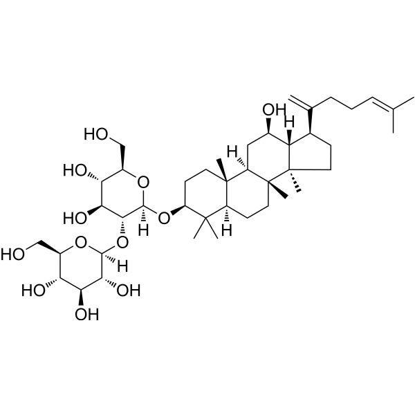
| Molecular Formula | C42H70O12 |
| Molecular Weight | 766.9980 |
| Exact Mass | 766.486 |
| CAS # | 494753-69-4 |
| PubChem CID | 11499198 |
| Appearance | White to light yellow solid powder |
| Density | 1.3±0.1 g/cm3 |
| Boiling Point | 854.5±65.0 °C at 760 mmHg |
| Flash Point | 470.6±34.3 °C |
| Vapour Pressure | 0.0±0.6 mmHg at 25°C |
| Index of Refraction | 1.589 |
| LogP | 6.7 |
| Hydrogen Bond Donor Count | 8 |
| Hydrogen Bond Acceptor Count | 12 |
| Rotatable Bond Count | 10 |
| Heavy Atom Count | 54 |
| Complexity | 1370 |
| Defined Atom Stereocenter Count | 19 |
| SMILES | CC(=CCCC(=C)[C@H]1CC[C@@]2([C@@H]1[C@@H](C[C@H]3[C@]2(CC[C@@H]4[C@@]3(CC[C@@H](C4(C)C)O[C@H]5[C@@H]([C@H]([C@@H]([C@H](O5)CO)O)O)O[C@H]6[C@@H]([C@H]([C@@H]([C@H](O6)CO)O)O)O)C)C)O)C)C |
| InChi Key | KWDWBAISZWOAHD-MHOSXIPRSA-N |
| InChi Code | InChI=1S/C42H70O12/c1-21(2)10-9-11-22(3)23-12-16-42(8)30(23)24(45)18-28-40(6)15-14-29(39(4,5)27(40)13-17-41(28,42)7)53-38-36(34(49)32(47)26(20-44)52-38)54-37-35(50)33(48)31(46)25(19-43)51-37/h10,23-38,43-50H,3,9,11-20H2,1-2,4-8H3/t23-,24-,25-,26-,27+,28-,29+,30+,31-,32-,33+,34+,35-,36-,37+,38+,40+,41-,42-/m1/s1 |
| Chemical Name | (2S,3R,4S,5S,6R)-2-[(2R,3R,4S,5S,6R)-4,5-dihydroxy-6-(hydroxymethyl)-2-[[(3S,5R,8R,9R,10R,12R,13R,14R,17S)-12-hydroxy-4,4,8,10,14-pentamethyl-17-(6-methylhepta-1,5-dien-2-yl)-2,3,5,6,7,9,11,12,13,15,16,17-dodecahydro-1H-cyclopenta[a]phenanthren-3-yl]oxy]oxan-3-yl]oxy-6-(hydroxymethyl)oxane-3,4,5-triol |
| HS Tariff Code | 2934.99.9001 |
| Storage |
Powder-20°C 3 years 4°C 2 years In solvent -80°C 6 months -20°C 1 month Note: This product requires protection from light (avoid light exposure) during transportation and storage. |
| Shipping Condition | Room temperature (This product is stable at ambient temperature for a few days during ordinary shipping and time spent in Customs) |
Biological Activity
| Targets |
- Ginsenoside Rk1 targets the Janus kinase 2/signal transducer and activator of transcription 3 (Jak2/Stat3) signaling pathway in lipopolysaccharide (LPS)-stimulated RAW264.7 cells. [2] - Ginsenoside Rk1 targets cell cycle regulatory proteins (cyclin B1, cdc2) and apoptosis-related proteins (Bax, Bcl-2, caspase-3) in MDA-MB-231 triple-negative breast cancer cells.[3] - Ginsenoside Rk1 exhibits activity against multiple molecular targets involved in inflammation, cancer, and oxidative stress (summarized in a systematic review), including nuclear factor-κB (NF-κB) and mitogen-activated protein kinases (MAPKs). [1] |
| ln Vitro |
Ginsenoside Rk1 (0–40 μM; 6 hours) reduces MCP-1 and TNF-α mRNA that are induced by lipopolysaccharide (LPS), and at 40 μM, it inhibits the expression of IL-1β. The phosphorylation of JAK2 and STAT3 (Tyr705 and Ser727) in RAW264.7 cells caused by LPS is inhibited by ginsenoside Rk1 (0-40 μM; 24 hours) in a dose-dependent way [2]. Cell viability was significantly reduced in response to ginsenoside Rk1 (0-160 μM; 48 hours) as compared to the control by 75.52 ± 2.51% (40 μM), 52.72 ± 2.54% (80 μM), 17.41 ± 2.94% (120 μM), and 12.63 ± 3.24% (160 μM) [3]. As compared to S phase and G2/M phase ratios, ginsenoside Rk1 (0-120 μM; 24 hours) enhances the G0/G1 phase ratio in MDA-MB-231 cells [3]. Reduced cell number, nuclear fragmentation, condensation, and the production of apoptotic bodies are a few of the dose-dependent ways that ginsenoside Rk1 (0-120 μM; 24 hours) increases the percentage of apoptotic cells [3]. - In LPS-stimulated RAW264.7 macrophages, Ginsenoside Rk1 (10–80 μM) dose-dependently inhibited the secretion of pro-inflammatory cytokines [tumor necrosis factor-α (TNF-α), interleukin-6 (IL-6)] and nitric oxide (NO). At 80 μM, TNF-α and IL-6 levels were reduced by ~65% and ~70%, respectively, and NO production was decreased by ~75%. Western blot showed Ginsenoside Rk1 (40–80 μM) downregulated LPS-induced phosphorylation of Jak2 and Stat3 [2] - In MDA-MB-231 cells, Ginsenoside Rk1 (20–100 μM) inhibited cell proliferation with an IC50 of 58.3 μM (MTT assay). Flow cytometry analysis revealed Ginsenoside Rk1 (40–80 μM) induced G2/M cell cycle arrest (cyclin B1 and cdc2 protein downregulation) and apoptosis (Bax/Bcl-2 ratio increase, caspase-3 activation). At 80 μM, the apoptotic rate was ~38% (vs. 3.2% in control) [3] - Ginsenoside Rk1 (5–40 μM) scavenged reactive oxygen species (ROS) in H2O2-induced PC12 cells, reducing ROS levels by ~50% at 40 μM, and protected cells from oxidative damage (cell viability increased by ~40% at 40 μM) [1] |
| ln Vivo |
- In a mouse acute inflammation model (intraperitoneal LPS injection, 5 mg/kg), oral administration of Ginsenoside Rk1 (20–80 mg/kg) for 7 days dose-dependently reduced serum TNF-α and IL-6 levels. At 80 mg/kg, serum TNF-α and IL-6 were decreased by ~55% and ~60%, respectively, compared to the LPS-only group. Histopathology showed Ginsenoside Rk1 (80 mg/kg) alleviated liver and lung inflammation (reduced neutrophil infiltration) [1] - In a MDA-MB-231 xenograft mouse model (female BALB/c nude mice), intraperitoneal injection of Ginsenoside Rk1 (20–60 mg/kg, every 2 days for 4 weeks) inhibited tumor growth. At 60 mg/kg, tumor volume and weight were reduced by ~50% and ~55%, respectively, vs. the vehicle group. Immunohistochemistry showed reduced Ki-67 (proliferation marker) and increased cleaved caspase-3 (apoptosis marker) in tumor tissues [3] |
| Enzyme Assay |
- For Jak2 kinase activity assay (Reference [2]): Recombinant human Jak2 kinase was incubated with ATP (100 μM), a biotinylated peptide substrate, and Ginsenoside Rk1 (10–80 μM) in kinase buffer (pH 7.5) at 37°C for 1 h. The phosphorylated peptide was captured by streptavidin-coated plates, detected with a phospho-specific antibody and HRP-conjugated secondary antibody. Absorbance at 450 nm was measured, and Jak2 activity was calculated as a percentage of the LPS-stimulated control [2] - For NO synthase (iNOS) activity assay (Reference [1]): RAW264.7 cells were treated with Ginsenoside Rk1 (10–80 μM) and LPS (1 μg/mL) for 24 h. Cells were lysed, and iNOS activity was measured by monitoring the conversion of L-arginine to L-citrulline (coupled with NADPH oxidation) at 340 nm. At 80 μM, Ginsenoside Rk1 inhibited iNOS activity by ~65% [1] |
| Cell Assay |
RT-PCR[2] Cell Types: RAW264.7 Cell Tested Concentrations: 10 μM, 20 μM, 40 μM Incubation Duration: 6 hrs (hours) Experimental Results: Inhibition of JAK2-dependent STAT3 activation in LPS-activated RAW264.7 cells. Western Blot Analysis[2] Cell Types: RAW264.7 Cell Tested Concentrations: 10 μM, 20 μM, 40 μM Incubation Duration: 6 hrs (hours) Experimental Results: Inhibition of JAK2-dependent STAT3 activation in LPS-activated RAW264.7 cells Cell Viability Assay[3 ] Cell Types: MDA-MB-231 Cell Tested Concentrations: 0 μM, 40 μM, 80 μM, 120 μM Incubation Duration: 48 hrs (hours) Experimental Results: Inhibited MDA-MB-231 cell proliferation in a dose- and time-dependent manner. Cell cycle analysis [3] Cell Types: MDA-MB-231 Cell Tested Concentrations: 0 μM, 40 μM, 80 μM, 120 μM Incubation Duration: 24 hrs (hours) Experimental Results: Induced G0/G1 phase arrest. Apoptosis analysis [3] Cell Types: MDA-MB-231 Cell Tested Concentrations: 0 μM, 40 μM, 80 μM, 120 μM Incubation Duration: 24 hrs (hours) Experimental Results: Induced apoptosis of MDA-MB-231 cells. - For RAW264.7 cytokine detection (Reference [2]): RAW264.7 cells were seeded in 24-well plates (5×10⁴ cells/well) and treated with Ginsenoside Rk1 (10–80 μM) for 1 h, then stimulated with LPS (1 μg/mL) for 24 h. Culture supernatants were collected, and TNF-α/IL-6 levels were measured by ELISA. Absorbance at 450 nm was read, and cytokine concentrations were calculated using standard curves [2] - For MDA-MB-231 cell cycle/apoptosis assay (Reference [3]): Cells were seeded in 6-well plates (2×10⁵ cells/well) and treated with Ginsenoside Rk1 (40–80 μM) for 48 h. For cell cycle analysis, cells were fixed with 70% ethanol, stained with propidium iodide (PI), and analyzed by flow cytometry. For apoptosis, cells were stained with Annexin V-FITC/PI and detected by flow cytometry. Protein expression (cyclin B1, Bax, caspase-3) was analyzed by Western blot (30 μg protein per lane) [3] - For PC12 cell oxidative stress assay (Reference [1]): PC12 cells were seeded in 96-well plates (1×10⁴ cells/well) and pre-treated with Ginsenoside Rk1 (5–40 μM) for 2 h, then exposed to H2O2 (200 μM) for 24 h. ROS levels were measured using a fluorescent probe (DCFH-DA) at 488 nm excitation. Cell viability was assessed by MTT assay [1] |
| Animal Protocol |
- For mouse inflammation model (Reference [1]): Male ICR mice (20–25 g) were divided into 4 groups (n=6/group): control, LPS (5 mg/kg, i.p.), LPS + Ginsenoside Rk1 (20 mg/kg, p.o.), LPS + Ginsenoside Rk1 (80 mg/kg, p.o.). Ginsenoside Rk1 was dissolved in 0.5% carboxymethyl cellulose (CMC-Na) and administered daily for 7 days; LPS was injected on day 7. 6 h post-LPS, mice were euthanized, serum was collected for cytokine detection, and liver/lung tissues were fixed for histopathology [1] - For MDA-MB-231 xenograft model (Reference [3]): Female BALB/c nude mice (4–6 weeks old) were subcutaneously injected with MDA-MB-231 cells (1×10⁶ cells/mouse). When tumors reached ~100 mm³, mice were grouped (n=6/group): vehicle (0.1% DMSO + saline, i.p.), Ginsenoside Rk1 (20 mg/kg, i.p.), Ginsenoside Rk1 (60 mg/kg, i.p.). Injections were given every 2 days for 4 weeks. Tumor volume [(length×width²)/2] was measured every 3 days. Mice were euthanized, tumors were weighed, and tissues were processed for immunohistochemistry [3] |
| ADME/Pharmacokinetics |
- Ginsenoside Rk1 has low oral bioavailability (~3.2%) in rats due to poor absorption and extensive first-pass metabolism. After oral administration of Ginsenoside Rk1 (50 mg/kg) to rats, the maximum plasma concentration (Cmax) was 128.5 ng/mL, and the half-life (t1/2) was 2.8 h [1] - In human liver microsomes, Ginsenoside Rk1 was metabolized by cytochrome P450 enzymes (CYP3A4, CYP2C9), with a metabolic clearance rate of 18.6 μL/min/mg protein [1] |
| Toxicity/Toxicokinetics |
- Ginsenoside Rk1 showed no acute toxicity in mice at oral doses up to 2000 mg/kg (no mortality or abnormal behavior observed for 14 days) [1] - In a 28-day subchronic toxicity study in rats, Ginsenoside Rk1 (50–200 mg/kg, p.o.) had no significant effects on body weight, organ weight (liver, kidney), or serum biochemical parameters (ALT, AST, creatinine, urea nitrogen) [1] - No cytotoxicity was observed in normal human mammary epithelial cells (HMEC) treated with Ginsenoside Rk1 (up to 100 μM), with cell viability >90% [3] |
| References |
[1]. Ginsenoside Rk1 bioactivity: a systematic review. PeerJ. 2017 Nov 17;5:e3993. [2]. Ginsenoside Rk1 suppresses pro-inflammatory responses in lipopolysaccharide-stimulated RAW264.7 cells by inhibiting the Jak2/Stat3 pathway. Chin J Nat Med. 2017 Oct;15(10):751-757. [3]. Ginsenoside Rk1 induces cell cycle arrest and apoptosis in MDA-MB-231 triple negative breast cancer cells. Toxicology. 2019 Apr 15;418:22-31. |
| Additional Infomation |
- Ginsenoside Rk1 is a rare ginsenoside derived from heat processing (steaming or roasting) of Panax ginseng, with enhanced biological activity compared to major ginsenosides (e.g., Rg1, Rb1) [1] - The anti-inflammatory mechanism of Ginsenoside Rk1 involves inhibition of Jak2/Stat3 and NF-κB pathways, while its anti-cancer activity is mediated by G2/M cycle arrest and mitochondria-dependent apoptosis [1, 2, 3] - Ginsenoside Rk1 has potential therapeutic applications in inflammatory diseases (e.g., sepsis, arthritis) and triple-negative breast cancer, but clinical trials are still limited [1, 3] |
Solubility Data
| Solubility (In Vitro) | DMSO : ~100 mg/mL (~130.38 mM) |
| Solubility (In Vivo) |
Solubility in Formulation 1: ≥ 2.5 mg/mL (3.26 mM) (saturation unknown) in 10% DMSO + 40% PEG300 + 5% Tween80 + 45% Saline (add these co-solvents sequentially from left to right, and one by one), clear solution. For example, if 1 mL of working solution is to be prepared, you can add 100 μL of 25.0 mg/mL clear DMSO stock solution to 400 μL PEG300 and mix evenly; then add 50 μL Tween-80 to the above solution and mix evenly; then add 450 μL normal saline to adjust the volume to 1 mL. Preparation of saline: Dissolve 0.9 g of sodium chloride in 100 mL ddH₂ O to obtain a clear solution. Solubility in Formulation 2: ≥ 2.5 mg/mL (3.26 mM) (saturation unknown) in 10% DMSO + 90% (20% SBE-β-CD in Saline) (add these co-solvents sequentially from left to right, and one by one), clear solution. For example, if 1 mL of working solution is to be prepared, you can add 100 μL of 25.0 mg/mL clear DMSO stock solution to 900 μL of 20% SBE-β-CD physiological saline solution and mix evenly. Preparation of 20% SBE-β-CD in Saline (4°C,1 week): Dissolve 2 g SBE-β-CD in 10 mL saline to obtain a clear solution. Solubility in Formulation 3: ≥ 2.5 mg/mL (3.26 mM) (saturation unknown) in 10% DMSO + 90% Corn Oil (add these co-solvents sequentially from left to right, and one by one), clear solution. For example, if 1 mL of working solution is to be prepared, you can add 100 μL of 25.0 mg/mL clear DMSO stock solution to 900 μL of corn oil and mix evenly. (Please use freshly prepared in vivo formulations for optimal results.) |
| Preparing Stock Solutions | 1 mg | 5 mg | 10 mg | |
| 1 mM | 1.3038 mL | 6.5189 mL | 13.0378 mL | |
| 5 mM | 0.2608 mL | 1.3038 mL | 2.6076 mL | |
| 10 mM | 0.1304 mL | 0.6519 mL | 1.3038 mL |