Physicochemical Properties
| Molecular Formula | C16H12O5 |
| Molecular Weight | 284.2635 |
| Exact Mass | 284.068 |
| CAS # | 437-64-9 |
| PubChem CID | 5281617 |
| Appearance | Light yellow to yellow solid powder |
| Density | 1.4±0.1 g/cm3 |
| Boiling Point | 546.5±50.0 °C at 760 mmHg |
| Melting Point | 290-292°C |
| Flash Point | 209.7±23.6 °C |
| Vapour Pressure | 0.0±1.5 mmHg at 25°C |
| Index of Refraction | 1.669 |
| LogP | 2.36 |
| Hydrogen Bond Donor Count | 2 |
| Hydrogen Bond Acceptor Count | 5 |
| Rotatable Bond Count | 2 |
| Heavy Atom Count | 21 |
| Complexity | 424 |
| Defined Atom Stereocenter Count | 0 |
| InChi Key | JPMYFOBNRRGFNO-UHFFFAOYSA-N |
| InChi Code | InChI=1S/C16H12O5/c1-20-11-6-12(18)16-13(19)8-14(21-15(16)7-11)9-2-4-10(17)5-3-9/h2-8,17-18H,1H3 |
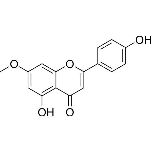
| Chemical Name | 5-hydroxy-2-(4-hydroxyphenyl)-7-methoxychromen-4-one |
| HS Tariff Code | 2934.99.9001 |
| Storage |
Powder-20°C 3 years 4°C 2 years In solvent -80°C 6 months -20°C 1 month Note: This product requires protection from light (avoid light exposure) during transportation and storage. |
| Shipping Condition | Room temperature (This product is stable at ambient temperature for a few days during ordinary shipping and time spent in Customs) |
Biological Activity
| Targets |
The targets of Genkwanin are associated with the miR-101/MKP-1/MAPK signaling pathway, including microRNA-101 (miR-101), mitogen-activated protein kinase phosphatase 1 (MKP-1), and mitogen-activated protein kinases (MAPKs, such as ERK, JNK, and p38). [1] |
| ln Vitro |
Analysis of cell viability revealed that at doses up to 50 μM, genkwanin had no effect on cell viability. LPS-induced NO generation is inhibited by genkwanin in a concentration-dependent manner. The expression of iNOS is limited to fetal stimulation. iNOS activity is not considerably impacted by genkwanin. The impact of genkwanin on the generation of proinflammatory cytokines was investigated. In LPS-stimulated RAW264.7 macrophages, genkwanin reduces TNF-a, IL-1b, and IL-6 production in a concentration-dependent manner. While having no effect on the integrity of NF-kB signaling, genkwanin strongly suppresses AP-1 signaling in buffer. It demonstrates that genkwanin has a fold effect on ERK1/2 phosphorylation but inhibits p38 and JNK phosphorylation in a concentration-dependent manner. After being first discovered as an immediate early gene, MKP-1 was found to be a bisphosphophosphatase that significantly affects p38 MAPK activity, JNK, and ERK1/2 activity. MKP-1 expression can be markedly increased by genkwanin, but not MKP-1 mRNA[1]. 1. In LPS-activated RAW264.7 macrophages, Genkwanin dose-dependently inhibited the production of proinflammatory cytokines. At concentrations of 10 μM, 20 μM, and 40 μM, it reduced the mRNA levels of TNF-α by approximately 30%, 55%, and 75%, respectively; IL-6 by approximately 25%, 50%, and 70%, respectively; and IL-1β by approximately 20%, 45%, and 65%, respectively, compared to the LPS-only group [1] 2. Genkwanin also suppressed nitric oxide (NO) production in LPS-activated RAW264.7 cells. Treatment with 20 μM and 40 μM Genkwanin decreased NO levels by about 40% and 65%, respectively, which was associated with reduced iNOS mRNA and protein expression [1] 3. Regarding the miR-101/MKP-1/MAPK pathway: Genkwanin (20 μM, 40 μM) downregulated LPS-induced miR-101 expression by approximately 40% and 60%, respectively; upregulated MKP-1 mRNA and protein levels by approximately 50% and 80% (at 40 μM); and inhibited the phosphorylation of ERK, JNK, and p38 MAPKs. Western blot results showed that 40 μM Genkwanin reduced p-ERK, p-JNK, and p-p38 levels by approximately 60%, 55%, and 50%, respectively, compared to LPS treatment alone [1] 4. MTT assay showed that Genkwanin at concentrations up to 40 μM had no significant cytotoxicity on RAW264.7 cells, with cell viability remaining above 90% [1] |
| Cell Assay |
1. RAW264.7 macrophage culture and treatment: RAW264.7 cells were cultured in complete medium containing 10% fetal bovine serum and 1% antibiotics at 37°C in a 5% CO2 incubator. When cells reached 70-80% confluence, they were seeded into 6-well plates (for mRNA/protein detection) or 96-well plates (for MTT/NO assay). After 24 hours of adherence, cells were pretreated with Genkwanin (10 μM, 20 μM, 40 μM) or DMSO (control) for 1 hour, then stimulated with LPS (1 μg/mL) for an additional 24 hours (for cytokine/NO detection) or 15 minutes (for MAPK phosphorylation detection) [1] 2. MTT cell viability assay: After treatment, 20 μL of MTT solution (5 mg/mL) was added to each well of the 96-well plate, and the plate was incubated at 37°C for 4 hours. The supernatant was discarded, and 150 μL of DMSO was added to dissolve the formazan crystals. The absorbance at 570 nm was measured using a microplate reader, and cell viability was calculated as the percentage of the control group [1] 3. NO detection assay: The supernatant of treated cells was collected, and an equal volume of Griess reagent was added. The mixture was incubated at room temperature for 10 minutes, and the absorbance at 540 nm was measured. NO concentration was calculated using a sodium nitrite standard curve [1] 4. Quantitative real-time PCR (qPCR) for cytokine/miR-101/MKP-1 mRNA: Total RNA was extracted from cells using an RNA extraction kit. For mRNA detection, cDNA was synthesized via reverse transcription, and qPCR was performed using specific primers for TNF-α, IL-6, IL-1β, iNOS, and MKP-1, with GAPDH as the internal reference. For miR-101 detection, specific reverse transcription primers were used, and U6 snRNA was the internal reference. The relative expression levels were calculated using the 2^(-ΔΔCt) method [1] 5. Western blot for MKP-1 and phosphorylated MAPKs: Total protein was extracted from cells, and protein concentration was determined using a protein assay kit. Equal amounts of protein were separated by SDS-PAGE, transferred to PVDF membranes, and blocked with 5% non-fat milk for 1 hour. Membranes were incubated with primary antibodies against MKP-1, ERK, p-ERK, JNK, p-JNK, p38, p-p38, and GAPDH (internal reference) overnight at 4°C, then with secondary antibodies for 1 hour at room temperature. The bands were visualized using an enhanced chemiluminescence kit, and band intensity was quantified using image analysis software [1] |
| References |
[1]. Genkwanin inhibits proinflammatory mediators mainly through the regulation of miR-101/MKP-1/MAPK pathway in LPS-activated macrophages. PLoS One. 2014 May 6;9(5):e96741. |
| Additional Infomation |
Genkwanin is a monomethoxyflavone that is apigenin in which the hydroxy group at position 7 is methylated. It has a role as a metabolite. It is a dihydroxyflavone and a monomethoxyflavone. It is functionally related to an apigenin. It is a conjugate acid of a genkwanin(1-). Genkwanin has been reported in Vernonia fasciculata, Aquilegia oxysepala, and other organisms with data available. 1. Genkwanin exerts its anti-inflammatory effect mainly through regulating the miR-101/MKP-1/MAPK pathway. LPS typically upregulates miR-101, which inhibits MKP-1 expression; reduced MKP-1 then leads to increased phosphorylation of ERK, JNK, and p38 MAPKs, ultimately promoting the production of proinflammatory mediators. Genkwanin reverses this process by downregulating miR-101, upregulating MKP-1, and inhibiting MAPK phosphorylation [1] 2. Genkwanin is a flavonoid compound, and its anti-inflammatory activity in LPS-activated macrophages suggests potential applications in the research of inflammatory diseases [1] |
Solubility Data
| Solubility (In Vitro) | DMSO : ~31.25 mg/mL (~109.93 mM) |
| Solubility (In Vivo) |
Solubility in Formulation 1: ≥ 0.5 mg/mL (1.76 mM) (saturation unknown) in 10% DMSO + 40% PEG300 + 5% Tween80 + 45% Saline (add these co-solvents sequentially from left to right, and one by one), clear solution. For example, if 1 mL of working solution is to be prepared, you can add 100 μL of 5.0 mg/mL clear DMSO stock solution to 400 μL PEG300 and mix evenly; then add 50 μL Tween-80 to the above solution and mix evenly; then add 450 μL normal saline to adjust the volume to 1 mL. Preparation of saline: Dissolve 0.9 g of sodium chloride in 100 mL ddH₂ O to obtain a clear solution. Solubility in Formulation 2: 0.5 mg/mL (1.76 mM) in 10% DMSO + 90% (20% SBE-β-CD in Saline) (add these co-solvents sequentially from left to right, and one by one), suspension solution; with ultrasonication. For example, if 1 mL of working solution is to be prepared, you can add 100 μL of 5.0 mg/mL clear DMSO stock solution to 900 μL of 20% SBE-β-CD physiological saline solution and mix evenly. Preparation of 20% SBE-β-CD in Saline (4°C,1 week): Dissolve 2 g SBE-β-CD in 10 mL saline to obtain a clear solution. Solubility in Formulation 3: ≥ 0.5 mg/mL (1.76 mM) (saturation unknown) in 10% DMSO + 90% Corn Oil (add these co-solvents sequentially from left to right, and one by one), clear solution. For example, if 1 mL of working solution is to be prepared, you can add 100 μL of 5.0 mg/mL clear DMSO stock solution to 900 μL of corn oil and mix evenly. (Please use freshly prepared in vivo formulations for optimal results.) |
| Preparing Stock Solutions | 1 mg | 5 mg | 10 mg | |
| 1 mM | 3.5179 mL | 17.5895 mL | 35.1791 mL | |
| 5 mM | 0.7036 mL | 3.5179 mL | 7.0358 mL | |
| 10 mM | 0.3518 mL | 1.7590 mL | 3.5179 mL |