GSK2850163 is a novel and potent inhibitor of inositol-requiring enzyme-1 alpha (IRE1α) which has IC50 values of 20 and 200 nM for IRE1α kinase and RNase activity, respectively. Activation of the inositol-requiring enzyme-1 alpha (IRE1a) protein caused by endoplasmic reticulum stress results in the homodimerization of the N-terminal endoplasmic reticulum luminal domains, autophosphorylation of the cytoplasmic kinase domains, and conformational changes to the cytoplasmic endoribonuclease (RNase) domains, which render them functional and can lead to the splicing of X-box binding protein 1 (XBP 1) mRNA.
Physicochemical Properties
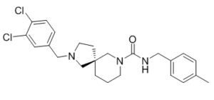
| Molecular Formula | C24H29CL2N3O |
| Molecular Weight | 446.41256403923 |
| Exact Mass | 445.17 |
| Elemental Analysis | C, 64.57; H, 6.55; Cl, 15.88; N, 9.41; O, 3.58 |
| CAS # | 2121989-91-9 |
| Related CAS # | GSK2850163 (S enantiomer);2309519-81-9;GSK2850163 hydrochloride;2319838-09-8 |
| PubChem CID | 73291790 |
| Appearance | Colorless to yellow solid powder |
| LogP | 4.9 |
| Hydrogen Bond Donor Count | 1 |
| Hydrogen Bond Acceptor Count | 2 |
| Rotatable Bond Count | 4 |
| Heavy Atom Count | 30 |
| Complexity | 583 |
| Defined Atom Stereocenter Count | 1 |
| SMILES | CC1=CC=C(C=C1)CNC(=O)N2CCC[C@@]3(C2)CCN(C3)CC4=CC(=C(C=C4)Cl)Cl |
| InChi Key | YFDASBFQKMHSSJ-XMMPIXPASA-N |
| InChi Code | InChI=1S/C24H29Cl2N3O/c1-18-3-5-19(6-4-18)14-27-23(30)29-11-2-9-24(17-29)10-12-28(16-24)15-20-7-8-21(25)22(26)13-20/h3-8,13H,2,9-12,14-17H2,1H3,(H,27,30)/t24-/m1/s1 |
| Chemical Name | (5R)-2-[(3,4-dichlorophenyl)methyl]-N-[(4-methylphenyl)methyl]-2,7-diazaspiro[4.5]decane-7-carboxamide |
| Synonyms | GSK2850163; GSK-2850163; GSK 2850163 |
| HS Tariff Code | 2934.99.9001 |
| Storage |
Powder-20°C 3 years 4°C 2 years In solvent -80°C 6 months -20°C 1 month |
| Shipping Condition | Room temperature (This product is stable at ambient temperature for a few days during ordinary shipping and time spent in Customs) |
Biological Activity
| Targets |
IRE1α (IC50 = 20 nM); IRE1α (IC50 = 200 nM)
The target of GSK2850163 is the endoribonuclease domain of human inositol-requiring enzyme 1α (IRE1α), a key mediator of the unfolded protein response (UPR) during endoplasmic reticulum (ER) stress. For human IRE1α endoribonuclease activity, the half-maximal inhibitory concentration (IC₅₀) is 0.1 μM (FRET-based assay) [1] . It does not inhibit the kinase domain of IRE1α (IC₅₀ > 10 μM) or other unrelated ribonucleases (e.g., RNase A, RNase T1) at concentrations up to 10 μM, demonstrating high domain and enzyme selectivity [1] |
| ln Vitro |
GSK2850163 is a new activator of inositol-requiring enzyme 1 alpha (IRE1α) that reduces RNase activity and IRE1α multiplication activity with IC50 values of 20 and 200 nM, respectively. In a dose-dependent way, GSK2850163 can lessen the rise in IRE1α autophosphorylation. GSK2850163 concentrations were able to raise the decreased activation of XBP 1. Ron (IC50=4.4 μM) and FGFR1 V561M (IC50=17 μM), two other mediators, are mildly inhibited by GSK2850163 [1]. 1. Inhibition of IRE1α endoribonuclease activity: GSK2850163 concentration-dependently inhibits the endoribonuclease activity of recombinant human IRE1α catalytic domain, with an IC₅₀ of 0.1 μM. At 1 μM, it achieves >90% inhibition of IRE1α-mediated cleavage of a fluorescently labeled XBP1 (X-box binding protein 1) RNA substrate (FRET assay). The inhibition is reversible, as dialysis of the drug-enzyme complex restores ~85% of endoribonuclease activity [1] 2. Suppression of XBP1 mRNA splicing in ER-stressed cells: HeLa cells treated with thapsigargin (1 μM, ER stress inducer) for 4 hours showed significant induction of XBP1 mRNA splicing (XBP1u → XBP1s). Pretreatment with GSK2850163 (0.1–1 μM) for 1 hour dose-dependently inhibited XBP1 splicing: 0.1 μM (30% inhibition), 0.5 μM (65% inhibition), 1 μM (92% inhibition) (RT-PCR analysis). No effect on XBP1 splicing was observed in unstressed cells [1] 3. Regulation of ER stress-responsive gene expression: In thapsigargin-induced HeLa cells, GSK2850163 (1 μM) downregulated the expression of UPR target genes downstream of IRE1α: CHOP (C/EBP homologous protein, 45% reduction), BiP (binding immunoglobulin protein, 38% reduction), and EDEM1 (ER degradation-enhancing α-mannosidase-like protein 1, 42% reduction) (quantitative real-time PCR) [1] 4. Inhibition of ER stress-induced apoptosis: HeLa cells exposed to thapsigargin (1 μM) for 24 hours showed 42% apoptotic cells (Annexin V/PI staining). Pretreatment with GSK2850163 (1 μM) reduced apoptotic cells to 15%, with a corresponding decrease in cleaved caspase-3 and cleaved PARP levels (Western blot). No inherent cytotoxicity was observed in unstressed cells treated with GSK2850163 up to 10 μM [1] 5. Selectivity for IRE1α endoribonuclease: GSK2850163 (10 μM) did not inhibit the kinase activity of IRE1α (ATPase assay) or the catalytic activity of unrelated ribonucleases (RNase A, RNase T1) and kinases (e.g., ERK1, JNK2), confirming high domain and enzyme selectivity [1] 6. Conformational regulation of IRE1α: X-ray crystallography and hydrogen-deuterium exchange mass spectrometry (HDX-MS) confirmed that GSK2850163 binds to the ATP-binding pocket of the IRE1α kinase domain, inducing a long-range conformational change that allosterically impairs the endoribonuclease domain’s ability to bind and cleave RNA substrates [1] |
| Enzyme Assay |
1. FRET-based IRE1α endoribonuclease activity assay: - Recombinant human IRE1α catalytic domain (kinase + endoribonuclease domains) was diluted in assay buffer (20 mM Tris-HCl pH 7.5, 150 mM NaCl, 5 mM MgCl₂, 1 mM DTT) to a final concentration of 50 nM [1] - Serial dilutions of GSK2850163 (0.001–10 μM) or vehicle were pre-incubated with IRE1α for 20 minutes at room temperature. A FRET-labeled XBP1 RNA substrate (5’-FAM-labeled, 3’-TAMRA-labeled, 26-mer) was added to a final concentration of 200 nM [1] - The reaction mixture was incubated at 37°C for 60 minutes, and fluorescence intensity (excitation 485 nm, emission 535 nm) was measured using a microplate reader. Cleavage of the RNA substrate reduces FRET efficiency, increasing FAM fluorescence [1] - The percentage inhibition of endoribonuclease activity was calculated relative to vehicle control, and IC₅₀ values were derived from dose-response curves [1] 2. Surface Plasmon Resonance (SPR) binding assay: - Recombinant human IRE1α kinase domain was immobilized on a CM5 sensor chip via amine coupling to achieve a surface density of ~900 resonance units (RU) [1] - Serial dilutions of GSK2850163 (0.01–100 μM) in running buffer (20 mM Tris-HCl pH 7.5, 150 mM NaCl, 5 mM MgCl₂, 0.05% surfactant P20) were injected over the chip surface at a flow rate of 30 μL/min [1] - Association and dissociation phases were recorded, and sensorgrams were fitted to a 1:1 Langmuir binding model to calculate the dissociation constant (KD = 0.08 μM) [1] 3. ATPase activity assay (IRE1α kinase domain selectivity): - Recombinant human IRE1α catalytic domain (50 nM) was incubated with GSK2850163 (0.01–10 μM) in assay buffer containing 1 mM ATP and a luciferase-based ATP detection reagent [1] - Luminescence intensity (reflecting remaining ATP) was measured at 0 and 60 minutes of incubation at 37°C. No significant reduction in ATP hydrolysis was observed, confirming lack of kinase domain inhibition [1] |
| Cell Assay |
1. XBP1 mRNA splicing assay (RT-PCR): - HeLa cells were seeded into 6-well plates at a density of 2×10⁵ cells/well and cultured overnight in DMEM supplemented with 10% fetal bovine serum [1] - Cells were pretreated with GSK2850163 (0.1–1 μM) or vehicle for 1 hour, then stimulated with thapsigargin (1 μM) to induce ER stress. Cells were harvested 4 hours post-stimulation [1] - Total RNA was extracted, and complementary DNA (cDNA) was synthesized from 1 μg RNA. RT-PCR was performed using primers specific for unspliced (XBP1u) and spliced (XBP1s) XBP1 mRNA. PCR products were separated on a 2% agarose gel and visualized by ethidium bromide staining [1] - Band intensity was quantified by densitometry, and the XBP1s/XBP1u ratio was calculated to assess splicing efficiency [1] 2. Western blot analysis of ER stress and apoptosis markers: - HeLa cells were seeded into 6-well plates and pretreated with GSK2850163 (1 μM) for 1 hour, followed by thapsigargin (1 μM) stimulation for 24 hours [1] - Cells were lysed in RIPA buffer containing protease and phosphatase inhibitors. Equal amounts of protein (30 μg/lane) were separated by SDS-PAGE, transferred to PVDF membranes, and probed with primary antibodies against CHOP, BiP, cleaved caspase-3, cleaved PARP, and β-actin (loading control) [1] - HRP-conjugated secondary antibodies were used, and protein bands were visualized by chemiluminescence. Band intensity was quantified by densitometry [1] 3. Apoptosis assay (Annexin V/PI double staining): - HeLa cells were seeded into 12-well plates at 1×10⁶ cells/well and treated as described for Western blot analysis [1] - Cells were harvested, washed twice with cold PBS, and stained with Annexin V-FITC and PI for 30 minutes at 4°C in the dark [1] - Apoptotic cells (Annexin V⁺/PI⁻ for early apoptosis; Annexin V⁺/PI⁺ for late apoptosis) were quantified by flow cytometry [1] 4. Quantitative real-time PCR for UPR target genes: - Total RNA from thapsigargin-induced HeLa cells (treated with GSK2850163 or vehicle) was extracted, and cDNA was synthesized [1] - Real-time PCR was performed using gene-specific primers for CHOP, BiP, EDEM1, and GAPDH (housekeeping gene). Relative gene expression levels were calculated using the 2^(-ΔΔCt) method [1] |
| Toxicity/Toxicokinetics |
1. In vitro cytotoxicity: GSK2850163 at concentrations up to 10 μM has no inherent cytotoxicity in unstressed HeLa cells, with cell viability >95% compared to vehicle control (MTT assay) [1] |
| References |
[1]. Long-Range Inhibitor-Induced Conformational Regulation of Human IRE1α Endoribonuclease Activity. Molecular Pharmacology December 2015, 88 (6) 1011-1023. |
| Additional Infomation |
1. GSK2850163 is a potent, selective, allosteric inhibitor of the IRE1α endoribonuclease domain, developed to study the role of IRE1α-mediated UPR in ER stress-related diseases [1] 2. Mechanism of action: GSK2850163 binds to the ATP-binding pocket of the IRE1α kinase domain (allosteric site), inducing a long-range conformational change that disrupts the dimerization and catalytic activity of the distal endoribonuclease domain. This blocks IRE1α-mediated XBP1 mRNA splicing and downstream UPR signaling, without affecting the kinase domain’s ATPase activity [1] 3. Structural basis: X-ray crystallography shows that GSK2850163 binds to the IRE1α kinase domain in a distinct conformation, stabilizing an inactive state of the endoribonuclease domain. Hydrogen-deuterium exchange mass spectrometry (HDX-MS) confirms that binding induces conformational changes spanning the kinase-endoribonuclease interdomain region [1] 4. Research applications: GSK2850163 is widely used as a tool compound to investigate the pathological roles of IRE1α in diseases associated with dysregulated ER stress (e.g., neurodegenerative diseases, diabetes, cancer) and to validate IRE1α as a therapeutic target [1] 5. Chemical properties: It belongs to the pyrazolo[1,5-a]pyrimidine chemical class, with a molecular weight of 410.4 g/mol. The chemical structure is optimized for selective binding to the IRE1α kinase domain and allosteric regulation of endoribonuclease activity [1] |
Solubility Data
| Solubility (In Vitro) |
DMSO: ~67.5 mg/mL (~151.2 mM) Ethanol: ~50 mg/mL (~112.0 mM) |
| Solubility (In Vivo) |
Solubility in Formulation 1: ≥ 2.25 mg/mL (5.04 mM) (saturation unknown) in 10% DMSO + 40% PEG300 + 5% Tween80 + 45% Saline (add these co-solvents sequentially from left to right, and one by one), clear solution. For example, if 1 mL of working solution is to be prepared, you can add 100 μL of 22.5 mg/mL clear DMSO stock solution to 400 μL PEG300 and mix evenly; then add 50 μL Tween-80 to the above solution and mix evenly; then add 450 μL normal saline to adjust the volume to 1 mL. Preparation of saline: Dissolve 0.9 g of sodium chloride in 100 mL ddH₂ O to obtain a clear solution. Solubility in Formulation 2: ≥ 2.25 mg/mL (5.04 mM) (saturation unknown) in 10% DMSO + 90% Corn Oil (add these co-solvents sequentially from left to right, and one by one), clear solution. For example, if 1 mL of working solution is to be prepared, you can add 100 μL of 22.5 mg/mL clear DMSO stock solution to 900 μL of corn oil and mix evenly. (Please use freshly prepared in vivo formulations for optimal results.) |
| Preparing Stock Solutions | 1 mg | 5 mg | 10 mg | |
| 1 mM | 2.2401 mL | 11.2005 mL | 22.4009 mL | |
| 5 mM | 0.4480 mL | 2.2401 mL | 4.4802 mL | |
| 10 mM | 0.2240 mL | 1.1200 mL | 2.2401 mL |