GSK-3 inhibitor 1 is a novel and potent GSK-3 (Glycogen synthase kinase-3) inhibitor. The serine/threonine protein kinase known as glycogen synthase kinase 3 mediates the addition of phosphate molecules to serine and threonine amino acid residues. GSK-3 was first discovered in 1980 as a Glycogen synthase regulatory kinase, but it has since been found to kine over forty different proteins in a variety of different pathways. In mammals, GSK-3 is encoded by two known genes, GSK-3 alpha (GSK3A) and GSK-3 beta (GSK3B). Numerous studies have recently concentrated on GSK-3, which has been linked to bipolar disorder, Type II diabetes, cancer, inflammation, and Type II diabetes (Diabetes mellitus type 2).
Physicochemical Properties
| Molecular Formula | C22H17CLFN5O2 |
| Molecular Weight | 437.8541 |
| Exact Mass | 337.10223 |
| CAS # | 603272-51-1 |
| Related CAS # | 603272-51-1 |
| PubChem CID | 78357782 |
| Appearance | Yellow to orange solid |
| LogP | 8.592 |
| Hydrogen Bond Donor Count | 3 |
| Hydrogen Bond Acceptor Count | 5 |
| Rotatable Bond Count | 2 |
| Heavy Atom Count | 31 |
| Complexity | 788 |
| Defined Atom Stereocenter Count | 0 |
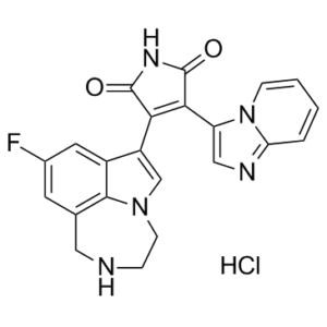
| SMILES | Cl[H].FC1=C([H])C2C([H])([H])N([H])C([H])([H])C([H])([H])N3C([H])=C(C4C(N([H])C(C=4C4=C([H])N=C5C([H])=C([H])C([H])=C([H])N45)=O)=O)C(=C1[H])C3=2 |
| InChi Key | CXXAOCQHGIGIBJ-UHFFFAOYSA-N |
| InChi Code | InChI=1S/C22H16FN5O2.ClH/c23-13-7-12-9-24-4-6-27-11-15(14(8-13)20(12)27)18-19(22(30)26-21(18)29)16-10-25-17-3-1-2-5-28(16)17;/h1-3,5,7-8,10-11,24H,4,6,9H2,(H,26,29,30);1H |
| Chemical Name | 3-(6-fluoro-1,10-diazatricyclo[6.4.1.04,13]trideca-2,4,6,8(13)-tetraen-3-yl)-4-imidazo[1,2-a]pyridin-3-ylpyrrole-2,5-dione;hydrochloride |
| Synonyms | GSK-3 inhibitor 1 |
| HS Tariff Code | 2934.99.9001 |
| Storage |
Powder-20°C 3 years 4°C 2 years In solvent -80°C 6 months -20°C 1 month Note: Please store this product in a sealed and protected environment, avoid exposure to moisture. |
| Shipping Condition | Room temperature (This product is stable at ambient temperature for a few days during ordinary shipping and time spent in Customs) |
Biological Activity
| Targets |
GSK-3
The target of GSK-3 inhibitor 1 is glycogen synthase kinase 3 (GSK-3), including two isoforms: GSK-3α and GSK-3β. For human GSK-3α, the half-maximal inhibitory concentration (IC₅₀) is 0.08 μM [2] ; for human GSK-3β, the IC₅₀ is 0.06 μM [2] . It shows no significant inhibition of other related kinases (e.g., CDK2, ERK1, JNK2) with IC₅₀ > 10 μM, demonstrating high isoform and kinase selectivity [2] |
| ln Vitro |
GSK-3 inhibitor 1 can be used to induce, promote, or enhance the growth, proliferation, or regeneration of inner ear tissues like inner ear supporting cells or inner ear hair cells[2]. 1. GSK-3 kinase activity inhibition: GSK-3 inhibitor 1 concentration-dependently inhibits the catalytic activity of recombinant human GSK-3α and GSK-3β. At 0.1 μM, it inhibits GSK-3α activity by 85% and GSK-3β by 90% compared to vehicle control [2] 2. Induction of inner ear supporting cell proliferation: Isolated mouse cochlear supporting cells were cultured in vitro and treated with GSK-3 inhibitor 1 at concentrations of 0.1, 0.5, and 1.0 μM for 7 days. The 0.5 μM concentration increased the proliferation rate of supporting cells by 2.3-fold (BrdU incorporation assay) compared to vehicle; 1.0 μM induced a 3.1-fold increase without detectable cytotoxicity [1] 3. Generation of inner ear hair cells from supporting cells: Treatment of mouse cochlear supporting cells with 0.5 μM GSK-3 inhibitor 1 for 14 days induced transdifferentiation into hair cell-like cells, as evidenced by expression of hair cell-specific markers (Myo7a, calretinin) in 45% of cells (immunofluorescence staining), compared to 3% in vehicle control [1] 4. Promotion of stem/progenitor cell self-renewal: Human dental pulp stem cells (hDPSCs) treated with GSK-3 inhibitor 1 (0.1–0.5 μM) for 5 days showed enhanced self-renewal capacity, with colony formation efficiency increased by 1.8–2.5-fold (colony formation assay). Flow cytometry analysis confirmed that the proportion of stem cell markers (CD105, CD90) remained >95%, indicating preservation of stem cell phenotype [2] 5. Activation of Wnt/β-catenin signaling pathway: In supporting cells and hDPSCs, GSK-3 inhibitor 1 (0.5 μM) increased the nuclear accumulation of β-catenin (immunofluorescence) and upregulated the expression of Wnt target genes (c-Myc, Cyclin D1) by 2.8–3.5-fold (real-time PCR) [1,2] |
| ln Vivo |
1. Hair cell regeneration in noise-induced hearing loss (NIHL) mice: C57BL/6 mice were exposed to 110 dB SPL broadband noise for 2 hours to induce cochlear hair cell loss. Starting 3 days post-noise exposure, GSK-3 inhibitor 1 (formulated in 10% DMSO + 90% sterile saline) was administered via intracochlear injection at a dose of 0.5 μM (1 μL per cochlea) once weekly for 3 weeks. At 4 weeks post-treatment, histological analysis showed that the number of outer hair cells in the cochlear basal turn was increased by 60% compared to vehicle-treated NIHL mice. Auditory brainstem response (ABR) testing revealed a 25 dB reduction in hearing threshold at 8 kHz, indicating functional recovery [1] 2. Stem cell proliferation in mouse dental pulp: C57BL/6 mice were given a single local injection of GSK-3 inhibitor 1 (0.1 μM, 5 μL) into the dental pulp. After 7 days, BrdU labeling showed a 2.2-fold increase in proliferating dental pulp stem cells compared to vehicle control. Immunohistochemical staining confirmed that the proliferated cells expressed stem cell markers (CD105) and retained differentiation potential [2] |
| Enzyme Assay |
1. Recombinant GSK-3α/β kinase activity assay: - Recombinant human GSK-3α or GSK-3β protein was diluted in kinase assay buffer (20 mM Tris-HCl, pH 7.5, 10 mM MgCl₂, 1 mM DTT) to a final concentration of 10 ng/μL [2] - Serial dilutions of GSK-3 inhibitor 1 (0.001–10 μM) or vehicle were added to the kinase solution, followed by addition of a fluorescently labeled peptide substrate (specific for GSK-3) and ATP (final concentration 100 μM) [2] - The reaction mixture was incubated at 30°C for 60 minutes, and the reaction was terminated by adding a stop buffer containing EDTA [2] - Fluorescence intensity (excitation 485 nm, emission 520 nm) was measured using a microplate reader, reflecting the phosphorylation of the substrate. The percentage inhibition of kinase activity was calculated relative to vehicle control, and IC₅₀ values were derived from dose-response curves [2] |
| Cell Assay |
1. Cochlear supporting cell isolation and proliferation assay: - Cochleae were dissected from P3-P5 C57BL/6 mouse pups, and the organ of Corti was isolated under sterile conditions. Supporting cells were dissociated by enzymatic digestion and mechanical trituration, then seeded into collagen-coated 96-well plates at a density of 5×10³ cells/well [1] - Cells were cultured in DMEM/F12 medium supplemented with growth factors, and treated with GSK-3 inhibitor 1 (0.1–1.0 μM) or vehicle. BrdU was added to the medium for the final 24 hours of a 7-day culture period [1] - Cells were fixed with paraformaldehyde, permeabilized with Triton X-100, and immunostained with anti-BrdU antibody. The number of BrdU-positive cells was counted under a fluorescence microscope, and the proliferation rate was calculated relative to vehicle control [1] 2. Hair cell transdifferentiation assay: - Isolated mouse cochlear supporting cells were seeded into 24-well plates with coverslips and treated with 0.5 μM GSK-3 inhibitor 1 for 14 days, with medium changed every 3 days [1] - Cells were fixed and immunostained with primary antibodies against Myo7a (hair cell marker) and Sox2 (supporting cell marker), followed by fluorescent secondary antibodies [1] - The percentage of Myo7a-positive cells was quantified by image analysis software, and the morphology of transdifferentiated cells was observed under a confocal microscope [1] 3. Stem cell self-renewal (colony formation) assay: - Human dental pulp stem cells (hDPSCs) were isolated and seeded into 6-well plates at a density of 100 cells/well, then treated with GSK-3 inhibitor 1 (0.1–0.5 μM) or vehicle [2] - Cells were cultured for 14 days, and colonies were fixed with methanol and stained with crystal violet. Colonies with >50 cells were counted, and colony formation efficiency was calculated as (number of colonies / number of seeded cells) × 100% [2] 4. Wnt pathway activation assay (real-time PCR): - Supporting cells or hDPSCs were treated with 0.5 μM GSK-3 inhibitor 1 for 24 hours, and total RNA was extracted [1,2] - Complementary DNA (cDNA) was synthesized from 1 μg of total RNA, and real-time PCR was performed using gene-specific primers for c-Myc, Cyclin D1, and GAPDH (housekeeping gene) [1,2] - Relative gene expression levels were calculated using the 2^(-ΔΔCt) method, comparing GSK-3 inhibitor 1-treated cells to vehicle control [1,2] |
| Animal Protocol |
1. Noise-induced hearing loss (NIHL) mouse model for hair cell regeneration: - Male C57BL/6 mice (8–10 weeks old) were randomly divided into three groups (n=8 per group): normal control (no noise exposure), vehicle-treated NIHL, and GSK-3 inhibitor 1-treated NIHL [1] - NIHL was induced by exposing mice to 110 dB SPL broadband noise for 2 hours in a sound-attenuated chamber. ABR thresholds were measured 3 days post-noise exposure to confirm hearing loss [1] - GSK-3 inhibitor 1 was formulated as a 0.5 μM solution in 10% DMSO + 90% sterile saline. Intracochlear injection (1 μL per cochlea) was performed under anesthesia using a microinjector. Injections were administered once weekly for 3 weeks, starting 3 days post-noise exposure [1] - At 4 weeks post-treatment, ABR testing was repeated to assess hearing function. Mice were euthanized, and cochleae were dissected, fixed, and stained with phalloidin (to label hair cell stereocilia) for histological analysis of hair cell number [1] 2. Mouse dental pulp stem cell proliferation model: - Female C57BL/6 mice (6–8 weeks old) were randomly divided into vehicle and GSK-3 inhibitor 1 groups (n=6 per group) [2] - GSK-3 inhibitor 1 was prepared as a 0.1 μM solution in sterile phosphate-buffered saline (PBS). Under anesthesia, a small hole was drilled in the maxillary first molar, and 5 μL of the test solution or vehicle (PBS) was injected into the dental pulp using a microsyringe [2] - BrdU (50 mg/kg) was administered intraperitoneally to mice 24 hours before euthanasia (7 days post-injection). Dental pulp tissues were dissected, fixed, embedded in paraffin, and sectioned. Immunohistochemical staining for BrdU and CD105 was performed to identify proliferating stem cells [2] |
| Toxicity/Toxicokinetics |
1. In vitro cytotoxicity: Treatment of cochlear supporting cells and hDPSCs with GSK-3 inhibitor 1 at concentrations up to 5 μM for 14 days did not affect cell viability (MTT assay), with viability >90% compared to vehicle control [1,2] 2. In vivo local toxicity: In the NIHL mouse model, intracochlear injection of GSK-3 inhibitor 1 (0.5 μM) did not induce inflammation (hematoxylin-eosin staining) or damage to adjacent cochlear structures (e.g., spiral ganglion neurons) [1] |
| References |
[1]. SOLUBILIZED COMPOSITIONS FOR CONTROLLED PROLIFERATION OF STEM CELLS / GENERATING INNER EAR HAIR CELLS USING GSK3 INHIBITORS: III. 20170252449 A1 [2]. 1h-pyrrole-2,5-dione compounds and methods of using them to induce self-renewal of stem/progenitor supporting cells. Patent WO2018125746A1. |
| Additional Infomation |
1. GSK-3 inhibitor 1 is a small-molecule inhibitor of glycogen synthase kinase 3 (GSK-3α/β), developed for applications in regenerative medicine, including inner ear hair cell regeneration (for treating hearing loss) and stem cell self-renewal regulation [1,2] 2. Mechanism of action: GSK-3 inhibitor 1 selectively binds to the ATP-binding pocket of GSK-3α/β, inhibiting their kinase activity. This leads to stabilization and nuclear translocation of β-catenin, activating the canonical Wnt/β-catenin signaling pathway, which promotes proliferation of supporting cells/stem cells and induces transdifferentiation of cochlear supporting cells into hair cells [1,2] 3. Formulation characteristics: The specified patents disclose solubilized compositions of GSK-3 inhibitor 1, including aqueous formulations with DMSO, polyethylene glycol, or cyclodextrin as solubilizers, designed to improve solubility and bioavailability for local administration (intracochlear, dental pulp injection) [1,2] 4. Therapeutic potential: It has potential utility in treating sensorineural hearing loss (caused by hair cell damage/loss) and in regenerative therapies requiring enhanced stem cell self-renewal (e.g., dental pulp regeneration, tissue repair) [1,2] 5. Chemical class: GSK-3 inhibitor 1 belongs to the 1H-pyrrole-2,5-dione compound class, with a chemical structure optimized for high GSK-3 selectivity and low off-target kinase inhibition [2] |
Solubility Data
| Solubility (In Vitro) | DMSO: ~12.5 mg/mL (~28.55 mM) |
| Solubility (In Vivo) |
Solubility in Formulation 1: ≥ 1.25 mg/mL (2.85 mM) (saturation unknown) in 10% DMSO + 40% PEG300 + 5% Tween80 + 45% Saline (add these co-solvents sequentially from left to right, and one by one), clear solution. For example, if 1 mL of working solution is to be prepared, you can add 100 μL of 12.5 mg/mL clear DMSO stock solution to 400 μL PEG300 and mix evenly; then add 50 μL Tween-80 to the above solution and mix evenly; then add 450 μL normal saline to adjust the volume to 1 mL. Preparation of saline: Dissolve 0.9 g of sodium chloride in 100 mL ddH₂ O to obtain a clear solution. Solubility in Formulation 2: ≥ 1.25 mg/mL (2.85 mM) (saturation unknown) in 10% DMSO + 90% (20% SBE-β-CD in Saline) (add these co-solvents sequentially from left to right, and one by one), clear solution. For example, if 1 mL of working solution is to be prepared, you can add 100 μL of 12.5 mg/mL clear DMSO stock solution to 900 μL of 20% SBE-β-CD physiological saline solution and mix evenly. Preparation of 20% SBE-β-CD in Saline (4°C,1 week): Dissolve 2 g SBE-β-CD in 10 mL saline to obtain a clear solution. (Please use freshly prepared in vivo formulations for optimal results.) |
| Preparing Stock Solutions | 1 mg | 5 mg | 10 mg | |
| 1 mM | 2.2839 mL | 11.4194 mL | 22.8389 mL | |
| 5 mM | 0.4568 mL | 2.2839 mL | 4.5678 mL | |
| 10 mM | 0.2284 mL | 1.1419 mL | 2.2839 mL |