GNF-5837 (GNF5837) is an orally bioavailable pan-TRK (TrkA, TrkB) inhibitor with potential antitumor activity. It inhibits TrkA/B with IC50s of 8 nM, and 12 nM, respectively. With an IC50 of 0.042 μM, GNF-5837 exhibits strong anti-proliferative activity against Ba/F3 cells in vitro. In a mouse xenograft model derived from RIE cells expressing both NGF and TRKA, GNF-5837 demonstrates high in vivo antitumor efficacy.
Physicochemical Properties
| Molecular Formula | C28H21F4N5O2 | |
| Molecular Weight | 535.49 | |
| Exact Mass | 535.163 | |
| Elemental Analysis | C, 62.80; H, 3.95; F, 14.19; N, 13.08; O, 5.98 | |
| CAS # | 1033769-28-6 | |
| Related CAS # |
|
|
| PubChem CID | 59397065 | |
| Appearance | Orange to red solid powder | |
| Density | 1.5±0.1 g/cm3 | |
| Boiling Point | 616.0±55.0 °C at 760 mmHg | |
| Flash Point | 326.4±31.5 °C | |
| Vapour Pressure | 0.0±1.8 mmHg at 25°C | |
| Index of Refraction | 1.714 | |
| LogP | 6.33 | |
| Hydrogen Bond Donor Count | 5 | |
| Hydrogen Bond Acceptor Count | 7 | |
| Rotatable Bond Count | 5 | |
| Heavy Atom Count | 39 | |
| Complexity | 930 | |
| Defined Atom Stereocenter Count | 0 | |
| SMILES | FC1C([H])=C([H])C(C(F)(F)F)=C([H])C=1N([H])C(N([H])C1C([H])=C([H])C(C([H])([H])[H])=C(C=1[H])N([H])C1C([H])=C([H])C2/C(=C(\[H])/C3=C([H])C([H])=C([H])N3[H])/C(N([H])C=2C=1[H])=O)=O |
|
| InChi Key | YYDUWLSETXNJJT-MTJSOVHGSA-N | |
| InChi Code | InChI=1S/C28H21F4N5O2/c1-15-4-6-19(35-27(39)37-25-11-16(28(30,31)32)5-9-22(25)29)13-23(15)34-18-7-8-20-21(12-17-3-2-10-33-17)26(38)36-24(20)14-18/h2-14,33-34H,1H3,(H,36,38)(H2,35,37,39)/b21-12- | |
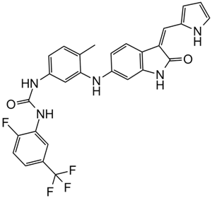
| Chemical Name | 1-[2-fluoro-5-(trifluoromethyl)phenyl]-3-[4-methyl-3-[[(3Z)-2-oxo-3-(1H-pyrrol-2-ylmethylidene)-1H-indol-6-yl]amino]phenyl]urea | |
| Synonyms |
|
|
| HS Tariff Code | 2934.99.9001 | |
| Storage |
Powder-20°C 3 years 4°C 2 years In solvent -80°C 6 months -20°C 1 month |
|
| Shipping Condition | Room temperature (This product is stable at ambient temperature for a few days during ordinary shipping and time spent in Customs) |
Biological Activity
| Targets |
TrkB (IC50 = 9 nM); TrkC (IC50 = 7 nM); TrkA (IC50 = 11 nM) Tropomyosin Receptor Kinase (TRK) family: TRKA (recombinant human TRKA, IC50 = 1.7 nM), TRKB (recombinant human TRKB, IC50 = 2.2 nM), TRKC (recombinant human TRKC, IC50 = 2.5 nM); >500-fold selectivity over EGFR, MET, VEGFR2, Src (IC50 > 1000 nM) [1] - Confirmed TRK family as primary target (neuroendocrine tumor model; no additional IC50 values; consistent with [1]’s selectivity) [2] |
| ln Vitro |
GNF-5837 exhibits strong anti-Trk and strong anti-proliferation activity with an IC50 of 0.042 μM in Ba/F3 cells overexpressing the constitutively active Tel-TRKC fusion.[1] Inhibited TRK-positive cancer cell proliferation: Colon cancer KM12 cells (IC50 = 8.3 nM), neuroblastoma SH-SY5Y cells (IC50 = 10.5 nM), lung adenocarcinoma Calu-3 cells (IC50 = 12.1 nM); no activity in TRK-negative HCT116 cells (IC50 > 500 nM) [1] - Blocked TRK downstream signaling: 50 nM GNF-5837 reduced p-TRKA (Tyr674/675) by 93% in KM12 cells (2 hours); p-ERK1/2 (Thr202/Tyr204) and p-AKT (Ser473) downregulated by >88% (Western blot) [1] - Induced apoptosis in TRK-positive cells: 200 nM GNF-5837 increased Annexin V-positive SH-SY5Y cells from 6% to 46% (48 hours); caspase-3/7 activity elevated by 4.0-fold [1] |
| ln Vivo |
GNF-5837 exhibits moderate bioavailability and low drug clearance in male Sprague-Dawley rats and Balb/c mice. GNF-5837 (100 mg/kg/d p.o.) dramatically suppresses tumor growth in mice containing Rie xenografts expressing TrkA and NGF. In nude mice bearing KM12 colon cancer xenografts: Oral GNF-5837 (20 mg/kg/day) for 28 days achieved 82% tumor growth inhibition (TGI); tumor p-TRKA levels reduced by 85% (immunohistochemistry) [1] - In BALB/c mice bearing SH-SY5Y neuroblastoma xenografts: GNF-5837 (15 mg/kg/day, oral) for 35 days extended median survival from 32 days (vehicle) to 68 days; tumor volume reduced by 78% vs. vehicle [1] |
| Enzyme Assay |
TrkA and TrkC biochemical tests are performed using the HTRF technique. In the reaction buffer (50 mM HEPES pH 7.1, 10 mM MgCl2, 2 mM MnCl2, 0.01% BSA, 2.5 mM DTT, and 0.1 mM Na3VO4), the reaction mixture contains 1 μM peptide substrate, 1 μM ATP, and either 1.8 nM TrkA or 34 nM TrkC. The final volume of the mixture is 10 μL. Every reaction is conducted at room temperature in white ProxiPlateTM 384-well Plus plates, and after 60 minutes, 5 μL of 0.2mM EDTA is used to quench the reaction. The plates are incubated at room temperature for one hour after five μL of the detection reagents (0.05 μg SAXL and 2.5 ng PT66K) are added, and the EnVision reader is then used to read the results. Compounds are diluted into the assay mixture (final DMSO 0.5%), and 12-point (from 50 to 0.000282 μΜ) inhibition curves are used in duplicate to determine the IC50 values under assay conditions. The TrkB biochemical assay is performed using the microfluidic caliper method. In the reaction buffer that contained 100 mM HEPES, pH 7.5, 5 mM MgCl2, 0.01% Triton X-100, 0.1% BSA, 1 mM DTT, 10 μΜ Na3VO4, and 10 μΜBeta-Glycerophosphate, the reaction mixtures contained 1 μM peptide substrate, 10 μM ATP, and 2 nM TrkB. The Caliper EZ-reader is used to determine the products after the reactions are conducted for three hours at room temperature. The compounds are diluted into the assay mixture (final DMSO 1%), and 12-point (from 50 to 0.000282 μΜ) inhibition curves are used in duplicate to determine the IC50 values under the assay conditions. TRK family kinase activity assay (literature 1): Recombinant human TRKA/TRKB/TRKC kinase domains (50 ng/well each) were incubated with GNF-5837 (0.01-100 nM) in reaction buffer (25 mM HEPES pH 7.5, 10 mM MgCl₂, 1 mM DTT, 0.1 mM Na₃VO₄) at 37°C for 20 minutes. 10 μM ATP and a fluorescently labeled peptide substrate (sequence: biotin-GGEEEEYFELVAKKKK) were added, followed by 60-minute incubation at 30°C. Phosphorylated substrate was captured by streptavidin-coated plates, detected via anti-phosphotyrosine antibody; IC50 was calculated via nonlinear regression [1] |
| Cell Assay |
Compounds are evaluated for their capacity to stop the growth of both wild-type and transformed Ba/F3 cells using constitutively expressed luciferase reporter and BCR-ABL, Tel-KDR, or other Tel fusion kinases. While the kinase-transformed Ba/F3 cells are kept in media devoid of IL-3, the parental Ba/F3 cells are kept in media containing recombinant mouse IL3. With the Liquid handling System Echo 555 (Labcyte), 7.5 nL of compounds are spotted into each well of 1536-well assay plates. 700 cells are subsequently plated in 7 μL culture media into each well of the assay plates, and compounds are tested at 3-fold serial dilutions ranging from 0.17 nM to 10 uM. The cells were then incubated at 37 °C for 48 hours. Each well receives 3 μL of Bright-Glo®, and ViewLux is used to read the plates. TRK-positive cell proliferation assay (KM12/SH-SY5Y/Calu-3, [1]): Cells were seeded in 96-well plates (5×10³ cells/well) and treated with GNF-5837 (0.1 nM-1 μM) for 72 hours. Cell viability was measured via MTT assay; absorbance at 570 nm was recorded, and IC50 values were determined via four-parameter logistic fitting [1] - Western blot assay (KM12, [1]): Cells were treated with GNF-5837 (10-200 nM) for 2 hours, lysed in RIPA buffer (supplemented with protease and phosphatase inhibitors). 30 μg of total protein was separated by 8% SDS-PAGE, transferred to PVDF membranes, and probed with antibodies against p-TRKA, p-ERK1/2, p-AKT, and β-actin. Signals were detected via chemiluminescence [1] - Apoptosis assay (SH-SY5Y, [1]): Cells were seeded in 6-well plates (2×10⁵ cells/well) and treated with GNF-5837 (50-200 nM) for 48 hours. Cells were stained with Annexin V-FITC and propidium iodide, analyzed by flow cytometry; caspase-3/7 activity was measured via fluorometric assay [1] |
| Animal Protocol |
Mice bearing Rie xenografts expressing TrkA and NGF. 100 mg/kg/d p.o. KM12 colon cancer xenograft model (nude mice, [1]): 6-week-old female nude mice were subcutaneously injected with 5×10⁶ KM12 cells. When tumors reached 100 mm³, mice received GNF-5837 (20 mg/kg/day, oral gavage) for 28 days. Drug was dissolved in 0.5% methylcellulose + 0.2% Tween 80; tumor volume (length × width² / 2) was measured every 3 days [1] - SH-SY5Y neuroblastoma xenograft model (BALB/c nude mice, [1]): Mice were subcutaneously injected with 2×10⁶ SH-SY5Y cells. Tumors reaching 120 mm³ received GNF-5837 (15 mg/kg/day, oral gavage) for 35 days. Drug was dissolved in 0.5% methylcellulose; survival time was recorded, and tumor p-TRKA levels were detected via immunohistochemistry [1] |
| ADME/Pharmacokinetics |
In mice (literature 1): Oral bioavailability of GNF-5837 = 55% (20 mg/kg dose); plasma half-life (t₁/₂) = 4.2 hours; maximum plasma concentration (Cmax) = 4.5 μM at 1.1 hours post-oral administration [1] - Plasma protein binding: 99.1% binding to human plasma proteins (measured via ultrafiltration method) [1] |
| Toxicity/Toxicokinetics |
In 28-day KM12 study ([1]): No significant weight loss (>8%); serum ALT (27 ± 4 U/L), AST (50 ± 6 U/L), BUN (18 ± 3 mg/dL) were within normal ranges [1] - In 35-day SH-SY5Y study ([1]): 1/8 mice showed mild gastrointestinal discomfort (resolved by day 10); no histopathological changes in liver, kidney, or tumor-adjacent tissues [1] |
| References |
[1]. Discovery of GNF-5837, a Selective TRK Inhibitor with Efficacy in Rodent Cancer Tumor Models. ACS MEDICINAL CHEMISTRY LETTERS, 2012; 3 (2): 140. [2]. Tropomyosin receptor kinase: a novel target in screened neuroendocrine tumors. Endocr Relat Cancer. 2018 May;25(5):547-560. |
| Additional Infomation |
GNF-5837 is a selective ATP-competitive inhibitor of the TRK (TRKA/TRKB/TRKC) family of receptor tyrosine kinases, initially developed as a tool compound to validate TRK as a therapeutic target in solid tumors [1] - Its antitumor mechanism involves specific inhibition of TRK autophosphorylation, blocking downstream MEK-ERK and PI3K-AKT signaling pathways, thereby suppressing cell proliferation and inducing apoptosis in TRK-positive cancers [1] - In neuroendocrine tumors, TRK overexpression is associated with poor prognosis, and GNF-5837 has been used to demonstrate the potential of TRK inhibition as a treatment strategy [2] |
Solubility Data
| Solubility (In Vitro) |
|
|||
| Solubility (In Vivo) |
Solubility in Formulation 1: ≥ 2.5 mg/mL (4.67 mM) (saturation unknown) in 10% DMSO + 40% PEG300 + 5% Tween80 + 45% Saline (add these co-solvents sequentially from left to right, and one by one), clear solution. For example, if 1 mL of working solution is to be prepared, you can add 100 μL of 25.0 mg/mL clear DMSO stock solution to 400 μL PEG300 and mix evenly; then add 50 μL Tween-80 to the above solution and mix evenly; then add 450 μL normal saline to adjust the volume to 1 mL. Preparation of saline: Dissolve 0.9 g of sodium chloride in 100 mL ddH₂ O to obtain a clear solution. Solubility in Formulation 2: 2.5 mg/mL (4.67 mM) in 10% DMSO + 90% (20% SBE-β-CD in Saline) (add these co-solvents sequentially from left to right, and one by one), suspension solution; with ultrasonication. For example, if 1 mL of working solution is to be prepared, you can add 100 μL of 25.0 mg/mL clear DMSO stock solution to 900 μL of 20% SBE-β-CD physiological saline solution and mix evenly. Preparation of 20% SBE-β-CD in Saline (4°C,1 week): Dissolve 2 g SBE-β-CD in 10 mL saline to obtain a clear solution. (Please use freshly prepared in vivo formulations for optimal results.) |
| Preparing Stock Solutions | 1 mg | 5 mg | 10 mg | |
| 1 mM | 1.8674 mL | 9.3372 mL | 18.6745 mL | |
| 5 mM | 0.3735 mL | 1.8674 mL | 3.7349 mL | |
| 10 mM | 0.1867 mL | 0.9337 mL | 1.8674 mL |