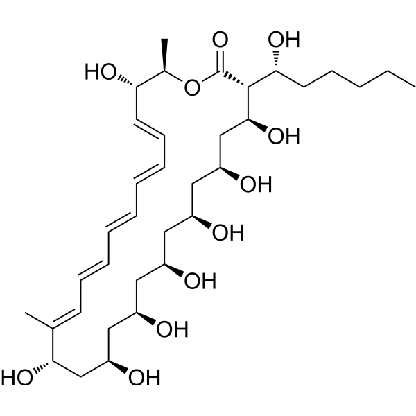
Physicochemical Properties
| Molecular Formula | C35H58O11 |
| Molecular Weight | 654.82842 |
| Exact Mass | 654.397 |
| CAS # | 480-49-9 |
| PubChem CID | 5351457 |
| Appearance | Light yellow to yellow solid powder |
| Density | 1.2±0.1 g/cm3 |
| Boiling Point | 913.9±65.0 °C at 760 mmHg |
| Melting Point | 163-180°C |
| Flash Point | 279.3±27.8 °C |
| Vapour Pressure | 0.0±0.6 mmHg at 25°C |
| Index of Refraction | 1.536 |
| LogP | -0.41 |
| Hydrogen Bond Donor Count | 9 |
| Hydrogen Bond Acceptor Count | 11 |
| Rotatable Bond Count | 5 |
| Heavy Atom Count | 46 |
| Complexity | 991 |
| Defined Atom Stereocenter Count | 0 |
| SMILES | CCCCCC(C1C(CC(CC(CC(CC(CC(CC(/C(=C\C=C/C=C/C=C/C=C/C(C(OC1=O)C)O)/C)O)O)O)O)O)O)O)O |
| InChi Key | IMQSIXYSKPIGPD-JUDYZCJOSA-N |
| InChi Code | InChI=1S/C35H58O11/c1-4-5-11-16-31(42)34-33(44)22-29(40)20-27(38)18-25(36)17-26(37)19-28(39)21-32(43)23(2)14-12-9-7-6-8-10-13-15-30(41)24(3)46-35(34)45/h6-10,12-15,24-34,36-44H,4-5,11,16-22H2,1-3H3/b7-6+,10-8+,12-9-,15-13+,23-14- |
| Chemical Name | (17Z,19Z,21E,23E,25E)-4,6,8,10,12,14,16,27-octahydroxy-3-(1-hydroxyhexyl)-17,28-dimethyl-1-oxacyclooctacosa-17,19,21,23,25-pentaen-2-one |
| HS Tariff Code | 2934.99.9001 |
| Storage |
Powder-20°C 3 years 4°C 2 years In solvent -80°C 6 months -20°C 1 month |
| Shipping Condition | Room temperature (This product is stable at ambient temperature for a few days during ordinary shipping and time spent in Customs) |
Biological Activity
| Targets |
- Filipin III targets unesterified cholesterol (in cell membranes and lysosomes, acting as a specific fluorescent probe for binding); it also exhibits antifungal activity by interacting with fungal membrane cholesterol. [1][4][5] |
| ln Vitro |
Use the TBS-T carrier Filipin III working solution for cell staining. 50 μg/mL of Filipin III was applied to NCI-60 cancer cells while they were in the dark. Use TBS-T to wash the cells after one hour after viral loading. After that, the coverslips were removed and put in a mounting media that contained water. Using the particle size algorithm in the MetaXpress image analysis software (Molecular Devices Corp., Sunnyvale, CA), freezing levels were quantified while images were being captured with an Olympus Provis fluorescence microscope that had a 40X oil objective [2 Huang Y, et al. assessed the migration in relation to TSPAN-GFP's cholesterol enrichment. TSPAN4-GFP-expressing NRK cells were stabilized at 37 °C using 2 μg/mL Filipin III. Following a 15-minute treatment period, TSPAN4-GFP intensity on migrasomes (ItM), filipin III intensity on RFs (IpR), and filipin III intensity on migrasomes (IpM) were measured using confocal microscopy Z-stack images. In order to achieve staining findings, Fibroblast was subjected to 25 μg/ml Filipin III and incubated in the dark for 45 minutes (20°C) as described by Kwiatkowska K, et al. [4]. Tissue staining: Filipin has a strong affinity for cholesterol, but it photobleaches quickly and only produces adapted natural fluorescence in response to brief excitations, making it challenging to detect with confocal panels [5]. Reid PC et al. identified cholesterol buildup in neurons using Filipin III. For two hours, NPC cerebellar tissue was stained with Filipin III (125 μg/ml) while being shielded from light from the front and bottom. Wash the slides together with 0.5 M Tris in clear FBS, then use fluorescent stains for the counterstain. Day 22 saw the development of cholesterol buildup in Purkinje neuron cells. Alternatively, NPC images were stained with Filipin III. On day 22, most neurons showed signs of cholesterol accumulation in the axon hillock area [5]. Novak A et al. employed Filipin III to identify cholesterol accumulation in malignancies. To make a working solution containing 50 μg/mL, dissolve Filipin III in dimethyl sub-solution/PBS (1:4). And place it in front of the tissue slice in the following image. half an hour. The loaded slides were rinsed, and then an aqueous fluorescent mounting media was applied to seal them for observation [6]. - Antifungal Activity: Filipin III, produced by Streptomyces filipinensis , inhibited the growth of Candida albicans (a pathogenic fungus). At concentrations of 5–10 μg/mL, it significantly suppressed Candida albicans proliferation; 10 μg/mL reduced fungal colony formation by ~60% compared to the control group. [1] - Cholesterol Visualization in Niemann-Pick Type C (NPC) Fibroblasts: NPC fibroblasts (with abnormal lysosomal cholesterol accumulation) were fixed, permeabilized, and stained with Filipin III (50 μg/mL). Fluorescence microscopy showed bright blue fluorescence (excitation: 355 nm; emission: 460 nm) in lysosomes, indicating specific binding of Filipin III to accumulated cholesterol. Normal fibroblasts showed minimal fluorescence. [4] - Cholesterol Staining in NPC1 Neuronal Cells: Primary neuronal cells from NPC1 mice (with cholesterol metabolism defects) were stained with Filipin III (25 μg/mL). Intense blue fluorescence was observed in the perinuclear region of NPC1 neurons, reflecting early cholesterol accumulation; wild-type neuronal cells showed negligible fluorescence. [5] |
| ln Vivo |
- Cholesterol Detection in NPC1 Mouse Brain: 1. Coronal sections (10 μm thick) of brains from NPC1 mice (6–8 weeks old) and wild-type mice were prepared. [5] 2. Sections were stained with Filipin III (30 μg/mL) after fixation and permeabilization. [5] 3. Fluorescence microscopy revealed strong blue fluorescence in neurons of the cerebral cortex and hippocampus of NPC1 mice, indicating significant cholesterol accumulation; wild-type mouse brain sections showed weak, diffuse fluorescence. [5] |
| Cell Assay |
- Cholesterol Staining Assay in NPC Fibroblasts: 1. NPC fibroblasts were seeded in 24-well plates (1×10⁴ cells/well) and cultured in DMEM medium (10% FBS) at 37°C (5% CO₂) for 48 hours until confluent. [4] 2. Cells were fixed with 4% paraformaldehyde for 15 minutes at room temperature, then permeabilized with 0.1% Triton X-100 for 10 minutes. [4] 3. Cells were incubated with Filipin III (50 μg/mL) in PBS for 30 minutes at 37°C, then washed 3 times with PBS to remove unbound probe. [4] 4. Fluorescence microscopy was used to image and analyze cholesterol distribution via blue fluorescence signals. [4] - Cholesterol Staining Assay in Primary Neuronal Cells: 1. Primary neurons were isolated from the cerebral cortex of NPC1 and wild-type mice, seeded on poly-L-lysine-coated coverslips (5×10³ cells/coverslip), and cultured in neurobasal medium (B27 supplement) for 7 days. [5] 2. Cells were fixed with 4% paraformaldehyde for 20 minutes, permeabilized with 0.2% Triton X-100 for 15 minutes, and blocked with 1% BSA for 30 minutes. [5] 3. Cells were incubated with Filipin III (25 μg/mL) in PBS for 45 minutes at 37°C, followed by 3 PBS washes. [5] 4. Fluorescence intensity was quantified using image analysis software to compare cholesterol levels between NPC1 and wild-type neurons. [5] |
| Animal Protocol |
- NPC1 Mouse Brain Tissue Preparation and Staining: 1. Animal preparation: NPC1 mice (6–8 weeks old, male) and wild-type C57BL/6 mice (same age/gender) were acclimated for 1 week (free access to food/water, 25°C, 12h light/dark cycle). [5] 2. Perfusion and fixation: Mice were anesthetized with sodium pentobarbital (intraperitoneal injection), transcardially perfused with cold PBS and 4% paraformaldehyde. Brains were removed and post-fixed in 4% paraformaldehyde for 24 hours, then transferred to 30% sucrose for cryoprotection. [5] 3. Sectioning: Brains were embedded in OCT compound, and 10 μm-thick coronal sections were cut with a cryostat and mounted on glass slides. [5] 4. Staining: Sections were incubated with Filipin III (30 μg/mL) in PBS for 1 hour at room temperature, washed 3 times with PBS, and mounted with anti-fade medium for fluorescence microscopy. [5] |
| References |
[1]. Functional analysis of filipin tailoring genes from Streptomyces filipinensis reveals alternative routes in filipin III biosynthesis and yields bioactive derivatives. Microb Cell Fact. 2015 Aug 7;14:114. [2]. Statin-induced mevalonate pathway inhibition attenuates the growth of mesenchymal-like cancer cells that lack functional E-cadherin mediated cell cohesion. Sci Rep. 2014 Dec 23;4:7593. [3]. Migrasome formation is mediated by assembly of micron-scale tetraspanin macrodomains. Nat Cell Biol. 2019 Aug;21(8):991-1002. [4]. Visualization of cholesterol deposits in lysosomes of Niemann-Pick type C fibroblasts using recombinant perfringolysin O. Orphanet J Rare Dis. 2014 Apr 28;9:64. [5]. A novel cholesterol stain reveals early neuronal cholesterol accumulation in the Niemann-Pick type C1 mouse brain. J Lipid Res. 2004 Mar;45(3):582-91. [6]. Role of the membrane cholesterol-glycosphingolipid complex as a ‘transistor’to regulate GSL receptor function and signaling of both lipids. bioRxiv, 2017: 137612. |
| Additional Infomation |
A complex of polyene antibiotics obtained from Streptomyces filipinensis. Filipin III alters membrane function by interfering with membrane sterols, inhibits mitochondrial respiration, and is proposed as an antifungal agent. Filipins I, II, and IV are less important. - Natural Source and Structure: Filipin III is a polyene macrolide compound biosynthesized by Streptomyces filipinensis . Its structure contains conjugated double bonds, which enable fluorescence emission and specific binding to cholesterol. [1] - Cholesterol-Binding Mechanism: Filipin III binds to unesterified cholesterol via hydrophobic interactions with the steroid core of cholesterol. This binding induces blue fluorescence, making it a gold standard probe for visualizing intracellular cholesterol distribution. [4][5] - Research Applications: Filipin III is widely used in studying cholesterol metabolism disorders (e.g., Niemann-Pick Type C disease) to detect abnormal cholesterol accumulation in cells and tissues, aiding disease diagnosis and mechanism research. [4][5] |
Solubility Data
| Solubility (In Vitro) | May dissolve in DMSO (in most cases), if not, try other solvents such as H2O, Ethanol, or DMF with a minute amount of products to avoid loss of samples |
| Solubility (In Vivo) |
Note: Listed below are some common formulations that may be used to formulate products with low water solubility (e.g. < 1 mg/mL), you may test these formulations using a minute amount of products to avoid loss of samples. Injection Formulations (e.g. IP/IV/IM/SC) Injection Formulation 1: DMSO : Tween 80: Saline = 10 : 5 : 85 (i.e. 100 μL DMSO stock solution → 50 μL Tween 80 → 850 μL Saline) *Preparation of saline: Dissolve 0.9 g of sodium chloride in 100 mL ddH ₂ O to obtain a clear solution. Injection Formulation 2: DMSO : PEG300 :Tween 80 : Saline = 10 : 40 : 5 : 45 (i.e. 100 μL DMSO → 400 μLPEG300 → 50 μL Tween 80 → 450 μL Saline) Injection Formulation 3: DMSO : Corn oil = 10 : 90 (i.e. 100 μL DMSO → 900 μL Corn oil) Example: Take the Injection Formulation 3 (DMSO : Corn oil = 10 : 90) as an example, if 1 mL of 2.5 mg/mL working solution is to be prepared, you can take 100 μL 25 mg/mL DMSO stock solution and add to 900 μL corn oil, mix well to obtain a clear or suspension solution (2.5 mg/mL, ready for use in animals). Injection Formulation 4: DMSO : 20% SBE-β-CD in saline = 10 : 90 [i.e. 100 μL DMSO → 900 μL (20% SBE-β-CD in saline)] *Preparation of 20% SBE-β-CD in Saline (4°C,1 week): Dissolve 2 g SBE-β-CD in 10 mL saline to obtain a clear solution. Injection Formulation 5: 2-Hydroxypropyl-β-cyclodextrin : Saline = 50 : 50 (i.e. 500 μL 2-Hydroxypropyl-β-cyclodextrin → 500 μL Saline) Injection Formulation 6: DMSO : PEG300 : castor oil : Saline = 5 : 10 : 20 : 65 (i.e. 50 μL DMSO → 100 μLPEG300 → 200 μL castor oil → 650 μL Saline) Injection Formulation 7: Ethanol : Cremophor : Saline = 10: 10 : 80 (i.e. 100 μL Ethanol → 100 μL Cremophor → 800 μL Saline) Injection Formulation 8: Dissolve in Cremophor/Ethanol (50 : 50), then diluted by Saline Injection Formulation 9: EtOH : Corn oil = 10 : 90 (i.e. 100 μL EtOH → 900 μL Corn oil) Injection Formulation 10: EtOH : PEG300:Tween 80 : Saline = 10 : 40 : 5 : 45 (i.e. 100 μL EtOH → 400 μLPEG300 → 50 μL Tween 80 → 450 μL Saline) Oral Formulations Oral Formulation 1: Suspend in 0.5% CMC Na (carboxymethylcellulose sodium) Oral Formulation 2: Suspend in 0.5% Carboxymethyl cellulose Example: Take the Oral Formulation 1 (Suspend in 0.5% CMC Na) as an example, if 100 mL of 2.5 mg/mL working solution is to be prepared, you can first prepare 0.5% CMC Na solution by measuring 0.5 g CMC Na and dissolve it in 100 mL ddH2O to obtain a clear solution; then add 250 mg of the product to 100 mL 0.5% CMC Na solution, to make the suspension solution (2.5 mg/mL, ready for use in animals). Oral Formulation 3: Dissolved in PEG400 Oral Formulation 4: Suspend in 0.2% Carboxymethyl cellulose Oral Formulation 5: Dissolve in 0.25% Tween 80 and 0.5% Carboxymethyl cellulose Oral Formulation 6: Mixing with food powders Note: Please be aware that the above formulations are for reference only. InvivoChem strongly recommends customers to read literature methods/protocols carefully before determining which formulation you should use for in vivo studies, as different compounds have different solubility properties and have to be formulated differently. (Please use freshly prepared in vivo formulations for optimal results.) |
| Preparing Stock Solutions | 1 mg | 5 mg | 10 mg | |
| 1 mM | 1.5271 mL | 7.6356 mL | 15.2711 mL | |
| 5 mM | 0.3054 mL | 1.5271 mL | 3.0542 mL | |
| 10 mM | 0.1527 mL | 0.7636 mL | 1.5271 mL |