Physicochemical Properties
| Molecular Formula | C23H25N7O2 |
| Molecular Weight | 431.490303754807 |
| Exact Mass | 431.20697307 |
| CAS # | 2304799-09-3 |
| Related CAS # | 2304799-09-3 |
| PubChem CID | 138454786 |
| Appearance | White to off-white solid |
| Hydrogen Bond Donor Count | 3 |
| Hydrogen Bond Acceptor Count | 6 |
| Rotatable Bond Count | 6 |
| Heavy Atom Count | 32 |
| Complexity | 661 |
| Defined Atom Stereocenter Count | 0 |
| InChi Key | XIMGYPHSYIMHAW-UHFFFAOYSA-N |
| InChi Code | InChI=1S/C23H25N7O2/c1-5-10-30-12-16(19-20(24)25-13-26-21(19)30)14-6-8-15(9-7-14)27-22(31)28-18-11-17(32-29-18)23(2,3)4/h5-9,11-13H,1,10H2,2-4H3,(H2,24,25,26)(H2,27,28,29,31) |
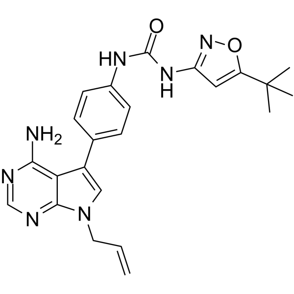
| Chemical Name | 1-[4-(4-amino-7-prop-2-enylpyrrolo[2,3-d]pyrimidin-5-yl)phenyl]-3-(5-tert-butyl-1,2-oxazol-3-yl)urea |
| Synonyms | FLT3-IN-4; FLT3-IN4; FLT3-IN 4 |
| HS Tariff Code | 2934.99.9001 |
| Storage |
Powder-20°C 3 years 4°C 2 years In solvent -80°C 6 months -20°C 1 month |
| Shipping Condition | Room temperature (This product is stable at ambient temperature for a few days during ordinary shipping and time spent in Customs) |
Biological Activity
| Targets |
FLT3-IN-4 targets Fms-like Tyrosine Receptor Kinase 3 (FLT3), including FLT3 wild-type (WT) with an IC50 of 0.5 nM, FLT3 internal tandem duplication (ITD) mutant with an IC50 of 0.3 nM, and FLT3 D835Y mutant with an IC50 of 0.4 nM[1] |
| ln Vitro |
FLT3-IN-4 (Compound 9u) exhibits strong inhibitory effects on MV4-11 and Molm-13 cells, with IC50 values of 0.089±0.001 nM and 0.022±0.003 nM, respectively[1]. FLT3-IN-4 exhibited potent antiproliferative activity against FLT3-driven leukemia cell lines: MV4-11 (FLT3 ITD) with an IC50 of 1.2 nM, MOLM-13 (FLT3 ITD) with an IC50 of 1.5 nM, and THP-1 (FLT3 WT) with an IC50 of 4.8 nM[1] - Western blot analysis showed that FLT3-IN-4 dose-dependently inhibited the phosphorylation of FLT3 (p-FLT3) and its downstream signaling molecules, including STAT5 (p-STAT5), ERK1/2 (p-ERK1/2), and AKT (p-AKT), in MV4-11 cells at concentrations ranging from 0.1 to 10 nM[1] - The compound induced apoptosis in MV4-11 cells, as evidenced by increased annexin V-positive cells and cleavage of caspase-3 and PARP after 24 hours of treatment at 1 nM[1] - FLT3-IN-4 suppressed colony formation of MV4-11 cells in soft agar assay, with a 50% inhibition at 0.8 nM[1] |
| ln Vivo |
In the MV4-11 (FLT3 ITD) xenograft mouse model, oral administration of FLT3-IN-4 at 30 mg/kg once daily for 14 days resulted in a tumor growth inhibition (TGI) rate of 85% compared to the vehicle control group[1] - Western blot analysis of tumor tissues from treated mice showed reduced p-FLT3, p-STAT5, and p-ERK1/2 levels, confirming target inhibition in vivo[1] - No significant body weight loss or obvious toxicological signs were observed in mice during the treatment period[1] |
| Enzyme Assay |
Kinase activity assay was performed by incubating FLT3-IN-4 with recombinant FLT3 kinase domain (WT, ITD, or D835Y mutant), ATP (at a concentration near the Km value of FLT3), and a specific peptide substrate. After incubation at 37°C for 60 minutes, the reaction was terminated, and the amount of phosphorylated substrate was detected using a fluorescence-based detection system. The IC50 value was calculated by plotting the inhibition rate against the compound concentration[1] |
| Cell Assay |
Antiproliferation assay: Leukemia cells (MV4-11, MOLM-13, THP-1) were seeded in 96-well plates at a density of 5×10³ cells per well and incubated overnight. FLT3-IN-4 was added at serial concentrations (0.001–100 nM), and the cells were cultured for 72 hours. Cell viability was measured using a colorimetric assay based on the reduction of a tetrazolium salt, and the IC50 values were calculated using GraphPad Prism software[1] - Western blot assay: MV4-11 cells were treated with FLT3-IN-4 at different concentrations (0.1, 1, 10 nM) for 4 hours. Cells were lysed in a buffer containing protease and phosphatase inhibitors, and the protein extracts were separated by SDS-PAGE. After transferring to a membrane, the blots were probed with primary antibodies against FLT3, p-FLT3, STAT5, p-STAT5, ERK1/2, p-ERK1/2, AKT, p-AKT, and β-actin (loading control), followed by incubation with a secondary antibody. The signals were detected using a chemiluminescent substrate[1] - Apoptosis assay: MV4-11 cells were treated with FLT3-IN-4 at 1 nM for 24 hours. Cells were harvested, washed with PBS, and stained with annexin V-FITC and propidium iodide (PI) according to the assay protocol. The percentage of apoptotic cells (annexin V-positive/PI-negative and annexin V-positive/PI-positive) was analyzed by flow cytometry[1] |
| Animal Protocol |
Xenograft model establishment: Female nude mice (6–8 weeks old) were subcutaneously inoculated with 5×10⁶ MV4-11 cells suspended in 100 μL of a mixture of PBS and Matrigel (1:1) into the right dorsal flank[1] - Treatment protocol: When the tumor volume reached approximately 100 mm³, mice were randomly divided into two groups (n=6 per group). The treatment group received oral administration of FLT3-IN-4 at 30 mg/kg once daily for 14 days. The vehicle control group received the same volume of the vehicle (5% DMSO, 30% PEG400, 65% normal saline)[1] - Tumor and body weight monitoring: Tumor volume was measured every 2 days using a caliper, calculated as (length × width²)/2. Body weight was recorded every 2 days to assess general toxicity[1] - Tissue collection: At the end of the treatment period, mice were euthanized, and tumor tissues were collected, frozen in liquid nitrogen, and stored at -80°C for subsequent Western blot analysis[1] |
| ADME/Pharmacokinetics |
Oral bioavailability (F) of FLT3-IN-4 in mice was 45% after a single oral dose of 10 mg/kg[1] - The plasma half-life (t1/2) was 6.2 hours, and the plasma clearance (CL) was 18 mL/min/kg[1] - The volume of distribution (Vd) was 1.2 L/kg, indicating moderate tissue distribution[1] - Peak plasma concentration (Cmax) was 890 ng/mL, achieved at 1.5 hours (Tmax) after oral administration[1] |
| Toxicity/Toxicokinetics |
In the 14-day in vivo toxicity study, oral administration of FLT3-IN-4 at 30 mg/kg did not cause significant body weight loss (weight change within ±5% compared to control)[1] - No obvious abnormalities were observed in gross pathological examination of major organs (liver, kidney, spleen, heart, lung) after treatment[1] - Plasma protein binding rate of FLT3-IN-4 was 82% in mouse plasma, measured by equilibrium dialysis[1] |
| References |
[1]. Identification of Pyrrolo[2,3- d]pyrimidine-Based Derivatives as Potent and Orally Effective Fms-like Tyrosine Receptor Kinase 3 (FLT3) Inhibitors for Treating Acute Myelogenous Leukemia. J Med Chem. 2019 Apr 25;62(8):4158-4173. |
| Additional Infomation |
FLT3-IN-4 is a pyrrolo[2,3-d]pyrimidine-based derivative designed as a potent and orally active FLT3 inhibitor for the treatment of acute myelogenous leukemia (AML)[1] - FLT3 mutations (especially ITD and D835Y) are common genetic abnormalities in AML, leading to constitutive activation of FLT3 signaling and promoting leukemic cell proliferation and survival[1] - FLT3-IN-4 showed high selectivity for FLT3 over other kinases (e.g., c-KIT, PDGFRα, EGFR) with IC50 values greater than 100 nM, reducing the potential for off-target effects[1] |
Solubility Data
| Solubility (In Vitro) | DMSO: ~25 mg/mL (~57.9 mM) |
| Solubility (In Vivo) |
Note: Listed below are some common formulations that may be used to formulate products with low water solubility (e.g. < 1 mg/mL), you may test these formulations using a minute amount of products to avoid loss of samples. Injection Formulations (e.g. IP/IV/IM/SC) Injection Formulation 1: DMSO : Tween 80: Saline = 10 : 5 : 85 (i.e. 100 μL DMSO stock solution → 50 μL Tween 80 → 850 μL Saline) *Preparation of saline: Dissolve 0.9 g of sodium chloride in 100 mL ddH ₂ O to obtain a clear solution. Injection Formulation 2: DMSO : PEG300 :Tween 80 : Saline = 10 : 40 : 5 : 45 (i.e. 100 μL DMSO → 400 μLPEG300 → 50 μL Tween 80 → 450 μL Saline) Injection Formulation 3: DMSO : Corn oil = 10 : 90 (i.e. 100 μL DMSO → 900 μL Corn oil) Example: Take the Injection Formulation 3 (DMSO : Corn oil = 10 : 90) as an example, if 1 mL of 2.5 mg/mL working solution is to be prepared, you can take 100 μL 25 mg/mL DMSO stock solution and add to 900 μL corn oil, mix well to obtain a clear or suspension solution (2.5 mg/mL, ready for use in animals). Injection Formulation 4: DMSO : 20% SBE-β-CD in saline = 10 : 90 [i.e. 100 μL DMSO → 900 μL (20% SBE-β-CD in saline)] *Preparation of 20% SBE-β-CD in Saline (4°C,1 week): Dissolve 2 g SBE-β-CD in 10 mL saline to obtain a clear solution. Injection Formulation 5: 2-Hydroxypropyl-β-cyclodextrin : Saline = 50 : 50 (i.e. 500 μL 2-Hydroxypropyl-β-cyclodextrin → 500 μL Saline) Injection Formulation 6: DMSO : PEG300 : castor oil : Saline = 5 : 10 : 20 : 65 (i.e. 50 μL DMSO → 100 μLPEG300 → 200 μL castor oil → 650 μL Saline) Injection Formulation 7: Ethanol : Cremophor : Saline = 10: 10 : 80 (i.e. 100 μL Ethanol → 100 μL Cremophor → 800 μL Saline) Injection Formulation 8: Dissolve in Cremophor/Ethanol (50 : 50), then diluted by Saline Injection Formulation 9: EtOH : Corn oil = 10 : 90 (i.e. 100 μL EtOH → 900 μL Corn oil) Injection Formulation 10: EtOH : PEG300:Tween 80 : Saline = 10 : 40 : 5 : 45 (i.e. 100 μL EtOH → 400 μLPEG300 → 50 μL Tween 80 → 450 μL Saline) Oral Formulations Oral Formulation 1: Suspend in 0.5% CMC Na (carboxymethylcellulose sodium) Oral Formulation 2: Suspend in 0.5% Carboxymethyl cellulose Example: Take the Oral Formulation 1 (Suspend in 0.5% CMC Na) as an example, if 100 mL of 2.5 mg/mL working solution is to be prepared, you can first prepare 0.5% CMC Na solution by measuring 0.5 g CMC Na and dissolve it in 100 mL ddH2O to obtain a clear solution; then add 250 mg of the product to 100 mL 0.5% CMC Na solution, to make the suspension solution (2.5 mg/mL, ready for use in animals). Oral Formulation 3: Dissolved in PEG400 Oral Formulation 4: Suspend in 0.2% Carboxymethyl cellulose Oral Formulation 5: Dissolve in 0.25% Tween 80 and 0.5% Carboxymethyl cellulose Oral Formulation 6: Mixing with food powders Note: Please be aware that the above formulations are for reference only. InvivoChem strongly recommends customers to read literature methods/protocols carefully before determining which formulation you should use for in vivo studies, as different compounds have different solubility properties and have to be formulated differently. (Please use freshly prepared in vivo formulations for optimal results.) |
| Preparing Stock Solutions | 1 mg | 5 mg | 10 mg | |
| 1 mM | 2.3176 mL | 11.5878 mL | 23.1755 mL | |
| 5 mM | 0.4635 mL | 2.3176 mL | 4.6351 mL | |
| 10 mM | 0.2318 mL | 1.1588 mL | 2.3176 mL |