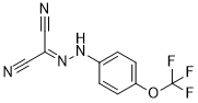
FCCP (full name: Carbonyl cyanide-4-(trifluoromethoxy)phenylhydrazone) is an ionophore and often referred to as a mitochondrial uncoupling agent. It is a mobile ion carrier that disrupts ATP synthesis by transporting hydrogen ions through a cell membrane before they can be used to provide the energy for oxidative phosphorylation. Unlike bafilomycin A1, which inhibits beta-amyloid production in cells expressing mutant but not wild-type APP, FCCP inhibited beta-amyloid production in both cell types. Moreover, the effects of FCCP were independent of alterations in total cellular APP levels or APP maturation, and the concentrations used did not alter either cellular ATP levels or cell viability.
Physicochemical Properties
| Molecular Formula | C10H5F3N4O | |
| Molecular Weight | 254.17 | |
| Exact Mass | 254.041 | |
| CAS # | 370-86-5 | |
| Related CAS # |
|
|
| PubChem CID | 3330 | |
| Appearance | Light yellow to yellow solid powder | |
| Density | 1.3±0.1 g/cm3 | |
| Boiling Point | 293.3±50.0 °C at 760 mmHg | |
| Melting Point | 174-175ºC (dec.)(lit.) | |
| Flash Point | 131.2±30.1 °C | |
| Vapour Pressure | 0.0±0.6 mmHg at 25°C | |
| Index of Refraction | 1.522 | |
| LogP | 3.65 | |
| Hydrogen Bond Donor Count | 1 | |
| Hydrogen Bond Acceptor Count | 8 | |
| Rotatable Bond Count | 3 | |
| Heavy Atom Count | 18 | |
| Complexity | 388 | |
| Defined Atom Stereocenter Count | 0 | |
| InChi Key | BMZRVOVNUMQTIN-UHFFFAOYSA-N | |
| InChi Code | InChI=1S/C10H5F3N4O/c11-10(12,13)18-9-3-1-7(2-4-9)16-17-8(5-14)6-15/h1-4,16H | |
| Chemical Name |
|
|
| Synonyms |
|
|
| HS Tariff Code | 2934.99.9001 | |
| Storage |
Powder-20°C 3 years 4°C 2 years In solvent -80°C 6 months -20°C 1 month |
|
| Shipping Condition | Room temperature (This product is stable at ambient temperature for a few days during ordinary shipping and time spent in Customs) |
Biological Activity
| ln Vitro |
In K695sw cells, FCCP (5 μM) led to a concentration-dependent reduction in Aβ and APPsβ production. The processing of wild-type APP is inhibited by FCCP. Cellular ATP levels were unaffected by FCCP at any of the amounts that were examined. Neither the secondary effects on oxidative phosphorylation nor the resultant decreased cell survival in K695sw cells affected the effect of FCCP on APP catabolism. In K695 cells, FCCP (5 μM or 500 nM), baf A1, and NH4Cl alter Tf-Tx and Tf-F cell fluorescence [1]. During brief in vitro culture, FCCP (200 nM) preserves and improves follicle integrity in cat ovarian tissue. Nonetheless, it appears that FCCP has no positive or negative effects on the cryopreservation of ovarian tissue [2]. 1. Modulation of amyloid precursor protein (APP) processing: FCCP (carbonyl cyanide p-(trifluoromethoxy)phenylhydrazone) dose-dependently altered APP processing in rat pheochromocytoma PC12 cells and human neuroblastoma SK-N-SH cells. At concentrations of 0.1-10 μM, it reduced the secretion of amyloid-β peptides (Aβ1-40 and Aβ1-42) by 30-50% (ELISA), with the most significant effect at 10 μM (Aβ1-40: 45% reduction; Aβ1-42: 50% reduction). It simultaneously increased the release of soluble APPα (sAPPα, α-secretase cleavage product) by 35% and decreased soluble APPβ (sAPPβ, β-secretase cleavage product) by 40% at 10 μM. Western blot analysis showed no change in total APP protein levels, but β-secretase (BACE1) activity was reduced by 30% and α-secretase (ADAM10) activity was increased by 25%, with no significant changes in their mRNA expression (qPCR) [1] 2. Preservation of follicle integrity during in vitro ovarian tissue culture: In domestic cat ovarian tissue fragments (1 mm³), FCCP pretreatment (0.1 μM, 37°C for 30 minutes) improved follicle survival and morphology during 7-day in vitro culture. The survival rate of follicles in the FCCP group was 75%, significantly higher than the control group (52%), and the proportion of morphologically normal follicles was 68% vs. 45% in controls. FCCP reduced reactive oxygen species (ROS) levels by 35% (DCFH-DA assay), stabilized mitochondrial membrane potential (ΔΨm, JC-1 staining), and decreased caspase-3 activity by 40% (Western blot). However, FCCP pretreatment did not improve follicle integrity after cryopreservation [2] 3. Induction of mitochondrial depolarization and activation of PINK1-Parkin pathway: FCCP (1-10 μM) dose- and time-dependently induced mitochondrial membrane potential depolarization in HeLa cells and mouse embryonic fibroblasts (MEFs), with a depolarization rate of 80% at 10 μM for 2 hours (JC-1 staining). It activated PINK1 autophosphorylation (Ser228/Ser402), increasing phosphorylated PINK1 levels by 3.5-fold (Western blot), and promoted Parkin phosphorylation at Ser65 (2.7-fold increase) and mitochondrial recruitment of Parkin (immunofluorescence co-localization with Tom20). The LC3-II/LC3-I ratio (mitophagy marker) was increased by 2.8-fold, indicating enhanced mitophagy [3] |
||
| ln Vivo |
|
||
| Enzyme Assay |
1. BACE1 activity inhibition assay: Prepare cell lysates from PC12 cells with protease inhibitors. Set up reaction mixtures containing 50 μg/mL lysate, 0.1-10 μM FCCP, and a fluorogenic BACE1 substrate (MCA-SEVNLDAEFK(Dnp)-RR-NH2) in assay buffer (50 mM NaAc, pH 4.5, 0.1% Triton X-100). Incubate at 37°C for 1 hour, then measure fluorescence intensity (excitation: 320 nm, emission: 405 nm). Calculate BACE1 activity inhibition percentage relative to vehicle control [1] 2. ADAM10 activity enhancement assay: Prepare SK-N-SH cell lysates. Reaction mixtures contain 50 μg/mL lysate, 0.1-10 μM FCCP, and a fluorogenic ADAM10 substrate (Mca-RPKPVE-Nval-WRK(Dnp)-NH2) in buffer (50 mM Tris-HCl, pH 7.5, 10 mM CaCl2, 0.1% BSA). Incubate at 37°C for 2 hours, detect fluorescence (excitation: 328 nm, emission: 393 nm), and calculate ADAM10 activity enhancement percentage [1] 3. PINK1 kinase activity assay: Purify recombinant human PINK1 protein. Set up reaction mixtures with 20 nM PINK1, 1-10 μM FCCP, 1 mM ATP, and a Parkin Ser65 peptide substrate in buffer (25 mM Tris-HCl, pH 7.5, 10 mM MgCl2, 1 mM DTT). Incubate at 30°C for 30 minutes, terminate the reaction, and detect phosphorylated peptide using a phosphorylation-specific antibody ELISA. Calculate PINK1 kinase activity relative to the no-drug control [3] |
||
| Cell Assay |
1. APP processing cell assay: Seed PC12 and SK-N-SH cells in 6-well plates (1×10⁶ cells/well) and incubate overnight. Treat with 0.1-10 μM FCCP for 24 hours. Collect supernatants to measure Aβ1-40, Aβ1-42, sAPPα, and sAPPβ by ELISA. Lyse cells for Western blot analysis of APP, BACE1, ADAM10, and GAPDH (loading control). Perform qPCR to quantify mRNA levels of APP, BACE1, and ADAM10 (no significant changes observed) [1] 2. Ovarian tissue in vitro culture assay: Cut domestic cat ovarian tissue into 1 mm³ fragments. Divide into control and FCCP pretreatment groups (0.1 μM, 37°C for 30 minutes). Culture in vitro for 7 days in medium containing 10% FBS and antibiotics, changing medium daily. Fix tissue, embed in paraffin, section, and stain with HE to evaluate follicle morphology, survival rate, and normal morphology proportion. Measure ROS levels with DCFH-DA probe (flow cytometry), mitochondrial membrane potential with JC-1 staining, and analyze apoptosis-related proteins (caspase-3, cleaved-caspase-3, Bcl-2, Bax) by Western blot [2] 3. Mitochondrial depolarization and PINK1 activation assay: Seed HeLa cells and MEFs in 6-well plates (5×10⁵ cells/well) and incubate overnight. Treat with 1-10 μM FCCP for 1-4 hours. Detect mitochondrial membrane potential by JC-1 staining (flow cytometry). Perform Western blot to analyze PINK1 (total and phosphorylated forms), Parkin (total and Ser65-phosphorylated forms), LC3, and Tom20 (mitochondrial marker). Observe co-localization of Parkin and Tom20 by immunofluorescence staining (confocal laser scanning microscopy) [3] |
||
| Animal Protocol |
|
||
| ADME/Pharmacokinetics |
Metabolism / Metabolites Organic nitriles are converted into cyanide ions through the action of cytochrome P450 enzymes in the liver. Cyanide is rapidly absorbed and distributed throughout the body. Cyanide is mainly metabolized into thiocyanate by either rhodanese or 3-mercaptopyruvate sulfur transferase. Cyanide metabolites are excreted in the urine. (L96) |
||
| Toxicity/Toxicokinetics |
Toxicity Summary Organic nitriles decompose into cyanide ions both in vivo and in vitro. Consequently the primary mechanism of toxicity for organic nitriles is their production of toxic cyanide ions or hydrogen cyanide. Cyanide is an inhibitor of cytochrome c oxidase in the fourth complex of the electron transport chain (found in the membrane of the mitochondria of eukaryotic cells). It complexes with the ferric iron atom in this enzyme. The binding of cyanide to this cytochrome prevents transport of electrons from cytochrome c oxidase to oxygen. As a result, the electron transport chain is disrupted and the cell can no longer aerobically produce ATP for energy. Tissues that mainly depend on aerobic respiration, such as the central nervous system and the heart, are particularly affected. Cyanide is also known produce some of its toxic effects by binding to catalase, glutathione peroxidase, methemoglobin, hydroxocobalamin, phosphatase, tyrosinase, ascorbic acid oxidase, xanthine oxidase, succinic dehydrogenase, and Cu/Zn superoxide dismutase. Cyanide binds to the ferric ion of methemoglobin to form inactive cyanmethemoglobin. (L97) 1. In vitro cytotoxicity in neural cells: FCCP (0.1-10 μM) showed no significant cytotoxicity to PC12 and SK-N-SH cells, with cell viability > 85% (MTT assay) after 24-hour treatment [1] 2. Toxicity in ovarian tissue: FCCP at 0.1 μM was non-toxic to cat ovarian tissue fragments, improving follicle survival. At 1 μM, it slightly reduced follicle survival rate to 60%, indicating mild toxicity at higher concentrations [2] 3. Cytotoxicity in HeLa/MEF cells: FCCP (1-10 μM) had no significant cytotoxicity to HeLa cells and MEFs, with cell viability > 80% (MTT assay) and apoptotic rate < 5% (Annexin V-FITC/PI staining) after 24-hour treatment [3] |
||
| References |
[1]. Novel effects of FCCP [carbonyl cyanide p-(trifluoromethoxy)phenylhydrazone] on amyloid precursor protein processing. J Neurochem. 1999 Apr;72(4):1457-65. [2]. Carbonyl cyanide 4-(trifluoromethoxy)phenylhydrazone (FCCP) pre-exposure ensures follicle integrity during in vitro culture of ovarian tissue but not during cryopreservation in the domestic cat model. J Assist Reprod Genet. 2016 Dec;33(12):1621-1631. Epub 2016 Sep 17. [3]. PINK1 is activated by mitochondrial membrane potential depolarization and stimulates Parkin E3 ligase activity by phosphorylating Serine 65. Open Biol. 2012 May;2(5):120080. |
||
| Additional Infomation |
Carbonyl cyanide p-trifluoromethoxyphenylhydrazone is a hydrazone that is hydrazonomalononitrile in which one of the hydrazine hydrogens is substituted by a p-trifluoromethoxyphenyl group. It has a role as an ionophore, an ATP synthase inhibitor and a geroprotector. It is a hydrazone, a nitrile, an organofluorine compound and an aromatic ether. It is functionally related to a hydrazonomalononitrile. Carbonyl cyanide p-trifluoromethoxyphenylhydrazone has been reported in Purpureocillium lilacinum and Microcoleus autumnalis with data available. Carbonyl cyanide-p-trifluoromethoxyphenylhydrazone is a chemical compound of cyanide. A proton ionophore that is commonly used as an uncoupling agent in biochemical studies. 1. Chemical and structural properties: FCCP (carbonyl cyanide p-(trifluoromethoxy)phenylhydrazone) is a synthetic protonophore uncoupler with the chemical name carbonyl cyanide 4-(trifluoromethoxy)phenylhydrazone. It is a yellow crystalline powder, soluble in DMSO (≥50 mg/mL) and ethanol (≥10 mg/mL), and slightly soluble in water [1, 2, 3] 2. Mechanism of action: FCCP acts as a proton carrier that penetrates mitochondrial membranes, uncoupling the proton gradient and inducing mitochondrial membrane potential depolarization, thereby inhibiting oxidative phosphorylation and increasing mitochondrial respiration rate. It modulates APP processing by inhibiting BACE1 and activating ADAM10, improves follicle integrity during in vitro culture by reducing ROS and inhibiting apoptosis, and activates the PINK1-Parkin pathway to promote mitophagy [1, 2, 3] 3. Research applications: A widely used tool compound for studying mitochondrial function (inducing depolarization and uncoupling). Potential research applications include neurodegenerative diseases (e.g., Alzheimer's disease, by reducing Aβ production), reproductive medicine (ovarian tissue in vitro culture), and Parkinson's disease-related mitophagy research [1, 2, 3] 4. Safety considerations: FCCP has potential cytotoxicity at high concentrations (>10 μM), which may induce cell apoptosis. It is a research tool compound with no clinical approval for therapeutic use [1, 2, 3] |
Solubility Data
| Solubility (In Vitro) |
|
|||
| Solubility (In Vivo) |
Solubility in Formulation 1: 2.5 mg/mL (9.84 mM) in 10% DMSO + 40% PEG300 + 5% Tween80 + 45% Saline (add these co-solvents sequentially from left to right, and one by one), suspension solution; with heating and sonication. For example, if 1 mL of working solution is to be prepared, you can add 100 μL of 25.0 mg/mL clear DMSO stock solution to 400 μL PEG300 and mix evenly; then add 50 μL Tween-80 to the above solution and mix evenly; then add 450 μL normal saline to adjust the volume to 1 mL. Preparation of saline: Dissolve 0.9 g of sodium chloride in 100 mL ddH₂ O to obtain a clear solution. Solubility in Formulation 2: 2.5 mg/mL (9.84 mM) in 10% DMSO + 90% (20% SBE-β-CD in Saline) (add these co-solvents sequentially from left to right, and one by one), suspension solution; with heating and sonication. For example, if 1 mL of working solution is to be prepared, you can add 100 μL of 25.0 mg/mL clear DMSO stock solution to 900 μL of 20% SBE-β-CD physiological saline solution and mix evenly. Preparation of 20% SBE-β-CD in Saline (4°C,1 week): Dissolve 2 g SBE-β-CD in 10 mL saline to obtain a clear solution. Solubility in Formulation 3: ≥ 2.5 mg/mL (9.84 mM) (saturation unknown) in 10% DMSO + 90% Corn Oil (add these co-solvents sequentially from left to right, and one by one), clear solution. For example, if 1 mL of working solution is to be prepared, you can add 100 μL of 25.0 mg/mL clear DMSO stock solution to 900 μL of corn oil and mix evenly. (Please use freshly prepared in vivo formulations for optimal results.) |
| Preparing Stock Solutions | 1 mg | 5 mg | 10 mg | |
| 1 mM | 3.9344 mL | 19.6719 mL | 39.3437 mL | |
| 5 mM | 0.7869 mL | 3.9344 mL | 7.8687 mL | |
| 10 mM | 0.3934 mL | 1.9672 mL | 3.9344 mL |