FB23-2 (also known as FB23-2; FB-23-2; FB 232) is a novel, potent and selective inhibitor of mRNA N6-methyladenosine (m6A) demethylase FTO that may have anticancer properties. It has a high level of in vivo inhibition against AML models and has an IC50 of 2.6 M for inhibiting FTO. By specifically inhibiting FTO's m6A demethylase activity, FB23-2 binds to FTO directly. Human acute myeloid leukemia (AML) cell line cells and primary blast AML cells exhibit markedly reduced proliferation and are more susceptible to differentiation/apoptosis in vitro, mimicking FTO depletion. Furthermore, xeno-transplanted mice treated with FB23-2 exhibit a marked inhibition of the development of both primary and cell-line-derived human AML cells. Collectively, our data imply that FTO is a druggable target and that inhibiting FTO with small molecules has the potential to be used to treat AML.
Physicochemical Properties
| Molecular Formula | C18H15CL2N3O3 |
| Molecular Weight | 392.2360 |
| Exact Mass | 391.05 |
| Elemental Analysis | C, 55.12; H, 3.85; Cl, 18.08; N, 10.71; O, 12.24 |
| CAS # | 2243736-45-8 |
| Related CAS # | 2243736-45-8 |
| PubChem CID | 138454779 |
| Appearance | White to light yellow solid powder |
| LogP | 4.9 |
| Hydrogen Bond Donor Count | 3 |
| Hydrogen Bond Acceptor Count | 5 |
| Rotatable Bond Count | 4 |
| Heavy Atom Count | 26 |
| Complexity | 486 |
| Defined Atom Stereocenter Count | 0 |
| InChi Key | ILHNIWOZZKIBNW-UHFFFAOYSA-N |
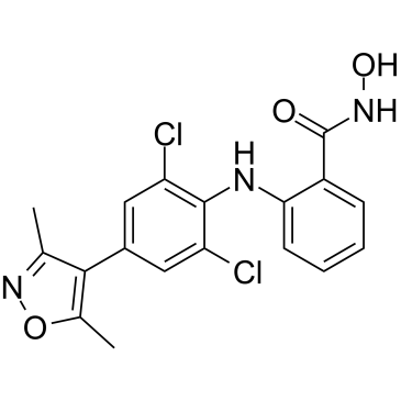
| InChi Code | InChI=1S/C18H15Cl2N3O3/c1-9-16(10(2)26-23-9)11-7-13(19)17(14(20)8-11)21-15-6-4-3-5-12(15)18(24)22-25/h3-8,21,25H,1-2H3,(H,22,24) |
| Chemical Name | 2-((2,6-dichloro-4-(3,5-dimethylisoxazol-4-yl)phenyl)amino)-N-hydroxybenzamide |
| Synonyms | FB 23-2; FB232; FB23-2; FB-23-2; FB 232; FB-232 |
| HS Tariff Code | 2934.99.9001 |
| Storage |
Powder-20°C 3 years 4°C 2 years In solvent -80°C 6 months -20°C 1 month Note: This product requires protection from light (avoid light exposure) during transportation and storage. |
| Shipping Condition | Room temperature (This product is stable at ambient temperature for a few days during ordinary shipping and time spent in Customs) |
Biological Activity
| Targets | FTO (IC50 = 2.6 μM) |
| ln Vitro |
FTO, a mRNA N6-methyladenosine (m6A) demethylase, was reported to promote leukemogenesis. Using structure-based rational design, we have developed two promising FTO inhibitors, namely FB23 and FB23-2, which directly bind to FTO and selectively inhibit FTO’s m6A demethylase activity. Mimicking FTO depletion, FB23-2 dramatically suppresses proliferation and promotes the differentiation/apoptosis of human acute myeloid leukemia (AML) cell line cells and primary blast AML cells in vitro.[1] Directly attaching to FTO, FB23-2 inhibits its m6A demethylase activity in a specific manner. Human acute myeloid leukemia (AML) cell line cells and primary blast AML cells are significantly suppressed in proliferation and are encouraged to differentiate/apoptose in vitro, simulating FTO depletion. [1] |
| ln Vivo |
In xeno-transplanted mice, FB23-2 significantly slows the development of human AML cell lines and primary cells.[1] FB23-2 suppresses leukemia progression and improves the survival of leukemic mice.[1] FB23-2 exhibits therapeutic efficacy in treating a patient-derived xeno-transplantation (PDX) AML mouse model[1] FB23-2 is safe in mice and displays a favorable pharmacokinetic profile.[1] |
| Enzyme Assay |
HPLC-based assay of the inhibition of m6A demethylation in RNA[1] In vitro ssRNA demethylations were performed with some modifications on the reported assay (Huang et al., 2015). The reactions, containing 0.25 μM FTOΔN31 or 3 μM ALKBH5ΔN66, 5 μM 15-mer ssRNA (5′-AUUGUCA(m6A)CAGCAGC-3′), 300 μΜ 2OG, 280 μΜ (NH4)2Fe(SO4)2, 2 mM L-ascorbic acid, and inhibitors at required concentrations in 50 mM Tris-HCl (pH 7.5 – 8.0), were incubated at 25 °C for 30 min. The reactions were terminated by heating for 5 min at 90 °C, and then the mixtures were subjected to digestion by nuclease P1 and alkaline phosphatase. The IC50 values were quantitated based on the inhibitory percentages of m6A demethylation in the presence of inhibitors at indicated concentrations, using nonlinear regression, dose-response fit on GraphPad Prism 5.0™. All reactions were performed in triplicate. Crystallization and structure determination of FTO/FB23 complex [1] Crystallizations were conducted with hanging-drop vapor-diffusion method at 18 °C. 8 mg/ml of FTOΔN31 protein was incubated with 5-folds FB23 and mixed with a reservoir solution containing 100 mM sodium citrate (pH 5.4), 11.5% (w/v) polyethylene glycol 3350, and 8% isopropanol. The crystals were cryo-protected using extra 20% (v/v) glycerol. Diffraction data were collected on the BL18U1 and BL17U1 beamline at the Shanghai Synchrotron Research Facility (SSRF). All X-ray data were processed using HKL2000 programs (Otwinowski and Minor, 1997), and converted to structure factors within the CCP4 program (Collaborative Computational Project, 1994). The structure was solved by molecular replacement in Phaser using the structure of FTO/MA complex (PDB code 4QKN) as the searching model. The model of structural complex FTO/FB23 was computationally refined with the program REFMAC5. Nuclear Magnetic Resonance (NMR) titration[1] Phosphate buffer (20 mM sodium phosphate (pH 7.4), 100 mM NaCl, 5% DMSO) was used for NMR data acquisition on a Bruker Avance III-600 MHz spectrometer equipped with a cryogenically cooled probe at 25 °C. Experimental samples contained 200 μM FB23 and FTO protein at 0 μM, 1 μM, 2 μM, and 3 μM, respectively. Cellular thermal shift assay (CETSA)[1] CETSA was conducted according to the protocol as previously described (Martinez Molina et al., 2013). NB4 and MONOMAC6 cells were collected and lysed in 50 mM Tris-HCl (pH 7.5), 150 mM NaCl, and 2 mM DTT. 50 μM FB23 or DMSO was added to the supernatant and incubated at 25 °C for 25 min. After denaturing at various temperatures for 5 min, samples were centrifuged, and the supernatants were analyzed by western blot. All experiments were performed in triplicate. |
| Cell Assay |
While MA9 and FLT3/NPM1 primary cells isolated from AML mice, five human AML cells (MA9.3ITD, MA9.3RAS, U937, ML2, and MV4-11), and human primary AML cells are treated with DMSO or 5 μM FB23-2 for 72 h for dot blot assay, NB4 and MONOMAC6 cells are treated with DMSO or FB23-2 at varying concentrations.[1] Cell proliferation assays 5,000 cells/well NB4, FTO KO NB4, and MONOMAC6 AML cells were seeded and treated with DMSO or FTO inhibitors for 72 hr. The cell proliferations were determined with CellTiter 96® AQueous Non-Radioactive Cell Proliferation Assay according to the manufacturer’s instructions. 10,000 cells/well human AML cells (MA9.3ITD, MA9.3RAS, U937, ML2, and MV4-11) and four primary cells from AML patients were seeded and subjected to FTO inhibitor treatment for 96 hr as indicated. 10,000 cells/well MA9 and FLT3/NPM1 primary cells isolated from AML mice and 5,000 cells/well shNS and shFTO NB4 cells were seeded and treated with FTO inhibitors for 24 hr, 48 hr, 72 hr, and 96 hr for proliferation determination.[1] Quantitation of FB23 and FB23-2 in AML cells NB4 and MONOMAC6 cells were treated with 10 μM FB23 or FB23-2 for 24 hr, respectively. Viable cells were distinguished with 0.1% trypan blue, counted and then harvested with PBS by several washings. Cells were diluted into 100 μl with 50% (v/v) water/methanol and followed by several shock freeze-thaw cycles. The supernatants were collected for analysis. The Ultimate 3000 system coupled with a TSQ Quantiva mass spectrometer was applied to determine the cellular concentration of compound FB23 and FB23-2. Analytes were separated on a XSELECt™ HSS T3 column (100 mm × 3.0 mm, 2.5 μm; Waters, USA). The mobile phases used for elution were (A) 0.1% (v/v) formic acid/water and (B) 0.1% (v/v) formic acid/acetonitrile. The mass spectrometer was operated in the negative MRM mode. Parent-to-product transitions were m/z 375.1→339.1, 375.1→298.1 for FB23, and m/z 390.3→318.0, 390.3→289.9 for FB23-2, respectively.[1] m6A dot blot assay NB4 and MONOMAC6 cells were treated with DMSO or FB23-2 at varying concentrations for 72 hr, while MA9 and FLT3/NPM1 primary cells isolated from AML mice, five human AML cell (MA9.3ITD, MA9.3RAS, U937, ML2, and MV4-11), and human primary AML cells were treated with DMSO or 5 μM FB23-2 for 72 hr for dot blot assay. Total RNA was separated with miRNeasy Mini Kit, and poly (A)+ RNA was further enriched with PolyATract mRNA isolation System IV in accordance with the manufacturer’s instructions. The RNA samples were diluted in RNA binding buffer, denatured at 65 °C for 5 min. Then one volume of 20 x SSC buffer was added into the RNA samples before dotted onto the Amersham Hybond-N+ membrane with Bio-Dot Apparatus. The RNA samples were cross-linked onto the membrane via UV irradiation. The membrane was stained with 0.02% methylene blue (MB) as loading control. After UV crosslinking and MB staining, the membrane was washed with PBST, blocked with 5% nonfat dry milk for 1 hr at room temperature and incubated with m6A antibody (1 : 2000) at 4 °C overnight. Finally, the membrane was then incubated with the HRP-conjugated goat anti-rabbit IgG and developed with Amersham ECL Prime Western Blotting Detection Reagent.[1] LC-MS/MS quantitation of m6A and m6Am in AML cells NB4 and MONOMAC6 cells were cultured with DMSO or 20 μM FB23-2 for 72 hr. mRNA was isolated in line with the dot blot assay, followed by the removal of contaminated rRNA with RiboMinus Transcriptome Isolation Kit. 300 ng mRNA was decapped with 5 units RppH with the thermopol buffer, then the products were digested by nuclease P1 for 1 hr at 42 °C. Subsequently, 1 unit of alkaline phosphatase and NH4HCO3 (100 mM) were added and incubated for another 1 hr at 37 °C. The Ultimate 3000 system coupled with a TSQ Quantiva mass spectrometer was applied to quantitate the cellular levels of A, m6A, and m6Am. Samples were centrifuged and loaded onto a XSELECt™ CSH™ C18 column (100 mm × 3.0 mm, 2.5 μm) and eluted by the gradient methanol. The parent-to-product transitions for A, m6A, and m6Am were 268.1/136.1, 282.1/150.1, and 296.2/150.1, respectively. |
| Animal Protocol |
Animal model[1] The NSGS mice were bred and subjected to the xeno-transplantation model. For the AML mouse model, 0.2 × 10⁶ MONOMAC6 cells were directly transplanted into NSGS mice via tail vein. After 10 days, FB23-2 (2 mg/kg/day) and DMSO vehicle were intraperitoneally injected into the mice for a continuous 10 days. The mice were euthanized by CO2 inhalation if they exhibited classical AML symptoms including hunched posture, paralysis, and reduced body weight. Meanwhile, the PB, spleen, and liver samples were collected for further analysis. PDX models were generated by injecting primary BM cells from AML patient Pt 2017_63 (2 X 10⁶ per mouse) into the tail veins of 6- to 8-week-old sublethally irradiated (2.5 Gy) NSGS mice (NOD.Cg-PrkdcscidIl2rgtm1WjlTg(CMV-IL3, CSF2, KITLG)1Eav/MloySzJ), which were purchased from The Jackson Laboratory. When recipient mice had 3–5% donor-derived AML cells in PB, 6 mg/kg/day FB23-2 was delivered by i.p. for 17 days, vehicle DMSO was administrated as control. Mice were weighed daily during treatment and doses were recalculated to make sure the mice received a consistent dose of 6 mg/kg/day. One day after the 17-day full treatment, mice were randomly picked up, and then PB cells were collected and analyzed for the engraftment of leukemia cells by FACS using anti-human-CD45 and anti-mouse-CD45. When the mice became moribund, BM cells were collected and analyzed for the engraftment of leukemia cells by FACS using anti-human-CD45 and anti-mouse-CD45. In addition, the LSCs population was determined as the human CD34+CD38−population. For second transplantation, the patient AML cells, collected from the spleen of primary NSGS mice which were transplanted with BM cells from AML patients and received FB23-2 or DMSO treatment, were transplanted into NSGS mice irradiated at 2.5 Gy. 8 weeks post transplantation, PB cells were collected for FACS analysis using anti-human-CD45 and anti-mouse-CD45 and the mice were continued to monitor for survival. |
| References |
[1]. Small-Molecule Targeting of Oncogenic FTO Demethylase in Acute Myeloid Leukemia. Cancer Cell. 2019 Apr 15;35(4):677-691.e10. |
Solubility Data
| Solubility (In Vitro) | DMSO: ~25 mg/mL (~63.7 mM) |
| Solubility (In Vivo) |
Solubility in Formulation 1: ≥ 2.08 mg/mL (5.30 mM) (saturation unknown) in 10% DMSO + 40% PEG300 + 5% Tween80 + 45% Saline (add these co-solvents sequentially from left to right, and one by one), clear solution. For example, if 1 mL of working solution is to be prepared, you can add 100 μL of 20.8 mg/mL clear DMSO stock solution to 400 μL PEG300 and mix evenly; then add 50 μL Tween-80 to the above solution and mix evenly; then add 450 μL normal saline to adjust the volume to 1 mL. Preparation of saline: Dissolve 0.9 g of sodium chloride in 100 mL ddH₂ O to obtain a clear solution. Solubility in Formulation 2: ≥ 2.08 mg/mL (5.30 mM) (saturation unknown) in 10% DMSO + 90% Corn Oil (add these co-solvents sequentially from left to right, and one by one), clear solution. For example, if 1 mL of working solution is to be prepared, you can add 100 μL of 20.8 mg/mL clear DMSO stock solution to 900 μL of corn oil and mix evenly. Solubility in Formulation 3: 10 mg/mL (25.49 mM) in 50% PEG300 50% Saline (add these co-solvents sequentially from left to right, and one by one), suspension solution; with ultrasonication. Preparation of saline: Dissolve 0.9 g of sodium chloride in 100 mL ddH₂ O to obtain a clear solution. (Please use freshly prepared in vivo formulations for optimal results.) |
| Preparing Stock Solutions | 1 mg | 5 mg | 10 mg | |
| 1 mM | 2.5495 mL | 12.7473 mL | 25.4946 mL | |
| 5 mM | 0.5099 mL | 2.5495 mL | 5.0989 mL | |
| 10 mM | 0.2549 mL | 1.2747 mL | 2.5495 mL |